What Causes Ptosis? Drooping Eyelid Causes Explained 2026
What causes ptosis ranges from a simple age-related stretching of the eyelid’s internal connective tissue to a life-threatening brain aneurysm compressing a cranial nerve, and identifying which cause is responsible depends on a specific set of accompanying signs that every person with a drooping eyelid should know. Ptosis is not a single disease; it is a physical sign produced by at least six distinct anatomical and physiological mechanisms.
The stakes here vary dramatically by cause. The National Eye Institute notes that while most ptosis in adults is benign and age-related, new-onset unilateral ptosis accompanied by a fixed dilated pupil and headache represents a neurological emergency with a narrow window for lifesaving intervention. That distinction is not communicated clearly enough in most publicly available health content.
This article explains every named cause of ptosis with the specific anatomical mechanism behind each one, ranks them by frequency and clinical urgency, covers how ptosis causes differ by age and sex, names the specific combinations of symptoms that require a 911 call, and walks through the diagnostic process from the first clinical examination to the final imaging or laboratory confirmation.
What Causes Ptosis
Ptosis (blepharoptosis, ICD-10: H02.4) is caused by dysfunction, weakness, or structural failure of the muscles and their nerve supply responsible for lifting the upper eyelid, with the most common cause in adults being age-related stretching of the levator aponeurosis and the most dangerous cause being compression of the oculomotor nerve by an intracranial aneurysm.
The eyelid is lifted by two muscles working in coordination. When either muscle fails, when its connective tissue attachments degrade, or when its nerve supply is disrupted, the eyelid drops to a position lower than its normal resting height. The degree of drooping ranges from 1 to 2 millimeters (barely noticeable) to complete closure of the eye.
Ptosis causes are classically divided into five anatomical categories. Each category reflects a distinct mechanism and requires a different diagnostic approach and urgency level.
| Category | Primary Mechanism | Examples | Urgency Level |
|---|---|---|---|
| Aponeurotic | Levator aponeurosis dehiscence or disinsertion | Involutional, post-surgical, contact lens-related | Elective |
| Neurogenic | Nerve supply disruption to levator or Müller’s muscle | Third nerve palsy, Horner syndrome | Variable: routine to emergency |
| Neuromuscular | Acetylcholine receptor dysfunction at neuromuscular junction | Myasthenia gravis | Urgent |
| Myogenic | Primary muscle disease of the levator | CPEO, myotonic dystrophy, congenital myopathy | Urgent to elective |
| Mechanical | Physical weight or scarring preventing eyelid elevation | Lid tumor, chalazion, scarring | Elective |
| Traumatic | Direct injury to levator muscle or aponeurosis | Blunt trauma, surgical complication | Variable |
Individual variation note: Congenital ptosis, present from birth, has a fundamentally different cause from all adult-onset categories. It typically reflects hypoplasia or fibrosis of the levator palpebrae superioris muscle during fetal development and requires early pediatric ophthalmology evaluation to prevent amblyopia (permanent vision impairment from deprivation of the visual stimulus during critical development).
Anatomy of Eyelid Elevation: the Levator Muscle and Müller’s Muscle
The upper eyelid is lifted by two distinct muscles with different nerve supplies, and understanding both is the foundation for understanding why different causes produce different types and degrees of ptosis.
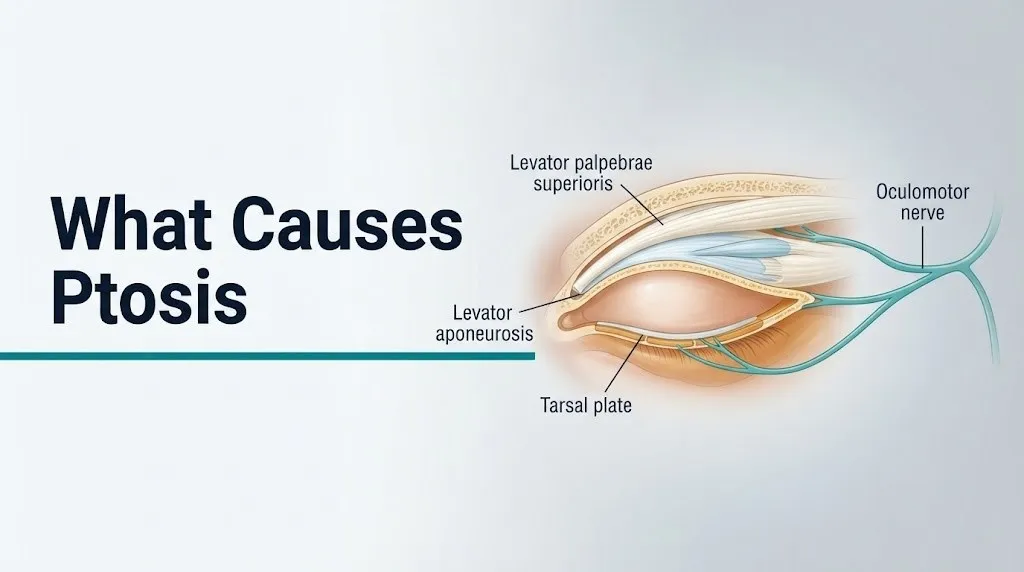
The levator palpebrae superioris is a striated voluntary muscle originating from the lesser wing of the sphenoid bone at the orbital apex. It runs anteriorly beneath the orbital roof and broadens into a wide, flat fibrous sheet called the levator aponeurosis. This aponeurosis inserts into the anterior face of the tarsal plate (the structural cartilaginous scaffold of the eyelid) and into the overlying skin, forming the visible upper eyelid crease. The levator provides approximately 12 to 14 millimeters of total eyelid elevation. It is innervated by the superior division of the oculomotor nerve (cranial nerve III).
The second eyelid elevator, Müller’s muscle (the superior tarsal muscle), is a smooth muscle lying directly behind the levator aponeurosis, connecting the inferior surface of the levator to the superior border of the tarsal plate. Unlike the voluntary levator, Müller’s muscle is innervated by postganglionic sympathetic fibers originating from the superior cervical ganglion and provides an additional 2 to 3 millimeters of eyelid elevation. It contributes to the resting tone that keeps the eyelid at its normal height even at rest.
Think of these two muscles like a two-stage pulley system on a garage door. The levator is the main spring doing most of the lifting. Müller’s muscle is the secondary tension cable keeping the door from sagging when the main spring relaxes. Cut the main spring, and the door falls completely. Reduce the tension cable, and the door drops only a few millimeters.
This two-muscle anatomy explains a clinically important distinction: third nerve palsy disrupts only the levator (the main spring) and produces severe or complete ptosis of 8 to 12 millimeters. Horner syndrome disrupts only Müller’s muscle (the tension cable) and produces a partial ptosis of just 2 to 3 millimeters. Recognizing this difference at the bedside guides the entire diagnostic workup.
Individual variation note: Eyelid crease height differs between anatomical groups. People of East Asian descent typically have a lower or absent eyelid crease due to the different level at which the levator aponeurosis inserts relative to the eyelid fat. This anatomical variation means that eyelid crease asymmetry, a standard clinical sign of aponeurotic ptosis, is less reliable in this population and should be interpreted with appropriate anatomical context.
Aponeurotic Ptosis: the Most Common Cause in Adults
Aponeurotic ptosis is the most frequent cause of drooping eyelids in adults over 40, produced by stretching, thinning, or frank tearing (dehiscence) of the levator aponeurosis where it attaches to the tarsal plate, reducing the mechanical efficiency of the levator palpebrae superioris despite the muscle itself remaining structurally normal.
The levator aponeurosis is a thin but tough fibrous sheet. Over decades of blinking (approximately 15,000 times per day), along with the mechanical stress of eyelid manipulation from contact lens insertion and removal, prior eyelid surgery, or prolonged eye rubbing, the aponeurosis gradually stretches. In many adults, the fibers physically disinsertion from the anterior tarsal plate, creating a gap between the contracting levator muscle and the structure it is meant to lift.
The clinical signature of aponeurotic ptosis is distinctive. The eyelid crease (the horizontal skin fold above the upper eyelid) is elevated or deepened compared to the other side, because the aponeurosis is pulling the skin upward even as it fails to pull the tarsus. The eyelid droops, but the crease is high. This pattern is the opposite of neurogenic or myogenic ptosis, where the crease is often in a normal or lower position.
According to a clinical review published in Survey of Ophthalmology, aponeurotic dehiscence accounts for the majority of acquired ptosis cases evaluated in oculoplastic surgical series, with long-term contact lens wear, particularly hard rigid gas-permeable lenses, identified as a documented accelerating factor in younger adults through repeated mechanical stress on the aponeurosis during lens insertion.
Individual variation note: People who have undergone cataract surgery with a lid speculum (the instrument that holds the eye open during surgery) have a well-documented elevated rate of post-operative aponeurotic ptosis. The mechanical stretching of the upper lid by the speculum during surgery can disinsert the aponeurosis from the tarsal plate. If ptosis develops within weeks of eye surgery, this is the most likely mechanism and should be discussed with the treating ophthalmologist.
Key Takeaway: Aponeurotic dehiscence is the most common cause of adult drooping eyelids, produces a characteristically elevated eyelid crease, and is frequently linked to contact lens use, prior eye surgery, or simple aging of the levator aponeurosis.
Congenital Ptosis: Causes in Newborns and Children
Congenital ptosis is present at birth or develops within the first year of life and is caused in most cases by incomplete development (dysgenesis) of the levator palpebrae superioris muscle, in which normal striated muscle fibers are replaced by fibrous or fatty tissue during fetal organogenesis.
The levator muscle forms from mesoderm during fetal development. In congenital ptosis, this developmental process is disrupted, leaving a levator muscle with reduced numbers of functional muscle fibers, replaced by fibrotic tissue that neither contracts properly nor relaxes fully. The clinical consequence of this fibrosis is a double defect: the eyelid doesn’t lift adequately on upgaze, but it also doesn’t lower fully on downgaze (called lid lag), because the fibrotic muscle can’t lengthen normally. This lid lag on downgaze is a distinguishing feature of congenital myogenic ptosis not seen in adult aponeurotic ptosis.
The most important clinical concern in congenital ptosis is amblyopia: permanent reduction in visual acuity in the affected eye from deprivation of the clear visual image during the critical period of visual cortex development (roughly birth to age 7 to 8 years). If a drooping upper lid covers the pupil, the affected eye receives a blurred or absent visual stimulus, and the visual cortex fails to develop normal connections with that eye.
The American Academy of Ophthalmology states that congenital ptosis severe enough to obstruct the visual axis requires surgical correction in the first months of life to prevent irreversible amblyopia, and that all children with unilateral congenital ptosis should be evaluated by a pediatric ophthalmologist regardless of apparent severity.
A less common but clinically important congenital variant is Marcus Gunn jaw-winking syndrome, a form of synkinesis in which the ptotic eyelid elevates involuntarily when the child chews or moves the jaw. This reflects an aberrant connection between the motor branch of the trigeminal nerve (which controls the pterygoid jaw muscles) and the levator palpebrae superioris, resulting from misdirected nerve fiber growth during fetal development.
Individual variation note: Infants born to mothers with myasthenia gravis can develop neonatal myasthenia gravis, a transient form caused by passive transfer of maternal anti-AChR antibodies across the placenta. These infants typically show ptosis, feeding difficulty, and hypotonia within the first days of life. The condition resolves within 2 to 6 weeks as maternal antibodies are cleared, but requires careful neonatal monitoring.
Neurogenic Ptosis: Third Nerve Palsy and the Oculomotor Nerve
Neurogenic ptosis caused by third nerve palsy (oculomotor nerve palsy) is the most clinically dangerous category of ptosis because one of its causes, a posterior communicating artery aneurysm, represents a neurosurgical emergency requiring immediate intervention to prevent subarachnoid hemorrhage and death.
The oculomotor nerve (cranial nerve III) exits the midbrain, passes between the posterior communicating artery and the superior cerebellar artery, travels through the cavernous sinus, and enters the orbit through the superior orbital fissure. Its superior division innervates the levator palpebrae superioris. Any lesion that damages CN III along this entire course can produce ptosis.
The specific feature that makes third nerve palsy an emergency is pupil involvement. The pupillomotor fibers (parasympathetic fibers that constrict the pupil) travel on the outer surface of CN III. An expanding aneurysm of the posterior communicating artery (PComA), as it presses against CN III from the outside, compresses these superficial pupillomotor fibers first. The result is the classic emergency triad:
- Ipsilateral complete or near-complete ptosis (levator denervated)
- Ipsilateral fixed, dilated pupil (pupillomotor fibers compressed, blocking pupillary constriction)
- Eye deviated “down and out” (superior, medial, and inferior recti denervated; only the lateral rectus and superior oblique, innervated by CN IV and VI, remain functional)
This triad, described by neurologists as a “pupil-involving third nerve palsy,” indicates an expanding PComA aneurysm until proven otherwise and requires immediate emergency brain imaging (CT angiography or MR angiography) and neurosurgical consultation.
Not all third nerve palsies are aneurysmal. In adults over 50 with diabetes or hypertension, microvascular ischemia of the CN III nerve trunk produces a “pupil-sparing” third nerve palsy: complete ptosis and ophthalmoplegia but a normally reactive pupil. The ischemia affects the inner nerve fibers (motor fibers to the extraocular muscles) while sparing the outer pupillomotor fibers. This presentation, though not an emergency, still requires urgent neuro-ophthalmic evaluation.
Individual variation note: In children, third nerve palsy is far less frequently caused by aneurysm and more often by trauma or post-viral inflammation. However, any child presenting with new complete ptosis and a fixed dilated pupil should be evaluated emergently because pediatric intracranial aneurysms, though rare, do occur.
Horner Syndrome: How Sympathetic Chain Disruption Causes Ptosis
Horner syndrome causes ptosis through disruption of the three-neuron cervical sympathetic chain that innervates Müller’s muscle, producing partial ptosis of 2 to 3 millimeters accompanied by miosis (constricted pupil) and, in first- and second-order lesions, anhidrosis (absent sweating on the ipsilateral face).
The sympathetic pathway to Müller’s muscle is one of the longest in the autonomic nervous system, running from the hypothalamus through the brainstem, down the cervical spinal cord, out through the T1 nerve root, up along the carotid arteries, and finally into the orbit along the ophthalmic branch of the trigeminal nerve. Lesions at any point along this three-neuron arc produce the same partial ptosis pattern, but the location of the causative lesion determines the associated symptoms and the urgency of investigation.
The three-neuron classification guides the diagnostic workup:
- First-order neuron lesions (hypothalamus to C8-T2): caused by brainstem stroke, syringomyelia, multiple sclerosis plaques, or Pancoast tumor of the lung apex. Anhidrosis affects the entire ipsilateral face and body.
- Second-order neuron lesions (T1 to superior cervical ganglion): caused by carotid artery dissection, Pancoast lung tumor, cervical rib, or neck trauma. Anhidrosis affects the face only. Carotid dissection is a particular emergency because it can progress to stroke.
- Third-order neuron lesions (superior cervical ganglion along the carotid to orbit): caused by cavernous sinus thrombosis, cluster headaches, or carotid cavernous fistula. No anhidrosis.
The Mayo Clinic notes that new-onset Horner syndrome accompanied by ipsilateral neck pain or recent neck trauma should prompt urgent imaging of the carotid artery to rule out carotid artery dissection, which carries a risk of stroke from thromboembolic clot traveling to the brain.
Individual variation note: Pancoast tumors (apical lung tumors) cause second-order Horner syndrome through invasion of the brachial plexus and sympathetic chain at the lung apex. These tumors are more prevalent in smokers. A middle-aged or older smoker who presents with new Horner syndrome and ipsilateral shoulder pain should have urgent chest imaging to exclude this diagnosis.
Key Takeaway: Horner syndrome produces only 2 to 3 mm of ptosis (from Müller’s muscle denervation) and is always accompanied by a small constricted pupil on the same side, making the combination of partial ptosis plus miosis the diagnostic fingerprint that should prompt urgent evaluation for carotid dissection or lung tumor.
Myasthenia Gravis and Neuromuscular Causes of Drooping Eyelids
Myasthenia gravis (MG) causes ptosis through autoimmune destruction of nicotinic acetylcholine receptors (AChR) at the neuromuscular junction of the levator palpebrae superioris, producing fatigable weakness that characteristically worsens throughout the day and improves after rest or sleep.
At a healthy neuromuscular junction, the motor nerve terminal releases acetylcholine vesicles, which bind to AChR on the muscle fiber’s motor endplate, triggering contraction. In myasthenia gravis, immunoglobulin G antibodies directed against the alpha subunit of the nicotinic AChR bind to and block these receptors, triggering complement-mediated destruction of the postsynaptic membrane. The number of functional receptors decreases. When the acetylcholine signal can no longer reliably trigger the endplate potential above threshold, the muscle weakens.
The defining clinical feature of myasthenic ptosis is fatigability. A patient with MG may have no ptosis when they first wake up but develop progressive eyelid drooping over the course of the morning. Sustained upgaze, which demands continuous levator contraction, reliably reproduces or worsens the ptosis within 30 to 60 seconds. This fatiguability distinguishes MG from all structural or mechanical causes of ptosis.
Approximately 50% to 70% of patients with myasthenia gravis present with ocular symptoms (ptosis and diplopia) as their initial manifestation, according to clinical series published in Ophthalmology. In about 10% to 15% of patients, the disease remains restricted to the ocular muscles (ocular myasthenia gravis) throughout its course. In the remainder, generalized disease develops, affecting swallowing, breathing, and limb strength, which constitutes a potential medical emergency (myasthenic crisis).
The ice pack test is a useful office-based test for suspected myasthenic ptosis: applying an ice pack to the closed eyelid for 2 minutes causes measurable improvement in ptosis. Cold temperature reduces acetylcholinesterase activity in the neuromuscular junction, allowing more acetylcholine to accumulate and partially restore neuromuscular transmission. An improvement of 2 millimeters or more is considered a positive result.
Individual variation note: Women under 40 have a higher prevalence of myasthenia gravis than men in the same age group, reflecting the general female predominance of autoimmune conditions during the reproductive years. Pregnant women with myasthenia gravis require careful monitoring because pregnancy can either worsen or temporarily improve the condition, and some medications used in MG management require dosing adjustment during pregnancy. A high-risk obstetrics and neurology team should jointly manage these patients.
Myogenic Ptosis: Muscle Disease and Eyelid Drooping
Myogenic ptosis results from primary disease of the levator palpebrae superioris muscle itself, not from nerve dysfunction, producing ptosis that is typically bilateral, slowly progressive, and accompanied by other evidence of systemic muscle disease.
Chronic progressive external ophthalmoplegia (CPEO) is one of the most recognized myogenic causes of ptosis. In CPEO, mitochondrial dysfunction (most commonly from large deletions in mitochondrial DNA) impairs energy production in the extraocular and eyelid muscles, which have among the highest energy demands of any skeletal muscle in the body. The result is a slowly progressive, typically symmetric bilateral ptosis developing over years to decades, accompanied by progressive limitation of eye movement in all directions. Because the eye movement limitation is symmetric, patients with CPEO often report no diplopia even with severe ophthalmoplegia, because both eyes move equally poorly.
Myotonic dystrophy type 1 (DM1) is a trinucleotide repeat disorder (CTG expansion in the DMPK gene on chromosome 19) producing multisystem muscle disease. Bilateral ptosis is among the earliest signs, appearing in young adulthood. The characteristic additional finding is facial muscle weakness, including a smooth, expressionless appearance and inability to bury the eyelashes on forced eyelid closure. Systemic features include cardiac arrhythmias, cognitive changes, cataracts, and insulin resistance.
The National Institute of Neurological Disorders and Stroke (NINDS) notes that CPEO can occur as an isolated mitochondrial myopathy or as part of Kearns-Sayre syndrome, which adds retinal pigmentary degeneration and cardiac conduction defects to the clinical picture and requires cardiac monitoring for potentially fatal arrhythmias.
Individual variation note: Oculopharyngeal muscular dystrophy (OPMD), caused by a GCG repeat expansion in the PABPN1 gene, produces late-onset ptosis (typically after age 45 to 50) combined with dysphagia. It is particularly prevalent in French-Canadian and Bukharan Jewish populations. A patient with late-onset bilateral ptosis and difficulty swallowing should be evaluated by a neurologist for OPMD before attributing the ptosis to simple aponeurotic aging.
Key Takeaway: Bilateral slowly progressive ptosis with limitation of eye movements in all directions points to myogenic ptosis from CPEO or myotonic dystrophy, and requires neurological evaluation because systemic complications including cardiac arrhythmias can be life-threatening in these conditions.
Mechanical Ptosis: Eyelid Masses, Scarring, and Structural Causes
Mechanical ptosis occurs when a physical load on the upper eyelid, from a tumor, cyst, scarring, or swelling, exceeds the levator muscle’s ability to lift the eyelid to its normal height, with the levator palpebrae superioris itself remaining neurologically and structurally intact.
Think of mechanical ptosis like placing a heavy book on top of a door spring mechanism that was working fine. The spring (levator) is undamaged. The door (eyelid) can’t open fully because the weight on top is simply too heavy.
Common causes of mechanical ptosis include:
- Eyelid tumors: both benign (large chalazion, neurofibroma in neurofibromatosis type 1) and malignant (sebaceous gland carcinoma, lymphoma of the orbit)
- Orbital tumors: lacrimal gland tumors or orbital pseudotumor pressing on the eyelid from above
- Severe dermatochalasis: excess upper eyelid skin and soft tissue from aging that physically weighs the eyelid down, a condition often confused with true levator ptosis
- Conjunctival scarring from chronic allergic eye disease, trachoma (Chlamydia trachomatis infection), chemical burns, or cicatrizing pemphigoid
- Amyloid deposits in the eyelid from systemic amyloidosis
Dermatochalasis is clinically important to distinguish from true ptosis because the treatments differ entirely. In dermatochalasis, the skin excess is surgically removed (upper blepharoplasty) but the levator does not require repair. In true aponeurotic ptosis, the skin may look similar but the margin reflex distance (MRD1, the distance from the corneal light reflex to the upper eyelid margin) is reduced, confirming that the eyelid itself, not just the skin, is in an abnormally low position.
The American Academy of Ophthalmology states that any ptosis associated with a palpable eyelid mass, proptosis (forward displacement of the globe), or resistance to retropulsion of the globe requires orbital imaging before surgical planning to exclude an orbital tumor as the primary cause.
Individual variation note: Children with neurofibromatosis type 1 (NF1) are at elevated risk for orbital plexiform neurofibroma, a benign nerve sheath tumor infiltrating the eyelid and orbit that causes mechanical ptosis and progressive disfigurement. Any child with a café-au-lait spot pattern and eyelid fullness or ptosis should be evaluated by a pediatric ophthalmologist and geneticist for NF1.
Traumatic Ptosis: Injury and Post-Surgical Eyelid Drooping
Traumatic ptosis results from direct mechanical disruption of the levator palpebrae superioris muscle, its aponeurosis, or its nerve supply, following blunt or penetrating orbital trauma, foreign body injury, or surgical manipulation of the eyelid and orbital structures.
Blunt orbital trauma, such as from a motor vehicle accident, a direct punch to the face, or an airbag deployment, can tear the levator aponeurosis from the tarsal plate (traumatic aponeurotic dehiscence) or directly contuse the levator muscle. Upper eyelid lacerations crossing the eyelid crease often transect the levator muscle or aponeurosis, requiring careful surgical exploration and repair to prevent permanent ptosis.
Post-surgical ptosis deserves specific discussion because it is substantially more common than most patients expect. Any eyelid or orbital surgery using a lid speculum (the spring-loaded instrument that holds the eye open during cataract surgery, corneal transplant, or vitreoretinal procedures) mechanically stretches the levator aponeurosis. Studies published in JAMA Ophthalmology have documented rates of new-onset ptosis after cataract surgery ranging from approximately 1% to 13%, depending on the type of speculum used and the duration of the procedure.
Post-surgical ptosis from speculum-induced aponeurotic dehiscence typically presents within days to weeks of the surgery. It is frequently temporary, improving as the aponeurotic stretch recovers over 3 to 6 months. Persistent ptosis beyond 6 months after surgery usually requires surgical repair.
To describe traumatic ptosis accurately to a provider at an evaluation:
- Note the exact timing: was drooping immediately present after the injury, or did it develop over days (suggesting edema first, then structural failure as swelling resolved)?
- Describe any penetrating injury, glass fragments, or forceful direct eyelid contact.
- Report any associated double vision (diplopia), change in eye position, or orbital pain, which suggest deeper orbital involvement.
- Bring any prior imaging (CT scan of the orbit) from the emergency evaluation.
- List all prior eye surgeries with their dates and the treating surgeon’s name.
Individual variation note: Athletes in contact sports (boxing, martial arts, ice hockey) who experience recurrent periorbital trauma accumulate aponeurotic microtrauma over years, potentially developing ptosis at a younger age than the general population. This presentation can be mistaken for age-related involutional ptosis if the sports history is not specifically elicited.
Medication-Induced and Botulinum Toxin Ptosis
Medication-induced ptosis is a reversible but often overlooked cause of drooping eyelids, produced when drugs interfere with neuromuscular transmission at the levator palpebrae superioris or directly suppress the levator muscle, with botulinum toxin type A being the most frequently implicated agent in the cosmetic and therapeutic medicine settings.
Botulinum toxin type A (marketed under brand names for clinical context: Botox, Dysport, Xeomin) works by cleaving SNAP-25, a SNARE complex protein essential for docking acetylcholine-containing vesicles to the presynaptic terminal membrane. Without functional SNAP-25, acetylcholine cannot be released into the neuromuscular junction. When botulinum toxin is injected into the forehead to treat glabellar lines, unwanted diffusion of the toxin across the orbital septum can reach the levator palpebrae superioris, producing iatrogenic ptosis. This is a dose-dependent complication occurring in approximately 1% to 5% of cosmetic botulinum toxin injections to the forehead and upper face, according to clinical complication data published in JAMA Ophthalmology.
Botulinum toxin ptosis typically develops 1 to 2 weeks after injection (the timeframe for neuromuscular blockade to take full effect) and resolves spontaneously over 2 to 4 months as the SNARE proteins regenerate. It is not permanent.
Other medication-related causes of ptosis include:
- Aminoglycoside antibiotics (gentamicin, tobramycin, neomycin): block presynaptic calcium channels, reducing acetylcholine release, which can precipitate or worsen ptosis in patients with subclinical or known myasthenia gravis
- High-dose systemic corticosteroids: cause glucocorticoid myopathy by inducing catabolism of type II skeletal muscle fibers, potentially weakening the levator in susceptible individuals
- Chlorpromazine and other phenothiazines: associated with ptosis through central sedation and alpha-adrenergic blockade affecting Müller’s muscle sympathetic tone
- Opioids: central sedation effects reduce voluntary levator tone
Individual variation note: Patients with known or suspected myasthenia gravis should specifically notify all treating physicians, anesthesiologists, and dentists before receiving aminoglycosides, certain cardiac medications (particularly quinidine and some calcium channel blockers), and neuromuscular blocking agents used during anesthesia, because these can precipitate myasthenic crisis with life-threatening respiratory failure.
How Age, Sex, and Health Conditions Affect Ptosis Risk
Age is the single strongest predictor of ptosis risk in the general population, with aponeurotic dehiscence accumulating progressively across decades of normal blinking and eyelid use, but biological sex, autoimmune disease history, and occupation all modify both the risk and the likely underlying cause.
Age determines the most probable cause. Adults under 40 with new ptosis are more likely to have myasthenia gravis, congenital aponeurotic variants, neurogenic causes, or contact lens-related aponeurotic dehiscence. Adults 40 to 65 develop predominantly aponeurotic ptosis, with myasthenia gravis remaining a diagnostic consideration. Adults over 65 have the highest prevalence of aponeurotic and mechanical ptosis from dermatochalasis, but neurogenic causes (microvascular third nerve palsy from diabetes or hypertension, Horner syndrome from carotid disease) become more prevalent with increasing vascular risk factor burden.
Biological sex influences cause probability in a specific way. Women aged 20 to 40 have a three-times higher incidence of myasthenia gravis than men in the same age range, according to epidemiological data published in Ophthalmology, reflecting the general female predominance of autoimmune diseases during the reproductive years. After age 60, the sex ratio in myasthenia gravis incidence equalizes and then favors men. In contrast, contact lens-induced aponeurotic ptosis has historically been more common in women, reflecting higher rates of contact lens use in women than men in population studies.
Pre-existing conditions that elevate ptosis risk include:
- Type 2 diabetes and hypertension (risk factors for microvascular third nerve palsy and Horner syndrome from carotid disease)
- Thyroid eye disease (can cause eyelid retraction mimicking contralateral ptosis, but can also cause true ptosis from myopathy or eyelid edema)
- Systemic autoimmune conditions (elevated risk for myasthenia gravis)
- Mitochondrial disease (elevated risk for CPEO)
Individual variation note: Pregnant women with pre-existing myasthenia gravis face a complex clinical picture. Pregnancy can either worsen or improve MG unpredictably, exacerbations are most common in the first trimester and in the postpartum period, and neonates born to MG mothers require monitoring for transient neonatal MG from transplacental antibody transfer. A neurologist and maternal-fetal medicine specialist should jointly manage myasthenic patients throughout pregnancy.
Key Takeaway: New ptosis in a woman under 40 should prompt evaluation for myasthenia gravis, while new ptosis in an adult over 50 with diabetes or hypertension and a fixed dilated pupil or eye movement limitation requires urgent neurological evaluation for third nerve palsy or Horner syndrome.
Emergency Causes of Ptosis: When a Drooping Eyelid Is Life-Threatening
Certain causes of ptosis require immediate emergency evaluation. Do not wait to see if the drooping resolves on its own.
Call 911 or go to the nearest emergency room immediately if a drooping eyelid is accompanied by:
- An ipsilateral fixed, dilated pupil (one pupil is significantly larger than the other and does not constrict in bright light), combined with ptosis on the same side, because this combination indicates a pupil-involving third nerve palsy from a posterior communicating artery aneurysm until proven otherwise, requiring emergency CT angiography or MR angiography and neurosurgical consultation
- Severe sudden headache described as “the worst headache of my life” accompanying new ptosis, because this combination suggests a subarachnoid hemorrhage from aneurysmal rupture
- Diplopia (double vision) with the eye deviated “down and out” on the same side as the drooping eyelid, the classic third nerve palsy motility pattern from CN III dysfunction
- Neck pain, jaw pain, or recent neck trauma combined with new partial ptosis and a small, constricted pupil on the same side (Horner syndrome pattern), because this indicates possible carotid artery dissection with risk of ipsilateral stroke
- Facial drooping, arm or leg weakness, slurred speech, or sudden confusion alongside a drooping eyelid, indicating a brainstem or cortical stroke involving the sympathetic or oculomotor pathways
- Progressive difficulty swallowing, speaking, or breathing alongside drooping eyelids, indicating a myasthenic crisis with potential respiratory failure
Urgent evaluation (same-day, within hours, by a neuro-ophthalmologist or ophthalmologist, not the emergency room but not delayed to a routine appointment) is required for:
- New ptosis with any limitation of eye movement but no pupil involvement
- New ptosis with neck or shoulder pain in a smoker (possible Pancoast tumor and Horner syndrome)
- Worsening fatigable ptosis with worsening swallowing, speaking, or breathing
These presentations can indicate a posterior communicating artery aneurysm, carotid artery dissection, brainstem stroke, or myasthenic crisis and require emergency medical assessment, not an urgent care appointment or a telehealth call.
How Doctors Diagnose What Causes Ptosis
The diagnostic evaluation of ptosis follows a systematic clinical examination to categorize the cause as aponeurotic, neurogenic, neuromuscular, myogenic, mechanical, or traumatic, followed by targeted testing to confirm the specific diagnosis.
The clinical examination includes:
- Margin reflex distance (MRD1): measured from the corneal light reflex to the upper eyelid margin in primary gaze. Normal MRD1 is 4 to 5 mm. Any value below 3.5 mm in an adult indicates true ptosis (not just dermatochalasis).
- Levator function: the excursion of the upper eyelid from maximum downgaze to maximum upgaze, with the frontalis muscle (forehead) manually neutralized. Normal is 12 to 17 mm. Less than 4 mm indicates severe levator dysfunction.
- Eyelid crease height: elevated crease suggests aponeurotic dehiscence; absent or low crease suggests congenital myogenic ptosis.
- Pupil examination: anisocoria (unequal pupils), pupil reactivity to light, and pupil size in dim vs. bright light distinguish third nerve palsy (fixed dilated pupil) from Horner syndrome (small pupil) from aponeurotic ptosis (no pupil abnormality).
- Fatigability testing: sustained upgaze for 60 seconds to elicit worsening of ptosis in suspected myasthenia gravis.
- Ice pack test: applied to closed eyelid for 2 minutes; improvement suggests myasthenia gravis.
- Phenylephrine 2.5% eye drops: instilled in the affected eye; improvement in ptosis by 2 mm or more indicates Müller’s muscle is functional and the patient is a candidate for a Müller’s muscle-conjunctival resection procedure.
Targeted confirmatory testing based on clinical findings:
| Suspected Cause | Confirmatory Test |
|---|---|
| Third nerve palsy (pupil-involving) | CT angiography or MR angiography of the brain |
| Horner syndrome | Cocaine or apraclonidine eye drops; MRI of brain, neck, and chest |
| Myasthenia gravis | Serum anti-AChR antibodies; anti-MuSK antibodies; single-fiber EMG |
| CPEO | Mitochondrial DNA deletion analysis; muscle biopsy |
| Myotonic dystrophy | Genetic testing for CTG repeat expansion in DMPK gene |
| Pancoast tumor | Chest CT with contrast |
Individual variation note: Elderly patients on multiple systemic medications may have several contributing factors to ptosis simultaneously (for example, aponeurotic dehiscence plus Horner syndrome from carotid disease plus mild myasthenia gravis). A thorough medication review and a complete ocular motility and pupil examination are particularly important in this population to avoid attributing all ptosis to a single benign cause when a dangerous concurrent condition is present.
Treatment Approaches for Ptosis by Cause
Treatment of ptosis is entirely cause-dependent: the correct intervention for aponeurotic ptosis (surgical reattachment of the levator aponeurosis) bears no resemblance to the correct intervention for myasthenic ptosis (acetylcholinesterase inhibitors and immunosuppression) or aneurysmal third nerve palsy (neurosurgical clipping or endovascular coiling of the aneurysm).
The treatment approach varies by cause:
- Aponeurotic ptosis: Surgical repair by an oculoplastic surgeon. Procedures include external levator aponeurosis repair (the most common, using a skin crease incision to reattach the aponeurosis to the tarsal plate) or Müller’s muscle-conjunctival resection (a posterior approach relying on Müller’s muscle for lift, requiring a positive phenylephrine test preoperatively).
- Congenital ptosis with poor levator function: Frontalis sling surgery, in which a silicone rod or autologous fascia lata is used to suspend the eyelid from the frontalis muscle, compensating for the absent levator function.
- Myasthenia gravis: Acetylcholinesterase inhibitors (named for clinical context: pyridostigmine, which prolongs acetylcholine action in the synaptic cleft); immunosuppressive therapy; thymectomy in appropriate candidates (the thymus is the site of aberrant immune tolerance induction in MG).
- Third nerve palsy from aneurysm: Emergent neurosurgical clipping or endovascular coiling of the PComA aneurysm. Ptosis typically resolves partially or fully after nerve decompression over weeks to months.
- Horner syndrome from carotid dissection: Urgent anticoagulation or antiplatelet therapy to prevent stroke, managed by a neurologist and vascular surgeon.
- Botulinum toxin ptosis: Watchful waiting as the neuromuscular blockade resolves over 2 to 4 months. Apraclonidine 0.5% eye drops (an alpha-adrenergic agonist that stimulates Müller’s muscle directly) may temporarily improve ptosis while awaiting recovery.
- CPEO: No treatment reverses the underlying mitochondrial dysfunction. Ptosis repair surgery is performed for comfort and visual field improvement but does not address the underlying myopathy.
The American Academy of Ophthalmology recommends that any ptosis causing a visual field deficit, head tilt posture, or amblyogenic threat in a child be addressed surgically in a timely manner, with the timing determined by the severity and the individual patient’s functional needs.
Individual variation note: Older adults undergoing ptosis repair surgery for aponeurotic dehiscence should be counseled that the eyelid crease height and eyelid contour are adjusted during the surgical procedure while the patient is awake and cooperative in the operating room. Patients taking anticoagulants (warfarin, direct oral anticoagulants) should inform their oculoplastic surgeon well in advance, as anticoagulation management for perioperative bleeding risk requires coordination with the prescribing physician.
Frequently Asked Questions About What Causes Ptosis
What is the most common cause of a drooping eyelid in adults?
The most common cause of a drooping eyelid in adults is aponeurotic dehiscence, in which the levator aponeurosis (the fibrous tissue connecting the levator muscle to the tarsal plate) stretches or tears away from its attachment.
This type of ptosis accumulates gradually with aging, contact lens use, prior eye surgery, or chronic eye rubbing, and typically presents with a drooping eyelid combined with an elevated or deepened eyelid crease on the affected side.
An oculoplastic surgeon can confirm the diagnosis through measurement of the margin reflex distance (MRD1) and levator function, and repair it surgically through the eyelid crease.
Can a drooping eyelid be a sign of a stroke or brain aneurysm?
A drooping eyelid combined with an enlarged, fixed, non-reactive pupil on the same side and eye deviation “down and out” is a recognized emergency sign of a posterior communicating artery aneurysm compressing the oculomotor nerve.
A brainstem stroke can also produce ptosis alongside facial weakness, limb weakness, slurred speech, double vision, and vertigo, depending on the location of the infarct.
Call 911 immediately if a drooping eyelid occurs suddenly alongside any of these features; this presentation requires emergency brain imaging and neurosurgical or neurological evaluation without delay.
What causes one eyelid to droop suddenly?
Sudden onset of unilateral ptosis (one eyelid drooping over hours rather than months) requires urgent evaluation because it is more likely to reflect a neurological cause than the gradual aponeurotic ptosis that develops slowly over years.
The most clinically urgent causes include pupil-involving third nerve palsy from a posterior communicating artery aneurysm, Horner syndrome from carotid artery dissection, and myasthenic crisis.
Any person who experiences sudden new unilateral ptosis should contact an ophthalmologist or neuro-ophthalmologist the same day, or call 911 if a fixed dilated pupil, severe headache, neck pain, or other neurological signs are present.
Can drooping eyelids be caused by a nerve problem?
Yes. Neurogenic ptosis from disruption of the oculomotor nerve (cranial nerve III) or the cervical sympathetic chain is one of the four major categories of ptosis cause.
Third nerve palsy from an intracranial aneurysm, microvascular ischemia, cavernous sinus lesion, or trauma produces ptosis by denervating the levator palpebrae superioris.
Horner syndrome from disruption of the cervical sympathetic chain at any of its three neuron levels produces partial ptosis (2 to 3 mm) by denervating Müller’s muscle, accompanied by a small constricted pupil on the same side.
Is ptosis in children different from ptosis in adults?
Ptosis in children is most commonly caused by congenital dysgenesis of the levator palpebrae superioris muscle, in which normal striated muscle fibers are incompletely formed and replaced by fibrous or fatty tissue during fetal development.
The primary concern in childhood ptosis that adults don’t face is amblyopia: if the drooping eyelid covers the pupil during the critical period of visual development (birth to approximately age 7 to 8), the affected eye may fail to develop normal visual acuity permanently.
Any child with a drooping eyelid should be evaluated by a pediatric ophthalmologist promptly, and surgical correction is performed as early as the first months of life when the visual axis is threatened.
When is a drooping eyelid a medical emergency?
A drooping eyelid is a medical emergency when it is accompanied by an ipsilateral fixed dilated pupil, severe sudden headache, eye deviation, neck pain after trauma, or any combination of limb weakness, facial drooping, or difficulty speaking.
These signs indicate either a posterior communicating artery aneurysm causing third nerve palsy, a carotid artery dissection causing Horner syndrome, or a brainstem stroke, all of which require emergency medical evaluation.
Call 911 or go to the nearest emergency room immediately for any of these combined presentations; a drooping eyelid alone that develops gradually over months without any of these features can be evaluated through a scheduled appointment with an ophthalmologist.
The most practically useful thing to take from this article is the pupil check. Whenever someone notices a drooping eyelid, check the pupils in a normally lit room. If the pupil on the drooping side is enlarged and non-reactive to light, that is a neurological emergency that requires a 911 call, not a morning of watching to see if it gets better.
For gradual ptosis that has developed over months without any pupil change, diplopia, or other neurological signs, schedule an evaluation with an ophthalmologist or oculoplastic surgeon. Bring a list of all your medications, a history of any eye surgery, and photographs of your eyes from several years ago if you have them, because comparing old photos helps the clinician establish the timeline.
You now have enough specific anatomical and clinical knowledge to walk into that appointment and describe your ptosis precisely: whether it’s worse at the end of the day or consistent all day, whether your eyelid crease has changed, whether you’ve had any eye surgery, and whether there is anything at all different about your pupils. That precision helps the clinician reach the correct cause faster.