Winged Scapula Causes: Why Your Shoulder Blade Protrudes
Winged scapula causes fall into two broad categories: nerve damage that paralyzes the muscles holding the shoulder blade flat against the thoracic wall, and muscle weakness or imbalance that reduces scapular stability without a named nerve injury. The most common single cause is palsy of the long thoracic nerve, which paralyzes the serratus anterior muscle and allows the scapula’s medial border and inferior angle to protrude posteriorly.
According to the American Academy of Orthopaedic Surgeons, scapular winging is an uncommon but clinically important condition that, when caused by true nerve palsy, can produce lasting shoulder dysfunction and arm elevation weakness if left unaddressed. The underlying cause directly determines the treatment pathway, which is why identifying the specific mechanism matters far more than simply noting the protrusion.
This article covers every named cause of scapular winging, from the most common nerve injuries to surgical complications, viral illness, genetic muscle disease, and postural dysfunction. You’ll also find out which clinical tests identify the cause, which populations face the highest risk, and exactly when to seek evaluation from a board-certified orthopedic surgeon or sports medicine physician.
Winged Scapula Causes: An Overview of the Main Mechanisms
Winged scapula causes can be grouped by whether they produce winging through peripheral nerve damage, primary muscle disease, or functional muscle imbalance without nerve involvement. Understanding which pathway is operating in a specific person changes both the urgency of evaluation and the appropriate management approach.
The scapula floats against the back of the rib cage. It has no direct bony joint with the thoracic wall apart from the indirect connections through the clavicle at the acromioclavicular and sternoclavicular joints. Every millimeter of its position is maintained by the pull of attached muscles. When any key stabilizing muscle loses sufficient strength or neural input, the scapula loses its anchor and drifts into abnormal position.
Neurogenic causes account for the majority of true scapular winging seen in clinical practice. A 2018 review published in the Journal of the American Academy of Orthopaedic Surgeons found that long thoracic nerve palsy is the most frequently identified cause, followed by spinal accessory nerve injury, with dorsal scapular nerve palsy representing a smaller but recognized subset.
Muscular and postural causes produce what is more precisely called scapular dyskinesis, a broad term covering any abnormal scapular motion or position. This category includes overhead athletes with periscapular muscle imbalance, people with thoracic kyphosis, and individuals with facioscapulohumeral muscular dystrophy.
Quick Tip:
- Not all shoulder blade protrusion is true winging from nerve damage. Functional dyskinesis can look similar from a distance.
- The two patterns differ: serratus anterior weakness causes medial border and inferior angle protrusion; trapezius weakness causes superior angle and lateral border protrusion.
- Adolescents with bilateral winging should be evaluated for facioscapulohumeral muscular dystrophy, regardless of how mild the protrusion appears.
What Is a Winged Scapula and Which Muscles Hold the Scapula Flat?
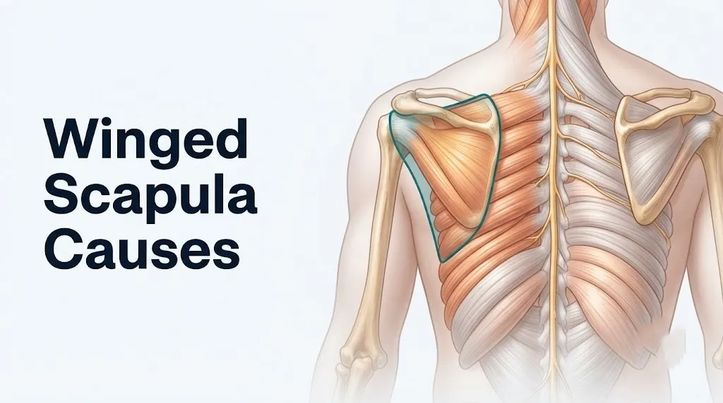
A winged scapula is a condition in which the medial border or inferior angle of the scapula protrudes away from the thoracic wall instead of lying flat, named for the resemblance to a folded bird wing when viewed from behind. The scapula is held against the rib cage entirely by the coordinated tension of several named muscles.
The primary stabilizing muscle is the serratus anterior. It originates from the outer surfaces of ribs 1 through 8 (and sometimes rib 9) and inserts along the entire costal (anterior) surface of the medial border of the scapula, from the superior to inferior angle. Its function is to protract the scapula and rotate it upward during arm elevation, while simultaneously pressing the medial border firmly against the thoracic wall. Think of the serratus anterior as the hand pressing the scapula flat from the front side of the blade. Remove that hand, and the blade tips backward.
The trapezius muscle (specifically its upper and lower fibers) provides a second stabilizing force, particularly for the superior angle and lateral border of the scapula. It supports the scapula against gravity during arm elevation and helps upwardly rotate the glenoid to face superiorly when the arm is raised overhead.
The rhomboid major and rhomboid minor run from the spinous processes of T2 through T5 and C7 through T1 to the medial border of the scapula. They retract the scapula and provide secondary stability to the medial border, though their predominant role is retraction rather than wall-pressing.
Individual variation note: Women who have undergone axillary lymph node dissection during breast cancer surgery have a documented risk of long thoracic nerve injury during that procedure, which can reveal itself as scapular winging weeks after surgery when the post-operative swelling resolves and the muscle weakness becomes functionally apparent.
Long Thoracic Nerve Damage: The Most Common Cause of Winged Scapula
Long thoracic nerve damage is the most common identifiable cause of winged scapula, occurring when this nerve is injured, compressed, or inflamed, leaving the serratus anterior muscle without motor innervation and allowing the medial scapular border to protrude. The long thoracic nerve carries no sensory fibers, which is why its damage often goes unnoticed until the visible winging or arm weakness appears.
The long thoracic nerve arises from the anterior rami of the C5, C6, and C7 nerve roots of the brachial plexus. It travels along the lateral chest wall in close proximity to the serratus anterior muscle, which it innervates along its entire length. Its superficial course along the lateral thorax makes it uniquely vulnerable to stretch, direct blows, and compression injuries compared to most other named peripheral nerves.
Because this nerve provides the only motor innervation to the serratus anterior, even partial damage produces visible functional consequences. When the serratus anterior is fully paralyzed, the medial border of the scapula protrudes prominently, particularly during forward flexion of the arm or when the patient pushes against a wall. When only partially damaged, the winging may appear only during specific movements.
The National Institute of Arthritis and Musculoskeletal and Skin Diseases (NIAMS) notes that recovery from long thoracic nerve palsy depends heavily on whether the nerve was stretched, compressed, or severed. Stretch and compression injuries often recover over 12 to 24 months with conservative management. Complete nerve laceration, as can occur during surgery, may require nerve grafting or muscle transfer.
Individual variation note: Young male athletes, particularly those in contact sports and weight lifting, are disproportionately affected by traumatic long thoracic nerve stretch injuries. This likely reflects both the greater forces sustained in these activities and the greater absolute muscle mass that can be rapidly lost when the nerve is injured.
Serratus Anterior Weakness and How It Produces Medial Border Winging
Serratus anterior weakness produces the classic winged scapula pattern of medial border and inferior angle protrusion, because without this muscle’s anterior pull on the costal surface of the scapula, the rhomboids and middle trapezius retract the scapular border away from the thoracic wall unopposed. This is the defining anatomical explanation that most online health resources fail to provide.
Under normal conditions, a constant tension balance exists between the serratus anterior pulling the medial border forward and the rhomboids pulling it backward. When the serratus anterior is weakened or paralyzed, the rhomboids win that tug-of-war by default. The medial border tilts posteriorly. The inferior angle protrudes outward. The appearance is most dramatic during the wall push-up test, where the patient places hands on a wall and performs a slow push-up while the examiner observes the posterior thorax.
Serratus anterior weakness can result from long thoracic nerve palsy, but it can also result from direct muscle trauma, myopathy, or, in less common cases, isolated serratus anterior atrophy without nerve injury. A 2020 review published in the Journal of Shoulder and Elbow Surgery noted that serratus anterior strength testing in forward flexion reliably distinguishes serratus anterior weakness from trapezius weakness: the former produces winging that worsens during forward flexion, while the latter produces winging most notable during abduction.
To describe serratus anterior weakness-related winging to a provider:
- Note whether the protrusion is visible at rest or only during movement.
- Specify which movements make it worse: forward arm flexion, pushing against a wall, or carrying objects.
- State whether any recent illness, injury, heavy lifting, or surgery preceded the onset.
- Describe any associated arm weakness, particularly difficulty raising the arm above shoulder height.
- Mention whether both shoulder blades are involved or only one.
Individual variation note: Adolescents presenting with progressive bilateral serratus anterior weakness without a history of injury or illness should be specifically evaluated for facioscapulohumeral muscular dystrophy, which can present with serratus anterior involvement before facial and shoulder girdle weakness become clinically obvious.
Key Takeaway: Serratus anterior weakness produces medial border protrusion that worsens during wall push-ups and forward arm flexion, and this pattern is distinct from the superior angle protrusion seen with trapezius weakness.
Spinal Accessory Nerve Injury and Trapezius Weakness Winging
Spinal accessory nerve injury causes a distinct winging pattern in which the superior angle and lateral scapular border protrude, the shoulder droops on the affected side, and arm abduction above 90 degrees becomes difficult, all because the trapezius muscle loses its motor innervation. This pattern differs visibly from serratus anterior winging and requires a separate clinical assessment.
The spinal accessory nerve (cranial nerve XI) provides the sole motor innervation to the upper, middle, and lower trapezius muscle. The nerve runs superficially through the posterior triangle of the neck, a triangular space bounded by the sternocleidomastoid muscle anteriorly, the trapezius posteriorly, and the clavicle inferiorly. Its superficial location in this triangle makes it highly vulnerable to iatrogenic injury during neck surgery and to direct blunt trauma to the posterior neck.
When the trapezius is denervated, the scapula can no longer be held up against gravity during arm elevation. The lateral border drops. The superior angle protrudes. Because the serratus anterior is still functional, the medial border stays relatively flat, which is the distinguishing physical feature compared to long thoracic nerve palsy.
Causes of spinal accessory nerve injury include neck dissection surgery (lymph node removal for head and neck cancers), carotid endarterectomy, penetrating trauma to the neck, direct blunt trauma to the posterior triangle, and, in rare cases, stretch injury from sudden head turning or shoulder depression.
Individual variation note: People who have undergone radical neck dissection for head and neck squamous cell carcinoma face the highest documented risk of spinal accessory nerve injury among all patient populations. The American Academy of Otolaryngology recommends modified neck dissection techniques specifically to reduce this risk, but injury rates remain clinically meaningful even with careful technique.
Dorsal Scapular Nerve Injury and Rhomboid Weakness as a Cause
Dorsal scapular nerve injury is a less commonly recognized cause of winged scapula that produces medial border protrusion through weakness of the rhomboid major, rhomboid minor, and levator scapulae muscles, though the winging pattern is generally less dramatic than that caused by serratus anterior paralysis. Dorsal scapular nerve injury is frequently underdiagnosed because its presentation can be subtle.
The dorsal scapular nerve arises primarily from the C5 nerve root and occasionally receives a contribution from C4. It penetrates the middle scalene muscle before descending along the medial border of the scapula, which is anatomically relevant because middle scalene hypertrophy or tightness (common in people with forward head posture and chronic neck tension) can compress the nerve along this course.
Rhomboid weakness alone produces a different functional deficit than serratus anterior weakness. The rhomboids retract the scapula and slightly tilt the inferior angle anteriorly. When they are weak, the scapula tends to be more protracted and downwardly rotated than normal, and the medial border can develop a subtle outward tilt. The winging is typically less prominent than serratus anterior winging and may only be visible to a trained examiner.
Clinically, dorsal scapular nerve entrapment or injury is worth considering in any patient with chronic medial scapular pain, a dull aching between the shoulder blade and spine, or subtle medial border protrusion without a clear history of trauma or viral illness. A 2017 case series published in the PM&R Journal highlighted dorsal scapular nerve entrapment as a frequently overlooked cause of chronic periscapular pain with subtle winging.
Individual variation note: People with chronic forward head posture and thoracic kyphosis are at elevated risk for dorsal scapular nerve compression due to persistent middle scalene tightness. This population skews toward desk workers in their 30s through 50s and people with longstanding sedentary occupational postures.
What Causes Winged Scapula from Direct Trauma or Injury
Direct trauma causes winged scapula by stretching, compressing, or contusing the long thoracic nerve where it runs along the lateral thorax, producing varying degrees of serratus anterior weakness that appear immediately or within days of the injury. The nerve’s superficial and predictable anatomical course makes it vulnerable to a specific set of injury mechanisms.
Blunt trauma to the anterolateral chest wall is the most straightforward traumatic mechanism. A direct blow to the lateral rib cage during a contact sport, a fall onto the shoulder, or a seat belt injury across the lateral thorax can bruise or stretch the long thoracic nerve where it traverses the lateral chest.
Stretch injury is a second traumatic mechanism, occurring when the neck is forced into lateral flexion away from the shoulder while the shoulder is simultaneously depressed. This traction stretches the long thoracic nerve across its length, producing a neurapraxia (conduction block without axon severing) that typically recovers over weeks to months. Wrestlers, rugby players, and football linemen are particularly exposed to this mechanism.
Carrying heavy loads over the shoulder or in backpacks produces a variant of stretch injury sometimes called backpack palsy. The weight depresses the shoulder girdle, stretching the long thoracic nerve between its fixed points at the brachial plexus and its distal distribution. Research published in Neurology has documented multiple cases of long thoracic nerve palsy following prolonged heavy backpack use, including in military personnel during extended field training.
| Trauma Type | Nerve at Risk | Mechanism | Typical Recovery |
|---|---|---|---|
| Blunt lateral chest blow | Long thoracic nerve | Direct contusion | Weeks to months (neurapraxia) |
| Neck-to-shoulder traction | Long thoracic nerve | Stretch/traction | Months (neurapraxia to axonotmesis) |
| Backpack compression | Long thoracic nerve | Prolonged shoulder depression | Months after load removal |
| Posterior neck blunt trauma | Spinal accessory nerve | Direct contusion | Variable; surgery sometimes needed |
| Penetrating neck trauma | Spinal accessory nerve | Laceration | Requires nerve repair if complete |
Individual variation note: Military recruits and long-distance hikers carrying loads exceeding 20 percent of body weight for extended durations face clinically recognized risk of backpack palsy affecting the long thoracic nerve. Proper pack fit, hip belt use to redistribute load, and rest breaks materially reduce this risk.
Key Takeaway: The long thoracic nerve’s superficial course along the lateral chest wall makes it vulnerable to blunt trauma, traction, and sustained compression, all of which can produce serratus anterior weakness and medial border winging.
Iatrogenic Causes of Winged Scapula After Surgery
Iatrogenic winged scapula, meaning winging caused by surgical injury to the long thoracic or spinal accessory nerve, is one of the most clinically important and frequently underrecognized causes, particularly in patients who have undergone breast surgery, chest surgery, or neck dissection. The onset of winging weeks after surgery often confuses both patients and non-specialist providers.
Long thoracic nerve injury during axillary lymph node dissection for breast cancer is a well-documented complication. The nerve lies close to the axillary vein and the lateral chest wall in the operative field. Even careful dissection can stretch or sever it. A study published in the Annals of Surgical Oncology found rates of long thoracic nerve injury ranging from 2 to 9 percent following axillary lymph node dissection, with the resulting serratus anterior weakness producing winging that becomes visible as post-operative swelling resolves.
Other surgical procedures that carry recognized long thoracic nerve risk include:
- Thoracotomy (open chest surgery for lung or esophageal procedures)
- First rib resection for thoracic outlet syndrome
- Intercostal nerve block procedures performed too laterally
- Port placement for video-assisted thoracic surgery (VATS)
- Mastectomy with lateral dissection
Spinal accessory nerve injury during neck dissection for head and neck cancers produces the trapezius-pattern winging described in the previous section. The nerve’s vulnerability during lymph node clearance from the posterior cervical triangle is well-established in the surgical literature.
Individual variation note: Women who develop scapular winging in the weeks following breast cancer surgery should specifically request evaluation of the long thoracic nerve, as this diagnosis is often delayed in this population when providers attribute shoulder asymmetry to post-operative swelling, radiation changes, or lymphedema rather than nerve injury.
Viral and Inflammatory Causes of Long Thoracic Nerve Palsy
Viral illness can cause winged scapula by triggering an inflammatory attack on the brachial plexus or its branches, a condition known as Parsonage-Turner syndrome (neuralgic amyotrophy), which can selectively damage the long thoracic nerve and produce serratus anterior paralysis, often with no preceding trauma. This is a cause that surprises most patients because the shoulder blade changes appear after what seemed like a routine illness.
Parsonage-Turner syndrome (ICD-10: G54.5) is an immune-mediated inflammatory neuropathy of the brachial plexus. It classically presents with sudden, severe shoulder or arm pain lasting days to weeks, followed by the rapid onset of muscle weakness and sometimes winging as the pain resolves. The long thoracic nerve is one of the most commonly affected branches, producing isolated serratus anterior weakness with medial border winging.
The syndrome is associated with a range of preceding triggers in published case series, including upper respiratory viral infections, surgical procedures, vaccinations, and intense exercise. The proposed mechanism involves molecular mimicry and immune-mediated inflammatory attack on peripheral nerve myelin sheaths, similar to mechanisms proposed for Guillain-Barré syndrome, though the evidence is based primarily on observational series and immunological case studies rather than controlled trials.
The Mayo Clinic notes that Parsonage-Turner syndrome can affect people of any age but most commonly presents in adults between ages 20 and 60, with a male predominance of approximately 2 to 1. Recovery is generally favorable but prolonged, with most patients showing meaningful recovery over 12 to 24 months, though some cases persist beyond three years.
Individual variation note: Patients who develop sudden severe shoulder pain followed by weakness in the absence of any injury should mention any recent illness, vaccination, or surgery to their evaluating provider. The pain-then-weakness pattern of Parsonage-Turner syndrome is frequently misattributed to a rotator cuff injury or cervical disc herniation before the correct diagnosis is made.
Key Takeaway: Parsonage-Turner syndrome can cause winged scapula without any injury by triggering immune-mediated damage to the long thoracic nerve, usually following a preceding illness or vaccination, and its pain-then-weakness sequence is a clinically distinctive pattern.
Facioscapulohumeral Muscular Dystrophy and Bilateral Winged Scapula
Facioscapulohumeral muscular dystrophy (FSHD) is a genetic muscle disease that produces bilateral winged scapula as one of its earliest and most characteristic features, caused by progressive degeneration of periscapular muscles including the serratus anterior, lower trapezius, and rhomboids. FSHD is one of the most important diagnoses to consider in any young person with bilateral, progressive winging and no clear history of injury or illness.
FSHD is the third most common form of muscular dystrophy, with an estimated prevalence of approximately 1 in 8,000 to 20,000 individuals according to the National Institute of Neurological Disorders and Stroke (NINDS). It is caused by a genetic expansion of the D4Z4 repeat region on chromosome 4q35, which leads to aberrant expression of the DUX4 transcription factor in muscle cells, triggering muscle cell death through a pathway involving activation of germline gene programs in adult muscle tissue.
Clinically, FSHD typically presents in the second or third decade of life. Weakness begins in the facial muscles and shoulder girdle, which is where the name comes from: face, scapula (shoulder blade), and humerus (upper arm). The serratus anterior is preferentially affected early, producing bilateral medial border protrusion that is often noted when the young person lifts their arms forward or is viewed from behind.
A key distinguishing clinical feature of FSHD is that the winging is bilateral and progressive. A 2014 study published in Neurology found that scapular fixation (surgical procedure to attach the scapula more firmly to the thoracic wall) improved functional arm elevation in a subset of FSHD patients when the winging was severe enough to compromise glenohumeral mechanics.
Individual variation note: FSHD shows earlier and more severe clinical expression in males with smaller D4Z4 repeat numbers. Females with FSHD may have a milder and later-onset phenotype due to partial compensation by hormonal and genetic modifier effects, which can delay diagnosis.
Scapular Dyskinesis and Postural Causes of Winging Without Nerve Damage
Scapular dyskinesis refers to altered scapular motion and position that can produce shoulder blade protrusion without any nerve injury or muscle disease, caused instead by muscle imbalance, thoracic kyphosis, or poor neuromuscular control of the periscapular muscles. This is the most common cause of the subtle winging that patients notice after prolonged desk work or reduced physical activity.
The distinction from neurogenic winging matters both clinically and practically. True nerve palsy produces fixed, structural weakness of a specific muscle. Dyskinetic winging involves muscles that are anatomically intact but inadequately activated, poorly coordinated, or inhibited by pain or posture-driven reflex patterns. The physical appearance may look similar to a lay observer, but the clinical examination and EMG findings differ.
Thoracic kyphosis, the rounding of the upper back, changes the mechanical relationship between the thoracic wall and the scapula. As the thorax curves forward, the rib cage moves away from its normal relationship with the flat scapular surface, and maintaining proper scapular contact requires proportionally greater serratus anterior activation. When serratus anterior strength or activation is insufficient to compensate for the altered rib cage geometry, subtle medial border protrusion develops without any nerve damage.
According to the Cleveland Clinic, scapular dyskinesis is extremely common in people with sedentary occupations, chronic neck and shoulder pain, and reduced upper body conditioning. It frequently co-occurs with shoulder impingement syndrome because the altered scapular position reduces the subacromial space and increases rotator cuff tendon compression during arm elevation.
Individual variation note: Adolescents and young adults who spend multiple hours daily in forward-flexed seated postures (studying, using smartphones, or at computers) show higher rates of clinically observable scapular dyskinesis than age-matched controls with more physically active daily routines, based on observational studies in sports medicine populations.
Overhead Athletes and Repetitive Strain as Causes of Winged Scapula
Overhead athletes, including competitive swimmers, tennis players, baseball pitchers, and gymnasts, develop winged scapula through repetitive long thoracic nerve microtrauma, serratus anterior fatigue, or periscapular muscle imbalance produced by the cumulative demands of overhead sport mechanics. This population represents the most common group affected by activity-related winging in a sports medicine clinical setting.
The long thoracic nerve is vulnerable during throwing, overhead striking, and swimming because the shoulder complex moves through extreme ranges of motion with high forces. Repeated scapular protraction against resistance, as occurs during the follow-through of a throw or the pull phase of swimming, subjects the long thoracic nerve to repetitive stretch along its lateral thoracic course.
Gymnasts face an additional risk mechanism: compression. Ring and bar exercises load the serratus anterior eccentrically (as the gymnast resists the protraction forces of the rings), and repeated high-load eccentric cycles can produce cumulative microtrauma to both the muscle itself and its nerve supply.
Research published in the American Journal of Sports Medicine has found that serratus anterior fatigue during prolonged overhead exercise significantly reduces scapular upward rotation efficiency, which is the movement that positions the shoulder socket correctly during arm elevation. When serratus anterior fatigues, the scapula loses proper upward rotation, the glenohumeral joint mechanics are disrupted, and both impingement and winging can develop simultaneously.
| Sport | Primary Mechanism | Nerve Most at Risk | Typical Presentation |
|---|---|---|---|
| Baseball pitching | Repeated long thoracic stretch in throwing | Long thoracic nerve | Gradual onset medial border winging |
| Competitive swimming | Repetitive serratus anterior overload | Long thoracic nerve | Bilateral or dominant-arm winging |
| Tennis | Overhead serve mechanics | Long thoracic nerve | Winging with overhead arm elevation |
| Gymnastics (rings/bars) | Eccentric serratus anterior loading | Long thoracic nerve | Medial border protrusion during support holds |
| Weight lifting | Sudden load with arm overhead | Long thoracic nerve | Acute onset after one-rep-maximum attempt |
Individual variation note: Female gymnasts who specialize in floor exercise and beam (with less ring and bar loading) have lower rates of long thoracic nerve palsy compared to male gymnasts, largely reflecting differences in apparatus demands rather than any inherent biological susceptibility.
Key Takeaway: Overhead athletes develop winging through repetitive long thoracic nerve microtrauma or serratus anterior fatigue, not typically through a single dramatic injury, which means onset is gradual and often missed until functional shoulder mechanics are already compromised.
Risk Factors That Make Winged Scapula More Likely
The primary risk factors for winged scapula include occupational overhead activity, contact or overhead sport participation, prior neck or chest surgery, recent viral illness, personal or family history of facioscapulohumeral muscular dystrophy, and carrying heavy loads over one shoulder. No single risk factor predicts winging with certainty, but combinations substantially increase probability.
Key risk factors by category:
- Occupational risks: repetitive overhead work (painters, plumbers, electricians working above shoulder height), prolonged unilateral shoulder loading (couriers, delivery workers), extensive keyboard and mouse use with elevated shoulders
- Sports and activity risks: overhead sport participation, contact sports with lateral chest or shoulder blows, heavy backpack use for extended periods
- Surgical history risks: any axillary, neck, thoracic, or chest wall surgery; prior radiation therapy to the axilla or posterior triangle of the neck
- Medical history risks: recent viral upper respiratory illness followed by sudden shoulder pain, prior Parsonage-Turner syndrome episode (recurrence rate estimated at 25 to 75 percent in affected individuals), autoimmune conditions that predispose to inflammatory neuropathy
- Genetic risks: family history of FSHD; adolescent or young adult with bilateral progressive shoulder girdle weakness
- Postural risks: severe thoracic kyphosis, chronic forward head posture, longstanding reduced upper body conditioning
Age and sex interact with risk category. Young males in contact or overhead sports represent the largest group presenting with traumatic long thoracic nerve palsy. Women with breast cancer surgical history represent the most clinically important iatrogenic risk group. Adolescents presenting with bilateral winging and no injury represent the highest-yield group for genetic evaluation.
The National Institute of Arthritis and Musculoskeletal and Skin Diseases notes that early identification of the underlying cause materially affects the likelihood of full recovery, making risk factor recognition a practical tool for prompting earlier evaluation.
How to Tell Which Cause Is Producing Your Winged Scapula
The pattern of winging, whether the medial border and inferior angle protrude or the superior angle and lateral border drop, is the single most useful physical clue to distinguishing between long thoracic nerve (serratus anterior) failure and spinal accessory nerve (trapezius) failure before any formal testing is done. This distinction can be made by a trained observer without any equipment.
Identifying the likely cause based on presentation pattern:
- Look at the scapula at rest. Medial border protrusion suggests serratus anterior weakness. Superior angle protrusion and ipsilateral shoulder droop suggest trapezius weakness.
- Ask the person to raise their arm forward in front of them to 90 degrees. If the medial border protrudes more with this movement, serratus anterior weakness is more likely. If the shoulder cannot reach 90 degrees and the superior angle rises abnormally, trapezius weakness is the more probable cause.
- Ask the person to press both hands against a flat wall and slowly perform a push-up. Classic medial border winging that increases with this wall push-up test strongly suggests long thoracic nerve palsy and serratus anterior weakness.
- Ask about the history. Sudden onset after viral illness suggests Parsonage-Turner syndrome. Onset after surgery suggests iatrogenic nerve injury. Bilateral progressive onset in a young person suggests FSHD. Gradual onset in an overhead athlete suggests repetitive microtrauma.
- Note whether both shoulder blades are affected. Bilateral winging should prompt evaluation for FSHD. Unilateral winging in the context of recent surgery or illness points toward nerve injury.
The wall push-up test and forward flexion elevation test, described in the Journal of Bone and Joint Surgery clinical review on scapular winging, remain the most reliable bedside assessments for distinguishing these two major cause categories before EMG results are available.
Individual variation note: People with obesity or significant paraspinal muscle bulk may show attenuated winging on visual inspection even when serratus anterior weakness is substantial, because surrounding soft tissue obscures the medial border protrusion. Clinical strength testing and EMG are more reliable than visual inspection alone in this population.
How Winged Scapula Is Diagnosed with EMG and Clinical Tests
Winged scapula is diagnosed through a combination of clinical history, physical examination including the wall push-up test and arm elevation testing, and electromyography (EMG) with nerve conduction velocity (NCV) study, which together confirm whether a named nerve is damaged, which nerve is affected, and the severity of the injury. EMG is the gold standard for confirming neurogenic winging and guiding prognosis.
Electromyography (EMG) records electrical activity within a muscle, distinguishing between normal muscle at rest (electrically silent), muscle under volitional activation (normal motor unit potentials), and denervated muscle (spontaneous fibrillation potentials and positive sharp waves). When the long thoracic nerve is damaged, the serratus anterior shows denervation changes on EMG that confirm the diagnosis and estimate the injury severity.
Nerve conduction velocity (NCV) study complements EMG by measuring the speed and amplitude of electrical conduction along named nerve segments. The long thoracic nerve is challenging to study with standard NCV techniques due to its length and depth in some portions, which is one reason that EMG of the serratus anterior muscle is often more diagnostically informative than NCV for this specific nerve.
Additional diagnostic tools used in specific circumstances include:
- MRI of the brachial plexus: useful when Parsonage-Turner syndrome is suspected, demonstrating increased signal in affected nerve fascicles and surrounding muscles in the early inflammatory phase
- Ultrasound of the long thoracic nerve: emerging technique that allows direct visualization of nerve swelling, continuity, or scarring; increasingly used in sports medicine and neurology practices as ultrasound resolution improves
- Genetic testing: ordered when FSHD is suspected, targeting the D4Z4 repeat contraction on chromosome 4q35
- Chest X-ray or CT: useful in evaluating for structural causes of nerve compression such as cervical rib, Pancoast tumor, or thoracic outlet anomaly
Individual variation note: In adolescents with suspected FSHD, genetic testing should be pursued before EMG in most cases, as the EMG pattern of myopathic changes can be subtle early in FSHD and a negative or borderline EMG does not rule out the diagnosis.
When to Seek Medical Evaluation for Winged Scapula
Any new-onset scapular winging accompanied by arm weakness, difficulty raising the arm above shoulder height, or sudden severe shoulder pain preceding the winging warrants evaluation by a primary care physician within one to two weeks, with urgent referral to a board-certified orthopedic surgeon, sports medicine physician, or neurologist if nerve palsy is suspected. Delay in diagnosis directly affects recovery outcomes for neurogenic causes.
The urgency of evaluation depends on the presenting pattern:
| Presentation | Urgency | Provider to Contact |
|---|---|---|
| Winging noticed gradually, no pain, no weakness | Routine | Primary care physician within 2 to 4 weeks |
| Winging with mild arm weakness, recent onset | Prompt | Primary care physician within 1 to 2 weeks; EMG referral |
| Sudden severe shoulder pain followed by winging | Urgent | Board-certified orthopedic surgeon or neurologist within days |
| Winging after recent surgery | Prompt | Surgical team and orthopedic consultation within 1 to 2 weeks |
| Bilateral progressive winging in adolescent | Prompt | Primary care physician and pediatric neurology referral |
| Winging with neck mass, voice change, or dysphagia | Urgent | Emergency department or urgent care same day |
| Winging following high-energy trauma with shoulder deformity | Emergency | Emergency department immediately |
Winged scapula itself does not constitute a life-threatening emergency in most presentations. The one important exception is high-energy trauma to the shoulder or neck that produces winging alongside vascular signs: an expanding neck hematoma, absent radial pulse, pallor or coldness of the hand, or loss of sensation in the arm. These signs suggest brachial plexus avulsion or vascular injury and require immediate emergency department evaluation.
Anyone whose scapular winging is accompanied by progressive bilateral arm weakness, difficulty swallowing, voice changes, or rapid progression over weeks should be evaluated urgently to rule out neurological conditions beyond a simple isolated nerve palsy.
Individual variation note: People over age 60 with new-onset unilateral winging, particularly if associated with unintended weight loss, night sweats, or cervical or axillary lymph node enlargement, should be evaluated for Pancoast tumor and lymphoma, which can compress or infiltrate the brachial plexus and its branches. These are rare causes but warrant consideration in this age and presentation context.
Key Takeaway: Sudden severe shoulder pain that resolves and leaves arm weakness and scapular winging behind should be evaluated urgently as possible Parsonage-Turner syndrome, not watched at home, because early diagnosis guides both prognosis and management planning.
Frequently Asked Questions About Winged Scapula Causes
What is the most common cause of a winged scapula?
The most common identifiable cause of winged scapula is long thoracic nerve palsy, which paralyzes the serratus anterior muscle and allows the medial border of the scapula to protrude posteriorly.
Long thoracic nerve palsy can result from direct trauma, repetitive stretch injury, viral illness triggering Parsonage-Turner syndrome, or surgical injury during chest or breast procedures.
A board-certified orthopedic surgeon or sports medicine physician can confirm the diagnosis with EMG and nerve conduction velocity testing.
Can a winged scapula go away on its own?
Whether a winged scapula resolves without intervention depends entirely on its cause: neurapraxic long thoracic nerve injuries (where the nerve is intact but temporarily blocked) often recover over 12 to 24 months without surgery, while complete nerve lacerations and muscular dystrophy-related winging do not resolve spontaneously.
Scapular dyskinesis caused by postural muscle imbalance can improve with structured physical therapy targeting serratus anterior and periscapular muscle strengthening.
Anyone with persistent winging beyond three months should seek evaluation by a board-certified orthopedic surgeon or neurologist rather than continuing to wait for spontaneous resolution.
What nerve damage causes winged scapula?
Long thoracic nerve damage (arising from C5, C6, C7 nerve roots) is the most common nerve injury causing winged scapula by paralyzing the serratus anterior muscle.
Spinal accessory nerve (cranial nerve XI) injury causes a second, distinct winging pattern by paralyzing the trapezius, producing superior angle protrusion and ipsilateral shoulder droop.
Dorsal scapular nerve injury can also cause subtle winging through rhomboid weakness, though this pattern is less dramatic and frequently overlooked.
Can poor posture cause a winged scapula?
Poor posture can cause scapular dyskinesis, a pattern of altered scapular position that may include medial border protrusion resembling true winging, without any nerve damage or muscle disease.
Thoracic kyphosis and forward head posture change the geometric relationship between the thoracic wall and scapula, requiring greater serratus anterior activation to maintain scapular contact; when activation is insufficient, subtle protrusion develops.
Posture-related winging generally responds to targeted physical therapy addressing thoracic mobility and periscapular muscle strength, unlike neurogenic winging which requires nerve recovery before muscles can be rehabilitated.
Is a winged scapula serious?
The seriousness of a winged scapula depends on its cause: true neurogenic winging from nerve palsy can produce lasting shoulder dysfunction, arm elevation limitation, and secondary rotator cuff injury if untreated.
Dyskinetic winging from posture or muscle imbalance is generally less serious and more reversible, but it can contribute to shoulder impingement and chronic shoulder pain if left unaddressed.
Any winged scapula that is new, painful, accompanied by arm weakness, or followed by recent surgery or illness should be evaluated by a primary care physician or orthopedic specialist rather than assumed to be a minor postural issue.
How do doctors test for a winged scapula?
Doctors typically begin with the wall push-up test and arm elevation assessment to observe the winging pattern and identify which muscle group is failing.
Electromyography (EMG) and nerve conduction velocity (NCV) study are the definitive tests for confirming neurogenic winging, identifying the specific nerve injured, and estimating injury severity.
When facioscapulohumeral muscular dystrophy is suspected, genetic testing for the D4Z4 repeat contraction on chromosome 4q35 is the most definitive diagnostic step, often more informative than EMG in early stages of the disease.
Understanding what is causing your winged scapula changes everything about how it should be managed. A postural serratus anterior activation deficit and a surgical long thoracic nerve laceration may look similar from across the room, but they follow entirely different clinical pathways. Getting the right diagnosis matters, and the starting point is a proper clinical examination, not a home self-assessment.
If your winging appeared after a viral illness, following surgery, after a single heavy training session, or in the context of progressive bilateral weakness, book an appointment with your primary care physician this week. Bring notes on exactly when the protrusion started, what preceded it, and which movements make it worse. Request a referral to a board-certified orthopedic surgeon, sports medicine physician, or neurologist depending on your clinical picture.
The sooner a nerve injury is identified and managed, the better the chance of meaningful recovery. Nerves regenerate slowly, approximately 1 to 3 millimeters per day, which means months matter when it comes to outcome.