What Are the Main Toe Pain Causes? Explained for 2026
Toe pain causes span a wide clinical spectrum, from a simple ingrown toenail to an acute gout attack, a stress fracture, or an early diabetic foot infection that can progress to limb-threatening tissue loss if left untreated. Identifying which cause is driving your pain requires understanding where in the toe the pain occurs, what triggers it, and what associated signs (swelling, redness, numbness, skin changes) accompany it.
The scale of the problem is larger than most people expect. According to the National Institute of Arthritis and Musculoskeletal and Skin Diseases (NIAMS), gout alone affects approximately 9.2 million adults in the United States, and foot and toe pain collectively represents one of the most frequent musculoskeletal complaints in primary care settings. The consequences of misidentifying a serious cause as a minor ache range from delayed fracture treatment to uncontrolled infection.
This article covers the 12 most clinically important causes of toe pain with the specific anatomical mechanism behind each, the populations at highest risk for each condition, how each is diagnosed, and the specific presentations that require emergency evaluation rather than a scheduled appointment.
Toe Pain Causes: An Overview of the Most Common Reasons Toes Hurt
The most common toe pain causes in adults are gout, bunions, ingrown toenails, Morton’s neuroma, hammertoe deformity, sesamoiditis, stress fractures, and arthritis, with diabetic neuropathy, Raynaud’s phenomenon, and skin or joint infections rounding out the clinically important list.
No single pain pattern reliably identifies one cause over another without clinical assessment. Pain that is explosively severe and present at rest suggests gout or septic arthritis. Pain with a burning, electric quality between the third and fourth toes suggests Morton’s neuroma. Gradual pain at the tip of a toe worsening with shoes suggests an ingrown toenail or hammertoe pressing against the shoe box.
| Cause | Primary Location | Pain Character | Key Associated Sign |
|---|---|---|---|
| Gout | First MTP joint | Explosive, constant, severe | Sudden onset, often at night |
| Bunion | First MTP joint medial side | Aching, worse with shoes | Bony prominence, hallux deviation |
| Morton’s neuroma | Third/fourth interspace | Burning, electric, radiating | Mulder’s click on palpation |
| Ingrown toenail | Hallux lateral or medial edge | Sharp, worse with pressure | Nail piercing skin, possibly infected |
| Hammertoe | PIP joint of lesser toes | Aching, friction | Toe bent downward, corn on dorsum |
| Sesamoiditis | Plantar first MTP joint | Dull ache, worse with push-off | Tenderness under the great toe |
| Stress fracture | Any metatarsal or phalanx | Dull ache worsening with activity | Point tenderness on bone |
| Arthritis (OA/RA) | MTP joints, PIP joints | Morning stiffness, chronic ache | Bilateral, gradual, associated systemic signs in RA |
| Diabetic neuropathy | Diffuse, often all toes | Burning, tingling, or absent | History of diabetes, skin changes |
| Septic arthritis | Single joint, any toe | Severe, constant, warm | Fever, inability to move joint |
The clinical value of this table is in what it shows across causes: a single acutely painful, swollen, warm joint in a person with no prior history most likely represents gout or septic arthritis, not a bunion or hammertoe. That distinction determines the urgency of the evaluation needed.
The Anatomy of the Toes and Why Location Matters for Diagnosis
The anatomical location of toe pain, whether at the joint, the tip, the nail, the plantar surface, or between the toes, is the single most reliable initial clue to its cause, because the specific structures at each location have distinct vulnerabilities to different disease processes.
Each toe consists of three bones called phalanges: the proximal phalanx, the middle phalanx, and the distal phalanx, with the exception of the great toe, which has only a proximal and distal phalanx. The joints between these bones are the proximal interphalangeal (PIP) joints and distal interphalangeal (DIP) joints. The joints between the metatarsal bones and the base of the proximal phalanges are the metatarsophalangeal (MTP) joints, numbered first through fifth from the great toe outward.
Two small sesamoid bones sit plantar to the first MTP joint, embedded in the flexor hallucis brevis tendon. They protect the first MTP joint from compressive forces during walking and push-off. Pain directly under the ball of the great toe, worsened by any loading activity, points specifically to sesamoid pathology because no other structure occupies that precise anatomical location.
Between the metatarsal heads run the common digital nerves (also called plantar interdigital nerves), which supply sensation to adjacent sides of neighboring toes. The third and fourth intermetatarsal space is where Morton’s neuroma most commonly forms because this space is narrower and the nerve passes beneath a thicker transverse metatarsal ligament, making it more susceptible to compression.
Think of the forefoot like a narrow apartment corridor with plumbing (blood vessels), electrical wiring (nerves), and structural walls (bones and joints) packed closely together. When anything in that corridor swells, even slightly, everything else in the space gets compressed. That is why a single inflamed MTP joint from gout can produce pain that radiates to surrounding toes, and why a neuroma between two metatarsal heads produces electric sensations in both adjacent toes rather than just one.
Older adults lose the natural fat padding of the forefoot over decades, a process called plantar fat pad atrophy, which increases direct compressive loading on the metatarsal heads and the nerves between them. This is one reason Morton’s neuroma and metatarsalgia are more prevalent in adults over 60.
Gout as a Cause of Toe Pain
Gout causes toe pain by depositing monosodium urate crystals in the synovial fluid and soft tissue of the first metatarsophalangeal (MTP) joint, triggering the NLRP3 inflammasome in joint macrophages and producing a surge of interleukin-1 beta (IL-1 beta) that drives one of the most intensely painful acute inflammatory responses in clinical medicine.
The mechanism begins with hyperuricemia, a chronically elevated serum uric acid level above 6.8 mg/dL (the saturation threshold for monosodium urate in physiological saline). When uric acid concentrations in the joint fluid exceed this threshold, needle-shaped urate crystals precipitate out of solution. The first MTP joint is targeted preferentially because it is the most peripheral joint in the body, and urate crystallization is favored by lower temperatures, which are present at the extremities.
When neutrophils and macrophages in the joint space encounter urate crystals, the crystals activate the NLRP3 inflammasome, a multiprotein complex inside the immune cell that processes pro-IL-1 beta into its active secreted form. The resulting IL-1 beta surge drives intense neutrophilic infiltration into the joint, producing the explosive swelling, heat, redness, and pain of an acute gout attack. The classic presentation is pain so severe that the weight of a bedsheet on the affected toe is intolerable.
According to the American College of Rheumatology (ACR), gout affects men at rates approximately three to four times higher than women until menopause, because estrogen promotes renal uric acid excretion. After menopause, female gout rates rise substantially as this hormonal protection declines. Postmenopausal women on thiazide diuretics (used for hypertension) are at particularly elevated gout risk because thiazides directly compete with uric acid for renal tubular secretion, raising serum urate levels.
Serum uric acid testing is the first-line laboratory evaluation for a suspected gout attack, though a normal uric acid level during an acute attack does not exclude gout because urate redistributes from serum into the joint during the inflammatory event.
Key Takeaway: Gout attacks the first MTP joint specifically because lower peripheral body temperature favors urate crystal formation there, and the explosive pain of a gout attack is driven by IL-1 beta release from the NLRP3 inflammasome, not by direct bacterial infection, which is why it can resolve spontaneously without antibiotics.
Bunions and Hallux Valgus Causing Big Toe Pain
A bunion, also called hallux valgus, causes big toe pain by producing progressive lateral deviation of the great toe and medial displacement of the first metatarsal head, creating abnormal joint mechanics at the first MTP joint, bursal inflammation, and friction against footwear at the medial prominence of the metatarsal head.
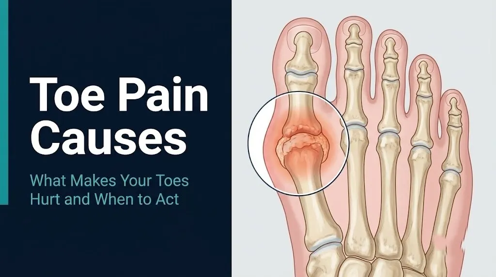
The structural deformity of a bunion involves three simultaneous changes in the first ray. The first metatarsal deviates medially (toward the midline), the hallux deviates laterally (toward the second toe), and the first MTP joint becomes subluxed, meaning the normal joint congruency is lost. The medial capsule and the abductor hallucis tendon become chronically stretched, while the adductor hallucis tendon on the lateral side pulls the toe progressively further into valgus alignment.
The medial metatarsal bursa, a fluid-filled sac that develops at the bony prominence in response to chronic friction, contributes significantly to the pain and swelling. The bunion itself is not the bone growing, as is commonly believed, but rather the exposed and inflamed medial metatarsal head becoming prominent as the toe’s alignment shifts.
Women develop bunions at rates approximately eight to ten times higher than men, according to data reviewed by the American Academy of Orthopaedic Surgeons (AAOS). Narrow-toed footwear that forces the toes into a constricted, laterally deviated position is the primary environmental factor, though genetic predisposition to first metatarsal hypermobility or ligamentous laxity determines who develops severe deformity even with appropriate footwear. A mother with severe hallux valgus substantially increases her daughter’s lifetime risk.
Bunion pain is typically worse with shoe wear that compresses the medial forefoot, often relieved by going barefoot or wearing wide-toed shoes. This pattern directly distinguishes bunion pain from gout pain, which is present and severe at rest regardless of footwear.
Morton’s Neuroma and Nerve-Related Toe Pain
Morton’s neuroma causes toe pain by producing perineural fibrosis around the common digital nerve (plantar interdigital nerve), most often in the third intermetatarsal space between the third and fourth toes, where chronic compression by the adjacent metatarsal heads during walking produces nerve thickening, fibrosis, and pain.
The term “neuroma” is technically a misnomer because this condition is not a true nerve tumor. It is a benign proliferation of perineural connective tissue around the interdigital nerve, driven by repetitive mechanical compression. The transverse metatarsal ligament, which bridges the metatarsal heads on the plantar side, compresses the nerve from below during toe dorsiflexion in push-off, and the metatarsal heads compress it from the sides during weight-bearing, particularly in narrow or high-heeled footwear.
The pain is characteristically electric, burning, or like walking on a pebble or marble, located between and around the affected toes. The Mulder’s click test, in which a clinician compresses the forefoot from side to side while pressing upward between the metatarsal heads, reproduces a palpable and sometimes audible click with pain reproduction, and has a clinically useful sensitivity for Morton’s neuroma.
According to the Mayo Clinic, Morton’s neuroma is significantly more common in women than in men, partly due to narrower forefoot anatomy and partly due to higher rates of compressive footwear use. High-heeled shoes shift body weight forward onto the forefoot, increasing the compressive load on the intermetatarsal nerves with every step. Runners who run in poorly fitted shoes with a narrow toe box develop this condition at elevated rates compared to recreational walkers.
Nerve conduction velocity (NCV) studies are not typically required for diagnosis but may be ordered by a neurologist or podiatrist to rule out a more proximal nerve entrapment when the presentation is atypical or when symptoms do not localize clearly to the forefoot.
Ingrown Toenail Pain and Infection
An ingrown toenail causes toe pain when the lateral or medial edge of the nail plate of the great toe (most commonly) grows into or presses against the nail fold (the soft tissue bordering the nail), producing a penetrating wound that becomes a portal of entry for skin bacteria.
The first stage of ingrown toenail pain is purely mechanical: the sharp nail edge presses against the nail fold, causing localized pressure pain that worsens when anything compresses the toe from the side. In the second stage, the nail penetrates the skin of the nail fold, creating a small wound. Bacteria normally present on the skin surface, primarily Staphylococcus aureus and gram-negative organisms, enter through this breach and produce paronychia, a localized nail fold infection with redness, warmth, swelling, and often purulent discharge.
Quick Tip:
- Cutting toenails straight across rather than curved at the corners is the primary preventive practice for ingrown toenails, because curved cutting leaves a sharp lateral spike that grows into the nail fold as the nail advances.
- Tight footwear that compresses the toes laterally is the second most common contributing factor, as it pushes the nail fold tissue against the nail edge.
- People with diabetes should not attempt home treatment of an ingrown toenail by cutting or probing the nail fold because the risk of introducing deeper infection in a setting of impaired wound healing is too high.
The infection of an ingrown toenail in a person with diabetes is a genuinely different clinical situation than the same condition in a healthy adult. Impaired peripheral circulation and neuropathy in diabetic feet mean that what appears to be a superficial nail fold infection can extend to the bone (osteomyelitis) more rapidly than expected. Any diabetic patient with a red, swollen toe around a nail should be evaluated by a primary care physician or podiatrist within 24 hours rather than treated at home.
Key Takeaway: An ingrown toenail is a mechanical problem with a microbial complication, but in patients with diabetes, the complication can rapidly become deep tissue infection or osteomyelitis, which is why diabetic patients cannot apply the same “wait and see” approach that healthy adults can use.
Hammertoe and Claw Toe Causing Toe Pain
Hammertoe causes toe pain by producing a fixed or flexible flexion deformity at the proximal interphalangeal (PIP) joint of a lesser toe, causing the dorsal surface of the bent joint to rub against the shoe upper and the tip of the toe to press against the shoe sole, generating corns, calluses, and chronic mechanical pain at both contact points.
The deformity results from an imbalance between the intrinsic muscles of the foot (the lumbrical and interosseous muscles, which normally flex the MTP joint and extend the PIP and DIP joints) and the extrinsic long tendons (the flexor digitorum longus and extensor digitorum longus). When the intrinsic muscles are weakened or the MTP joint is pushed into extension (as occurs chronically in high-heeled shoes or with a tight toe box), the long flexors dominate at the PIP joint, pulling it into the characteristic downward flexion position.
A corn forms at the dorsal PIP joint prominence where repetitive friction against the shoe stimulates a thickened keratin response in the overlying skin. The corn itself becomes a focal point of pain distinct from the underlying joint deformity, and attempting to cut or remove a corn without addressing the underlying structural problem invariably results in recurrence.
Claw toe differs from hammertoe in that both the PIP and distal interphalangeal (DIP) joints are flexed while the MTP joint is hyperextended, giving the toe a claw-like appearance. Claw toe deformity across multiple toes simultaneously raises clinical suspicion for an underlying neurological condition (Charcot-Marie-Tooth disease, diabetic neuropathy, or other peripheral neuropathy) affecting the intrinsic foot musculature.
Hammertoe deformity is more prevalent in older women than any other demographic group, according to data from the AAOS, reflecting the combined effects of decades of compressive footwear and progressive ligamentous laxity. Early flexible deformities can often be managed with wider footwear and toe-strengthening exercises, while rigid fixed deformities require surgical correction.
Sesamoiditis and Turf Toe in Athletes
Sesamoiditis causes pain at the plantar surface of the first MTP joint by producing inflammation in or around the medial or lateral sesamoid bones, the two small bones embedded in the flexor hallucis brevis tendon beneath the great toe, typically from repetitive compressive loading during athletic activity.
Think of the sesamoid bones like the pulleys in a bicycle derailleur system. They redirect the pull of the flexor hallucis brevis tendon around the first metatarsal head, protecting the joint from direct compressive loads during push-off. In activities requiring repetitive toe push-off, including sprinting, jumping, dancing, and certain racket sports, these small bones absorb forces many times body weight per step. When cumulative load exceeds the tissue’s repair capacity, inflammatory changes develop in the sesamoid bone itself (sesamoiditis) or, in more severe cases, a true stress fracture of the sesamoid occurs.
Turf toe is a distinct condition caused by an acute hyperextension sprain of the first MTP joint, where the joint is forced into extreme dorsiflexion beyond its normal range. The plantar plate and the ligamentous capsule of the first MTP joint are torn, producing immediate pain, swelling, and loss of push-off strength. According to research published in Foot and Ankle International, turf toe is particularly prevalent in American football players on artificial turf surfaces, where the shoe-to-surface interface generates higher traction forces than natural grass, making the first MTP joint more susceptible to hyperextension injury.
The American Academy of Orthopaedic Surgeons notes that sesamoid stress fractures can be misidentified on plain X-rays as bipartite sesamoids, which are a normal anatomical variant in which the sesamoid bone is congenitally divided into two pieces. MRI is the most reliable imaging modality to distinguish an acute stress fracture from a bipartite sesamoid when the clinical presentation does not resolve with conservative management.
Female ballet dancers carry elevated risk for sesamoid stress fractures because their performance requires repetitive en pointe positioning that loads the first MTP joint plantar surface beyond what most other activities demand.
Key Takeaway: Sesamoiditis and turf toe both affect the first MTP joint but through different mechanisms: sesamoiditis is a repetitive overload injury of the plantar sesamoid bones, while turf toe is an acute ligamentous sprain from a single hyperextension event, and distinguishing between them requires either MRI or clinical correlation with the injury history.
Stress Fractures Causing Toe Pain
Stress fractures of the toes and metatarsals cause toe pain when cumulative mechanical loading on a bone exceeds the rate at which bone remodeling can repair microdamage, producing a crack that initially appears as a focal area of bone marrow edema before progressing to a visible fracture line on imaging.
Unlike acute traumatic fractures, which occur from a single high-energy force, stress fractures develop gradually from repetitive submaximal loads. In the foot, the second and third metatarsals are the most commonly affected bones, because they bear disproportionate load transfer when the first ray is hypermobile (as in hallux valgus) or when gait abnormalities shift weight medially. The fifth metatarsal is the site of the clinically important Jones fracture, a stress fracture at the junction of the metatarsal base and shaft that carries an elevated risk of non-union due to poor blood supply at that location.
Pain from a metatarsal stress fracture is characteristically dull and aching at rest early in the course, worsening with any weight-bearing activity and progressively limiting the ability to walk. Point tenderness directly over the involved bone, reproduced by pressing a single fingertip along the shaft, is the most reliable clinical finding and distinguishes stress fracture pain from the more diffuse plantar tenderness of plantar fasciitis.
Research published in the Journal of Bone and Joint Surgery has confirmed that female runners, particularly those with the female athlete triad (now termed relative energy deficiency in sport, RED-S), which involves low energy availability, menstrual irregularity, and low bone mineral density, are at substantially elevated stress fracture risk compared to male runners at equivalent training volumes. Postmenopausal women with osteoporosis are similarly at elevated risk for metatarsal stress fractures from activities that most younger adults tolerate without injury.
Any athlete who reports insidious onset of forefoot pain that has been present for more than two weeks and is not improving with rest should be evaluated by a primary care physician or board-certified orthopedic surgeon with weight-bearing foot radiographs and, if those are negative, MRI of the affected area.
Arthritis as a Cause of Chronic Toe Pain
Arthritis causes chronic toe pain when either cartilage breakdown in osteoarthritis (OA) or synovial membrane inflammation in rheumatoid arthritis (RA) affects the metatarsophalangeal, proximal interphalangeal, or distal interphalangeal joints of the toes, progressively reducing joint space and producing persistent aching stiffness and deformity.
Osteoarthritis of the first MTP joint produces a specific condition called hallux rigidus, characterized by progressive loss of great toe dorsiflexion due to osteophyte formation around the joint and cartilage erosion. Walking requires approximately 65 degrees of great toe dorsiflexion at push-off; when hallux rigidus limits this range, the body compensates by rolling off the medial edge of the foot, which strains the medial ankle and produces secondary knee and hip pain in some patients. The pain is aching and constant, worse with activity, and accompanied by a palpable bony ridge across the dorsal MTP joint.
Rheumatoid arthritis most commonly affects the MTP joints of the lesser toes symmetrically and bilaterally, producing synovitis-driven joint destruction that is distinctly different in character from OA. The synovial inflammation of RA is driven by tumor necrosis factor alpha (TNF-alpha) and interleukin-6 produced by activated synovial fibroblasts and macrophages, creating a pannus tissue that erodes articular cartilage and subchondral bone. Morning stiffness lasting more than 30 to 60 minutes, symmetrical joint involvement, and systemic symptoms (fatigue, low-grade fever) are the clinical features that distinguish RA-related toe joint pain from OA.
According to the NIAMS, psoriatic arthritis and reactive arthritis are two additional inflammatory arthritides that preferentially target the toe joints, often producing a “sausage toe” pattern called dactylitis where an entire toe swells diffusely rather than at a single joint. Dactylitis in any toe warrants evaluation by a rheumatologist for systemic inflammatory arthritis.
Adults over 65 with toe pain and morning stiffness lasting more than 45 minutes should be evaluated by a primary care physician with inflammatory markers (CRP, ESR, rheumatoid factor) and referred to a rheumatologist if those tests suggest systemic inflammatory arthritis.
Toe Pain Caused by Diabetes and Peripheral Neuropathy
Toe pain caused by diabetes reflects two simultaneous processes in most patients: peripheral neuropathy that alters or eliminates the normal pain sensation in the toes, and microvascular disease that reduces the blood supply needed for tissue repair, creating a situation where tissue damage progresses without the warning signal of pain that healthy individuals rely on.
Diabetic peripheral neuropathy results from chronic hyperglycemia damaging the myelin sheath and axons of peripheral nerves through the polyol pathway (excess glucose is converted to sorbitol by aldose reductase, accumulating in nerve cells) and through advanced glycation end-product (AGE) accumulation on nerve proteins. The result is a progressive distal-to-proximal “stocking-glove” pattern of sensory loss that begins in the toes. Early neuropathy causes burning, tingling, or electric pain in the toes; advanced neuropathy causes complete sensory loss, where wounds or infections produce no pain signal at all.
This is the clinical danger that no competitor article addresses: a diabetic patient with advanced peripheral neuropathy may have a deep tissue infection, an abscess, or early Charcot neuroarthropathy (a progressive, painless destruction of the foot joints from neuropathy-related loss of protective sensation) without feeling significant pain. The absence of pain in a diabetic patient’s toe is not reassuring; it may indicate that neuropathy is advanced enough to eliminate the warning signal.
According to the NIAMS, approximately 60 to 70% of people with diabetes develop some form of peripheral neuropathy, and diabetic foot complications represent the leading cause of non-traumatic lower limb amputations in the United States. Any person with diabetes who develops a toe wound, skin breakdown, redness, or swelling, even if minimally painful, should be evaluated by a primary care physician or podiatrist within 24 hours.
People with poorly controlled type 1 diabetes develop neuropathy earlier and more severely than those with well-controlled type 2 diabetes, reflecting the cumulative effect of hyperglycemia exposure on peripheral nerve integrity.
Key Takeaway: In diabetic patients with peripheral neuropathy, the absence of pain in a toe with visible skin changes (redness, swelling, wound, or discoloration) is clinically dangerous, not reassuring, because advanced neuropathy eliminates the pain signal while tissue destruction continues.
Raynaud’s Phenomenon and Vascular Causes of Toe Pain
Raynaud’s phenomenon causes toe pain, numbness, and color change through episodic vasospasm of the digital arteries supplying the toes, triggered by cold exposure or emotional stress, producing a characteristic triphasic color sequence (white, then blue, then red) as arterial spasm, deoxygenation, and reactive vasodilation occur in sequence.
The underlying mechanism involves an exaggerated sympathetic nervous system response to cold in the digital arterial smooth muscle. Cold exposure normally produces mild digital vasoconstriction in all humans. In Raynaud’s phenomenon, the vasoconstrictive response is dramatically amplified, fully occluding digital arterial flow. The affected toes turn white (pallor from ischemia), then blue or purple (cyanosis from deoxygenation of stagnant venous blood), then intensely red and painful (reactive hyperemia from vasodilation when spasm resolves). The pain typically occurs during the red phase as blood flow returns to oxygen-deprived tissue.
Primary Raynaud’s disease (idiopathic, not associated with a systemic condition) is more common in young women, particularly those aged 15 to 30 years, and is generally less severe than secondary Raynaud’s. Secondary Raynaud’s phenomenon occurs in the setting of connective tissue diseases including systemic sclerosis (scleroderma), systemic lupus erythematosus, and Sjögren’s syndrome. Secondary Raynaud’s carries a higher risk of digital ischemia, ulceration, and in severe cases, digital gangrene.
Women are affected by both primary and secondary Raynaud’s phenomenon at rates significantly higher than men, with primary Raynaud’s showing approximately a 9:1 female-to-male ratio in some studies, according to research in Arthritis and Rheumatology.
Any person experiencing recurrent Raynaud’s attacks, particularly if associated with joint pain, skin tightening, or difficulty swallowing, should be evaluated by a rheumatologist for an underlying connective tissue disease, as these symptoms together suggest secondary rather than primary Raynaud’s.
Toe Pain in Runners, Women, and Older Adults
Toe pain in runners most commonly involves Morton’s neuroma, sesamoiditis, metatarsal stress fractures, and black toenail (subungual hematoma), while toe pain in women disproportionately involves bunions, Raynaud’s phenomenon, and rheumatoid arthritis, and toe pain in older adults is most often driven by osteoarthritis, hallux rigidus, hammertoe deformity, and gout.
Runners present a specific overuse injury pattern driven by the repetitive forefoot loading of the running gait. The second metatarsal is the most commonly fractured bone in distance runners because its position at the center of the forefoot makes it a primary load-transfer point during push-off. Black toenail, the bluish-black discoloration from a subungual hematoma (blood pooling under the nail), occurs when the toe repetitively strikes the inside of the shoe’s toe box during downhill running or in shoes with insufficient toe room. It is not a serious injury on its own but can be confused with a melanoma under the nail if it does not move distally with nail growth over weeks.
During pregnancy, toe pain and swelling are common due to fluid retention causing generalized edema that compresses the interdigital nerves, a physiological state that resolves postpartum. Pregnant women with bilateral forefoot burning and numbness that developed in the third trimester most often have transient pregnancy-related nerve compression rather than Morton’s neuroma. Evaluation by an obstetric provider and reassurance is appropriate in the absence of other clinical red flags.
To describe your toe pain accurately to a primary care physician, podiatrist, or orthopedic surgeon:
- State the exact location: which toe, which side of the toe, which joint, or the plantar (bottom) versus dorsal (top) surface.
- Describe the pain character: sharp, burning, electric, dull ache, throbbing, or pressure.
- Note when the pain occurs: at rest, with walking, at night, or only with specific activities.
- Record whether both feet are affected or only one.
- List any associated signs: swelling, redness, skin color changes, numbness, or tingling.
- Bring a list of all current medications, including any that affect uric acid levels (diuretics, low-dose aspirin) or bone density (corticosteroids, proton pump inhibitors).
Big Toe Pain Versus Little Toe Pain: What Location Tells You
Big toe pain most commonly indicates gout, bunion, hallux rigidus, sesamoiditis, or turf toe, while little toe (fifth toe) pain most commonly results from a bunionette (tailor’s bunion at the fifth metatarsal head), a fifth metatarsal stress fracture or Jones fracture, corns, or hammertoe deformity affecting the fifth toe.
The first MTP joint’s anatomy makes it uniquely vulnerable to gout because of the peripheral temperature gradient (lower temperature at the great toe favors urate crystallization), to bunion formation from the biomechanical leverage of the first metatarsal, and to sesamoid pathology because of the plantar sesamoid apparatus specific to this joint. No other MTP joint has this combination of vulnerabilities, which is why big toe pathology is so clinically diverse.
The fifth toe carries its own distinct vulnerability at the base of the fifth metatarsal, where a sudden inversion injury of the ankle can avulse the styloid process (the bony prominence at the base of the fifth metatarsal) through contraction of the peroneus brevis tendon. This is called a pseudo-Jones fracture or avulsion fracture of the fifth metatarsal base and is distinct from the Jones fracture at the metatarsal diaphysis. The two fracture types are clinically important to distinguish because they carry different healing rates and treatment requirements.
Second toe pain is the presentation most associated with Freiberg’s infraction, a relatively rare condition in which the second metatarsal head undergoes avascular necrosis (bone death from loss of blood supply) due to repetitive compressive loading. Freiberg’s infraction is more common in adolescent females, where rapid growth may outstrip the vascular supply to the second metatarsal head. On plain X-rays, it appears as flattening and sclerosis of the second metatarsal head. MRI identifies it earlier, before radiographic changes appear.
Individuals who have recently changed footwear or significantly increased activity who develop second or third toe pain should have weight-bearing foot radiographs taken to exclude stress fracture or Freiberg’s infraction before assuming an overuse soft tissue cause.
How Toe Pain Is Diagnosed
Toe pain is diagnosed through a structured sequence of clinical history, physical examination, and targeted imaging or laboratory testing, with the specific tools chosen based on the suspected cause from the initial assessment.
The diagnostic workup for toe pain typically follows this sequence when a primary care physician, podiatrist, or orthopedic surgeon evaluates the complaint:
- Clinical history: Duration, onset pattern (acute vs. gradual), specific location, triggers (walking, cold exposure, activity, footwear), and associated symptoms (fever, skin changes, prior similar episodes, medication list) establish the differential diagnosis before any test is ordered.
- Physical examination: Visual inspection of alignment (bunion, hammertoe, claw toe), skin condition (corns, calluses, nail changes, redness, skin breakdown), and palpation of each joint and bone for point tenderness, swelling, warmth, and range of motion assessment.
- Weight-bearing foot radiographs: First-line imaging for suspected structural deformity, arthritis, stress fracture, or Jones fracture. Weight-bearing films are required because they show the joints under load, revealing pathology invisible on non-weight-bearing films.
- Serum uric acid and inflammatory markers: Ordered when gout or inflammatory arthritis is suspected. Serum uric acid, CRP, ESR, complete blood count (CBC), rheumatoid factor, and anti-cyclic citrullinated peptide (anti-CCP) antibody testing are the standard panel for suspected inflammatory arthritis.
- MRI of the foot: Ordered when X-rays are normal but clinical suspicion for stress fracture, Morton’s neuroma, Freiberg’s infraction, or osteomyelitis remains high. MRI is the most sensitive imaging tool for early bone marrow edema and soft tissue pathology.
- Joint aspiration (arthrocentesis): Performed when septic arthritis or crystal arthropathy (gout or pseudogout) cannot be distinguished clinically. The synovial fluid is examined under polarized light microscopy for the needle-shaped, negatively birefringent monosodium urate crystals of gout, which is the definitive confirmatory test.
- Nerve conduction velocity (NCV) study: Ordered by a neurologist or podiatrist when peripheral neuropathy is suspected as a cause of toe pain, numbness, or burning, to quantify the degree of nerve damage and its distribution.
Patients preparing for a diagnostic evaluation for toe pain should bring prior imaging, a complete medication list, and a written description of the pain pattern including when it started, what makes it better or worse, and any prior treatments tried.
Emergency Signs: When Toe Pain Requires Urgent or Emergency Care
Certain presentations of toe pain require immediate emergency evaluation. Do not wait to see if these resolve on their own.
Call 911 or go to the nearest emergency room immediately if you experience:
- Toe or foot pain with rapidly spreading redness that is advancing up the foot or lower leg over hours (this can indicate necrotizing fasciitis, a life-threatening bacterial infection of the fascial planes that carries a mortality rate of 25 to 35% and requires emergency surgical debridement)
- Toe or foot pain with fever above 38.5 degrees Celsius (101.3 degrees Fahrenheit), a single acutely hot and swollen joint that cannot bear any weight, particularly if you are immunocompromised or have diabetes (these are cardinal signs of septic arthritis requiring joint aspiration and intravenous antibiotics within hours to prevent permanent joint destruction)
- A toe with dark (black or purple) skin discoloration, particularly in a diabetic patient or someone with known peripheral vascular disease, that is not from trauma (this can indicate digital gangrene from ischemia requiring vascular surgery evaluation)
- Toe or foot pain with skin that appears to have a “dishwater” or brown fluid-filled appearance, severe pain out of proportion to visible skin changes, or crepitus (crackling sensation) when the skin is touched (these signs in combination suggest gas-forming bacteria in soft tissue, a necrotizing fasciitis variant requiring immediate surgical intervention)
- Fever with a known infected ingrown toenail in a diabetic patient, or any diabetic patient with a toe wound that has not healed in more than two weeks (these presentations can indicate osteomyelitis, which requires intravenous antibiotic therapy and frequently surgical debridement)
These presentations can indicate necrotizing fasciitis, septic joint infection, digital ischemia, or deep tissue osteomyelitis, and all require emergency surgical and medical assessment, not an urgent care appointment or a next-day phone call to a dental or primary care office.
Frequently Asked Questions About Toe Pain Causes
What is the most common cause of toe pain?
Gout, bunions, and ingrown toenails are among the most frequent causes of toe pain in adults seen in primary care settings, with gout affecting approximately 9.2 million Americans according to the National Institute of Arthritis and Musculoskeletal and Skin Diseases.
The most common cause in any individual depends on their age, sex, activity level, and medical history, which is why a physical examination is necessary to determine the specific diagnosis.
A primary care physician can begin the evaluation with a history, physical exam, and basic blood tests (serum uric acid, CRP) before determining whether referral to a podiatrist or rheumatologist is needed.
Why does my big toe hurt for no reason?
Sudden, spontaneous pain in the big toe without apparent injury is the classic presentation of an acute gout attack, caused by monosodium urate crystal deposition in the first metatarsophalangeal joint triggering an intense inflammatory response.
Gout attacks often begin at night, and the pain can be severe enough that the weight of bedding is intolerable, even though no injury occurred.
A serum uric acid level and evaluation by a primary care physician within one to two weeks of the episode is appropriate to confirm the diagnosis and discuss long-term urate-lowering therapy.
Can toe pain be a sign of something serious?
Toe pain can signal serious conditions including gout (which if untreated leads to chronic joint destruction), septic arthritis (a joint infection requiring emergency evaluation), diabetic foot infection (which can progress to osteomyelitis and amputation), or necrotizing fasciitis (a life-threatening soft tissue infection).
The signs that elevate toe pain from a routine complaint to an emergency include fever, rapidly spreading redness, dark skin discoloration, or a diabetic patient with any skin breakdown or wound on the toe.
Any of these red flag signs warrant same-day emergency evaluation rather than watchful waiting or a scheduled appointment.
Why do my toes hurt at night?
Toe pain that awakens someone from sleep or is worst at night is most consistent with gout (which typically begins during rest when peripheral body temperature drops and urate crystallization is favored), rheumatoid arthritis (which produces prolonged morning stiffness), or diabetic peripheral neuropathy (which often causes burning and electric pain at night when distracting daytime activities are absent).
Raynaud’s phenomenon can also produce toe pain at night if the bedroom temperature is cold enough to trigger arterial vasospasm.
Persistent nocturnal toe pain lasting more than two weeks warrants evaluation by a primary care physician who can order appropriate blood tests to distinguish these causes.
How do I know if my toe pain is gout or something else?
Gout produces an explosively painful, hot, swollen first metatarsophalangeal joint that reaches peak severity within 12 to 24 hours of onset, often beginning at night and often in a person with a history of purine-rich diet, alcohol use, or diuretic medication use.
Septic arthritis can mimic gout exactly, which is why a joint aspiration (arthrocentesis) with synovial fluid analysis under polarized light microscopy is the definitive test to distinguish them when fever or systemic illness accompanies the joint symptoms.
A serum uric acid level above 6.8 mg/dL supports gout, but a normal level during the acute attack does not exclude it, which is another reason joint fluid analysis is the gold standard.
When should I see a doctor for toe pain?
Toe pain with fever, rapidly spreading redness, or severe swelling in a single joint requires same-day emergency evaluation, not a scheduled appointment.
Diabetic patients with any toe wound, redness, or new swelling should be evaluated by a primary care physician or podiatrist within 24 hours because impaired healing and neuropathy make complications progress faster in this population.
Toe pain without red flag signs that does not improve with rest, appropriate footwear, and activity modification after two to four weeks warrants a scheduled evaluation with a primary care physician, podiatrist, or board-certified orthopedic surgeon for imaging and an accurate diagnosis.
Acting on What You Now Know
The range of causes behind toe pain is wider than most people expect, but the diagnostic logic for narrowing it down is specific and learnable. Location matters: first MTP joint points toward gout, bunion, or sesamoiditis. Between the third and fourth toes points toward Morton’s neuroma. A single acutely swollen joint with fever points away from gout and toward septic arthritis. Pain without any visible sign in a diabetic patient is not reassuring; it can mean neuropathy is eliminating the warning signal while damage progresses.
The next step after reading this is to match your specific pain pattern to the most likely cause using location, character, and timing, then determine which clinical threshold applies to you. If you have diabetes, red skin around a toe, or fever with joint pain, your threshold for same-day evaluation should be very low.
A primary care physician can begin this evaluation efficiently; a podiatrist or board-certified orthopedic surgeon (foot and ankle specialty) is the appropriate next step when imaging or specialist management is indicated.