Fungal Sinusitis Symptoms: Types, Stages & Red Flags
Fungal sinusitis symptoms range from chronic nasal congestion and thick nasal discharge in the non-invasive forms to black nasal crusting, facial swelling, and neurological changes in the life-threatening invasive forms, and the difference between these presentations is not a matter of degree but of entirely different pathological mechanisms. Understanding which type of fungal sinusitis is producing your symptoms determines whether you need a rhinology outpatient appointment or an emergency room tonight.
According to the Centers for Disease Control and Prevention, invasive fungal infections including those affecting the sinuses carry mortality rates exceeding 50 percent in neutropenic patients even with aggressive antifungal therapy, making early recognition of the correct symptom type the single most important variable in survival. For the non-invasive forms, which affect a much broader population including people with allergies and nasal polyps, the stakes are lower but the diagnostic delay is still measured in years rather than weeks in many documented cases.
This article covers all five clinical types of fungal sinusitis, explains the specific physiological mechanism producing each distinct symptom pattern, identifies the populations at highest risk for the most dangerous forms, and gives you a specific framework for assessing your urgency level based on what you are actually experiencing.
What Is Fungal Sinusitis and Why Does It Happen
Fungal sinusitis is inflammation of one or more of the paranasal sinuses caused by fungal organisms, and it occurs when the normal relationship between inhaled fungal spores and the sinonasal immune defense breaks down through either immune dysregulation, structural abnormality, or overwhelming fungal load.
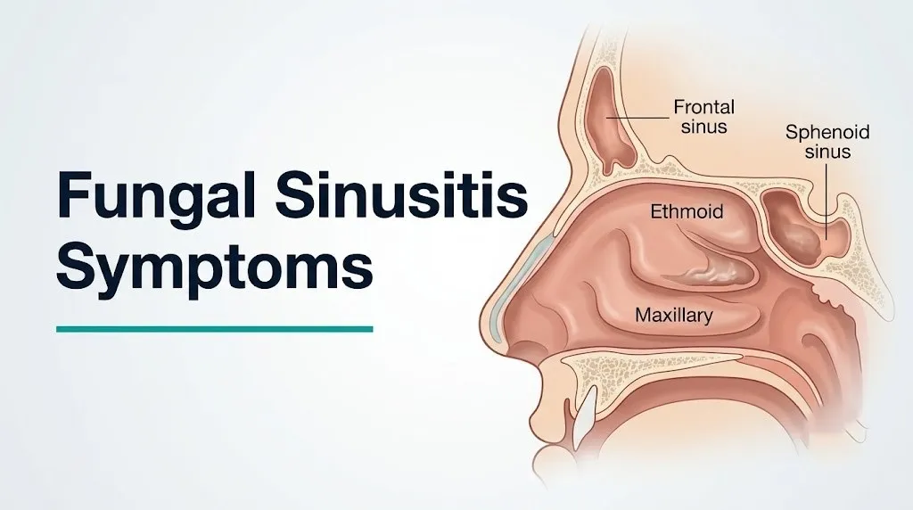
The paranasal sinuses are four paired air-filled cavities within the facial skeleton: the maxillary sinuses in the cheekbones, the ethmoid sinuses between the eyes, the frontal sinuses in the forehead, and the sphenoid sinuses at the skull base behind the nose. All are lined by pseudostratified ciliated columnar epithelium that traps inhaled particles and moves them toward the nasal cavity for clearance. Humans inhale hundreds to thousands of fungal spores daily without developing sinusitis, because the mucociliary clearance system and the innate immune response of nasal macrophages and neutrophils neutralize spores before they can germinate.
Fungal sinusitis develops when this defense fails. Five distinct failure modes produce five clinically distinct diseases:
- Allergic fungal sinusitis (AFS): immune hypersensitivity to fungal antigens in an atopic host
- Fungal ball (mycetoma): non-invasive fungal colonization of a single sinus without immune activation
- Acute invasive fungal sinusitis: rapid angioinvasive fungal infection in a severely immunocompromised host
- Chronic invasive fungal sinusitis: slow tissue invasion over months in a mildly immunocompromised host
- Chronic granulomatous invasive fungal sinusitis: Th1-mediated granulomatous response in a specific immunocompetent geographic population
The causative fungi differ by type. Dematiaceous molds (Bipolaris, Curvularia, Alternaria) are the primary pathogens in AFS. Aspergillus species predominate in fungal ball and chronic invasive disease. Mucorales (Mucor, Rhizopus, Cunninghamella) and Aspergillus fumigatus are the primary pathogens in acute invasive disease.
Individual variation note: Patients with cystic fibrosis experience colonization of their bronchial and sinonasal tracts by Aspergillus at higher rates than the general population due to their abnormal mucus rheology and impaired mucociliary clearance, but they more commonly develop allergic bronchopulmonary aspergillosis (ABPA) than invasive sinusitis unless they are also immunocompromised.
Fungal Sinusitis Symptoms: The Complete Clinical Picture
The complete symptom picture of fungal sinusitis depends entirely on which of the five clinical types is present, ranging from chronic nasal obstruction and facial pressure in non-invasive forms to black nasal crusting, periorbital swelling, cranial nerve palsies, and altered mental status in the invasive forms.
No single generic symptom list applies to all types. This is the clinical reality that most health content on this topic obscures by presenting one undifferentiated symptom list. The table below provides the framework for understanding which symptoms correspond to which type and at what urgency level:
| Type | Primary Symptoms | Immune Context | Urgency |
|---|---|---|---|
| Allergic fungal sinusitis | Chronic congestion, nasal polyps, thick multicolored mucus, unilateral facial pressure | Atopic, immunocompetent | Outpatient, weeks |
| Fungal ball | Unilateral facial pain, foul odor, low-grade pressure | Immunocompetent, often elderly | Outpatient, weeks |
| Chronic invasive | Progressive unilateral symptoms, proptosis over months, vision changes | Mildly immunocompromised | Urgent, days |
| Acute invasive | Rapid black nasal crust, facial pain, periorbital swelling, fever | Severely immunocompromised or DKA | Emergency room today |
| Chronic granulomatous invasive | Progressive unilateral nasal mass, proptosis, diplopia | Immunocompetent, specific geography | Urgent, days |
Individual variation note: Elderly patients over 70 with undiagnosed type 2 diabetes mellitus may present with what appears to be a fungal ball or chronic invasive sinusitis on their initial evaluation, but their occult hyperglycemic state places them at risk for rapid progression to an acute invasive course if their glucose and ketoacidosis status is not promptly assessed.
Nasal Discharge and Nasal Crust in Fungal Sinusitis
The character of nasal discharge and nasal crusting in fungal sinusitis is one of the most diagnostically informative symptoms because different fungal types produce chemically and visually distinct secretions that correlate with the underlying pathological mechanism.
In allergic fungal sinusitis, the characteristic discharge is thick, viscous, and described by patients as “peanut butter-like” or “axle grease-like” in consistency. The color ranges from tan to brown to dark green. This material is eosinophilic mucin: a dense accumulation of eosinophils, Charcot-Leyden crystals (diamond-shaped protein crystals from eosinophil degranulation), and fragmented, non-viable fungal hyphae, all suspended in abnormally viscous mucus. The mucin is not pus and contains no live organisms in sufficient numbers to cause tissue invasion. Its thickness and density impair the normal mucociliary clearance mechanism, leading to obstruction.
In fungal ball sinusitis, discharge is typically unilateral and may carry a foul, cheesy, or moldy odor from the anaerobic metabolic byproducts of the dense hyphal mass. Patients often describe an offensive smell that they can detect themselves. The discharge itself may not be visually dramatic.
The finding that carries the most urgent clinical significance is black nasal discharge or black nasal crusting. Black coloration in the nasal mucosa or on expelled nasal material indicates necrosis of the sinonasal tissue caused by fungal vascular invasion. The black color represents infarcted, gangrenous tissue. This is the characteristic appearance of acute invasive mucormycosis or acute invasive aspergillosis, and it is a clinical emergency requiring same-day evaluation.
The Centers for Disease Control and Prevention identifies black or bloody nasal discharge combined with facial swelling in a patient with known immunocompromise as a presentation that should trigger immediate emergency evaluation for invasive fungal disease.
Individual variation note: Post-nasal drip from AFS in patients who also have asthma may trigger lower airway bronchospasm, with patients reporting simultaneous sinus symptoms and wheezing that worsens in parallel. This overlap occurs because the same eosinophilic inflammatory process affecting the sinonasal mucosa can amplify lower airway eosinophilic inflammation through the unified airway hypothesis.
Facial Pain and Pressure in Fungal Sinusitis
Facial pain and pressure in fungal sinusitis are produced by increased pressure within the affected sinus cavities and by direct inflammatory or invasive involvement of the surrounding facial bone and soft tissue, with the specific location of pain mapping directly to the anatomical position of the affected sinus.
The paranasal sinuses occupy specific anatomical territories, and the pain from each maps predictably:
- Maxillary sinus involvement (the most common sinus affected in fungal ball): pain and pressure below the eye, across the cheek, and sometimes referred to the upper teeth. Pain from the upper molar or premolar region without a dental cause should raise clinical suspicion for maxillary sinus disease.
- Ethmoid sinus involvement (frequently affected in AFS): pain between and behind the eyes, with a sensation of deep orbital pressure.
- Frontal sinus involvement: pain across the forehead and above the eyebrows, often worse when bending forward (which increases hydrostatic pressure within the sinus).
- Sphenoid sinus involvement (a characteristic site in acute invasive aspergillosis): deep, central headache that is poorly localized, often described as a headache “behind the eyes” or “in the center of the head.” Sphenoid sinus involvement is anatomically adjacent to the cavernous sinus, the internal carotid artery, and multiple cranial nerve foramina, making it the most clinically dangerous sinus location.
Think of the sinuses like a series of sealed pressure chambers in the face. When fungal disease fills any chamber with thick mucin, a fungal ball, or swollen mucosa, the pressure inside that chamber rises. The surrounding bone conducts that pressure as pain. When the disease breaks through the chamber walls, the pain changes character: it becomes sharper, more localized, and is accompanied by new signs from whatever structure the infection has entered.
According to the International Forum of Allergy and Rhinology, facial pain that is unilateral, persistent, and unresponsive to two or more antibiotic courses is one of the strongest clinical indicators warranting nasal endoscopy and CT imaging to evaluate for fungal sinus disease.
Individual variation note: Patients with trigeminal neuralgia may experience facial pain that superficially resembles the pain distribution of maxillary or ethmoid sinus fungal disease. A dentist or primary care physician evaluating facial pain should always assess for sinus congestion, nasal discharge, and nasal obstruction on the same side as the pain before attributing it solely to neurological causes.
Key Takeaway: Black nasal crusting or discharge in any patient with diabetes, cancer treatment, or organ transplant is a clinical emergency, not a sinus symptom to monitor at home, because it indicates tissue necrosis from fungal vascular invasion.
Headache Patterns in Fungal Sinusitis
Headache in fungal sinusitis follows patterns that reflect which sinus is involved and whether the infection is producing pressure from within a cavity or invasive destruction of surrounding structures.
The most clinically concerning headache pattern in fungal sinusitis is the sphenoid sinus headache: a deep, central, poorly localizable ache located “behind the eyes” or “in the center of the skull” that is present at rest and does not respond to standard analgesics. The sphenoid sinuses sit at the skull base, separated from the cavernous sinus, the pituitary gland, and the optic chiasm by thin bony walls. Unlike the frontal or maxillary sinuses, the sphenoid sinus has no accessible anterior wall for drainage, making it less likely to decompress spontaneously and more prone to producing pressure-related neurological signs as disease advances.
A headache from AFS typically correlates with the pattern of polyp distribution: most often frontal or periorbital in location, worsening with head position changes, and present for months to years. It is rarely acute or severe enough to wake the patient from sleep. Patients often report that the headache feels like a permanent pressure rather than a sharp pain.
By contrast, the headache of acute invasive fungal sinusitis is rapid in onset, severe, and accompanied by fever and systemic signs within days of beginning. It does not stay confined to a predictable sinus distribution. As infection spreads from the sinuses toward the skull base, the headache becomes more diffuse and is joined by cranial nerve signs and meningismus (neck stiffness, photophobia) if meningeal involvement occurs.
According to clinical guidelines published by the Infectious Diseases Society of America, new severe headache in a neutropenic or organ transplant patient should be evaluated with CT or MRI of the sinuses and brain to exclude invasive fungal sinusitis before attributing the headache to other causes.
Individual variation note: Patients who have recently undergone sphenoid sinus surgery for any reason have altered anatomy and scarring that can make the headache pattern of recurrent or residual fungal disease atypical and more difficult to localize clinically without imaging.
Allergic Fungal Sinusitis Symptoms
Allergic fungal sinusitis symptoms are produced by a Type I IgE-mediated and Type III immune complex hypersensitivity reaction to fungal antigens in the sinonasal mucosa of atopic individuals, and they include chronic nasal obstruction, nasal polyposis, thick multicolored mucus discharge, facial pressure, and in advanced cases, proptosis from orbital expansion of inspissated eosinophilic mucin.
AFS is the most common form of fungal sinusitis in immunocompetent individuals, and it shares clinical features with chronic rhinosinusitis with nasal polyps (CRSwNP) to the point that the two are frequently confused. What distinguishes AFS is not the symptoms themselves but their underlying driver: the characteristic eosinophilic mucin, elevated serum IgE specific to the causative mold (most often Bipolaris, Curvularia, or Aspergillus), and a specific CT imaging pattern showing heterogeneous hyperattenuating material within the sinuses.
Key AFS symptoms:
- Chronic nasal obstruction, typically bilateral, present for months to years before diagnosis
- Nasal polyposis: grape-like protrusions of edematous sinonasal mucosa visible on nasal endoscopy and palpable as soft masses on anterior rhinoscopy
- Thick, discolored nasal discharge that ranges from tan to dark green or brown; the inspissated nature distinguishes it from typical bacterial mucopurulent discharge
- Loss of smell (anosmia or hyposmia) from polyp-related obstruction of the olfactory cleft between the superior turbinate and the nasal septum
- Facial pressure or fullness, typically bilateral and chronic rather than the acute severe pain of bacterial rhinosinusitis
- In advanced cases, visible facial asymmetry or proptosis (forward displacement of the eyeball) from expansion of the sinus cavity by accumulated eosinophilic mucin
Research published in the Journal of Allergy and Clinical Immunology found that patients with AFS have total serum IgE levels typically exceeding 1000 IU/mL, with specific IgE to the causative dematiaceous mold detectable on skin prick or ImmunoCAP testing in the majority of confirmed cases.
Individual variation note: AFS is more common in young adults living in warm, humid climates, where environmental dematiaceous mold spore counts are higher. In the United States, AFS is more frequently diagnosed in the Southeast and Southwest than in Northern states, reflecting the regional distribution of Bipolaris and Curvularia species in outdoor air.
Fungal Ball Sinusitis Symptoms
Fungal ball sinusitis symptoms are produced by a dense, non-invasive mass of fungal hyphae packed into a single sinus cavity, most frequently the maxillary sinus, and include unilateral facial pressure, unilateral post-nasal drip with a foul odor, and recurrent unilateral sinus infections that fail to respond to antibiotics.
Think of a fungal ball like a tightly packed nest of mycelial threads sitting inside the sinus cavity. The mass is not invading the sinus walls. It is simply occupying the space, blocking normal drainage through the ostiomeatal complex, and gradually growing within the sinus environment. There is no significant immune response: the host’s immune system is not producing a hypersensitivity reaction (as in AFS) and is not being overwhelmed (as in invasive disease). The problem is purely mechanical plus the byproducts of fungal metabolism.
Specific symptoms of fungal ball sinusitis:
- Unilateral facial pressure or fullness localized to the cheek or below the eye (maxillary distribution)
- Unilateral post-nasal drip with a persistent foul or musty odor that patients can detect themselves
- Recurrent “bacterial” sinus infections on one side that temporarily improve with antibiotics but recur within weeks, because antibiotics address secondary bacterial superinfection but not the primary fungal mass
- Occasional low-grade facial pain that is chronic and dull rather than acute and severe
- In some cases, no symptoms at all: fungal balls are discovered incidentally on CT scans obtained for unrelated reasons in approximately 10 to 20 percent of cases, according to retrospective imaging studies published in Otolaryngology: Head and Neck Surgery
Fungal ball does not produce fever, systemic symptoms, or neurological signs, because there is no tissue invasion and no significant immune activation. The absence of fever and systemic signs in a patient with unilateral sinus symptoms is one of the clinical features that suggests fungal ball rather than bacterial acute rhinosinusitis.
Individual variation note: Fungal balls of the maxillary sinus are more frequent in patients who have had prior dental treatment of the upper molar or premolar roots, because dental materials including zinc oxide and amalgam can migrate into the maxillary sinus through the sinus floor and provide a nidus for Aspergillus colonization. Patients with a history of upper molar root canal treatment who develop unilateral maxillary symptoms should specifically mention this history to an otolaryngologist.
Acute Invasive Fungal Sinusitis Symptoms
Acute invasive fungal sinusitis symptoms develop over days and include rapidly worsening unilateral facial pain, high fever, nasal congestion progressing to black nasal crust or eschar, periorbital swelling, and neurological signs, all driven by angioinvasive fungal hyphae producing vascular thrombosis and tissue infarction.
This is the form that kills. The pathological mechanism distinguishes it from every other type: Aspergillus fumigatus and Mucorales species possess the capacity to invade blood vessel walls (angioinvasion), producing thrombosis of the arteries and veins supplying the sinonasal mucosa. When the vascular supply to a segment of tissue is eliminated by fungal thrombosis, that tissue undergoes infarction and necrosis. Infarcted tissue turns black: the clinical hallmark of this disease.
The symptom sequence in acute invasive fungal sinusitis typically follows this timeline in severely immunocompromised patients:
- Days 1 to 2: Fever (temperature above 38.3°C/101°F) that does not respond to broad-spectrum antibiotics, combined with new unilateral nasal congestion
- Days 2 to 4: Facial pain worsens on the affected side; the nasal mucosa appears pale, dusky, or gray on examination (loss of normal pink mucosal color indicating ischemia)
- Days 3 to 5: Black eschar appears on the nasal septum, inferior turbinate, or palate; this represents infarcted necrotic tissue; the hard palate may develop a dark discoloration or a perforation in advanced cases
- Days 4 to 7: Periorbital swelling, proptosis (forward displacement of the eyeball), and visual changes as infection extends through the lamina papyracea into the orbit
- Days 5 to 10 or sooner: Cranial nerve signs, altered mental status, and other neurological changes as disease extends through the cribriform plate or via the cavernous sinus into the intracranial compartment
The Infectious Diseases Society of America states that acute invasive fungal sinusitis in neutropenic patients requires same-day initiation of antifungal therapy and urgent surgical debridement to have any meaningful impact on mortality.
Individual variation note: Patients undergoing induction chemotherapy for acute myeloid leukemia (AML) face the highest per-patient risk of acute invasive aspergillosis of any clinical population, because induction regimens produce prolonged profound neutropenia (neutrophil counts below 100 per microliter for 2 to 4 weeks) that eliminates the neutrophil-mediated defense against inhaled Aspergillus spores entirely.
Key Takeaway: In any immunocompromised patient, black or gray nasal mucosa visible on examination, or black material expelled from the nose, is a finding that requires emergency evaluation within hours, not days, because each day of delay in surgical debridement of acute invasive fungal sinusitis measurably worsens survival.
Chronic Invasive Fungal Sinusitis Symptoms
Chronic invasive fungal sinusitis produces symptoms that develop over months rather than days, including unilateral facial swelling, progressive orbital symptoms, and cranial nerve involvement, and it can mimic a sinonasal tumor in its slow, mass-like expansion through tissue.
Unlike the acute invasive form, chronic invasive fungal sinusitis does not produce dramatic rapid tissue necrosis. The fungal hyphae invade mucosal and bony tissue slowly, producing a granulation tissue response without the angiotropic thrombosis that characterizes the acute form. There is no black eschar. Instead, the tissue becomes thickened, vascular, and progressively destructive in a pattern that radiographically and clinically resembles a malignancy.
Patients typically present after weeks to months of worsening symptoms:
- Progressive unilateral nasal obstruction and discharge with a gradual rather than rapid onset
- Facial swelling that develops slowly and may initially be attributed to chronic sinusitis or dental disease
- Unilateral periorbital swelling and proptosis developing over weeks as infection extends from the ethmoid or sphenoid sinus into the orbital fat
- Progressive visual disturbance as the optic nerve is compressed at the orbital apex
- Facial numbness or altered sensation in the distribution of the trigeminal nerve (forehead, cheek, jaw) as perineural invasion occurs
The absence of fever and systemic toxicity in chronic invasive disease is what delays diagnosis: patients and clinicians do not perceive the clinical urgency that is apparent in the acute invasive form. By the time orbital or intracranial involvement is recognized, the infection has often been present for three to six months.
Chronic invasive fungal sinusitis predominantly affects patients with poorly controlled diabetes mellitus without ketoacidosis, patients on long-term moderate-dose corticosteroid therapy, and patients with AIDS with CD4 counts below 200 cells per microliter, according to case series published in Clinical Infectious Diseases.
Individual variation note: Patients taking tumor necrosis factor-alpha (TNF-alpha) inhibitors (such as adalimumab or infliximab) for rheumatoid arthritis or inflammatory bowel disease have an increased risk of chronic invasive aspergillosis affecting the sinuses, because TNF-alpha is a key cytokine in the macrophage-mediated defense against Aspergillus. Rheumatologists prescribing these agents should screen for prior fungal exposure and consider antifungal prophylaxis in high-exposure settings.
Orbital and Vision Symptoms of Fungal Sinusitis
Orbital and vision symptoms in fungal sinusitis indicate that infection has extended from the paranasal sinuses through the thin bony orbital walls into the orbital contents, producing symptoms that include periorbital swelling, proptosis, double vision, reduced visual acuity, and complete vision loss if the optic nerve is compressed or infarcted.
The ethmoid sinuses are separated from the medial orbital wall by the lamina papyracea, a paper-thin bony plate that provides minimal resistance to fungal invasion. The sphenoid sinus shares its posterior wall with the orbital apex, the narrow posterior compartment containing the optic nerve, the superior orbital fissure with its cranial nerve contents, and the ophthalmic artery. Extension into the orbital apex produces the clinical syndrome of orbital apex syndrome: simultaneous involvement of cranial nerves II (optic), III (oculomotor), IV (trochlear), and VI (abducens) with the first branch of the trigeminal nerve (V1).
Observable orbital symptoms by anatomical stage of involvement:
- Medial orbital involvement (from ethmoid extension): periorbital swelling of the medial eyelid, tenderness over the medial orbital rim, early proptosis
- Orbital fat involvement: proptosis becomes more prominent; resistance to retropulsion of the eyeball is increased on examination
- Orbital apex syndrome: ptosis (drooping eyelid), fixed dilated pupil, loss of all extraocular movements, severe periorbital pain, and reduced visual acuity occurring together
- Complete vision loss: optic nerve infarction from fungal thrombosis of the ophthalmic artery or direct nerve compression; this finding is an absolute emergency
The presence of any orbital symptom in a patient with fungal sinusitis, regardless of type, represents intracranial extension risk and requires immediate ophthalmological and neurosurgical assessment alongside infectious disease and otolaryngology consultation.
According to case series published in Otolaryngology: Head and Neck Surgery, the time from first orbital sign to intracranial extension in acute invasive fungal sinusitis can be as short as 24 to 48 hours in profoundly neutropenic patients.
Individual variation note: Children with immune thrombocytopenia or primary immunodeficiencies such as chronic granulomatous disease are at risk for invasive Aspergillus sinusitis with orbital extension at ages when clinicians least expect a life-threatening fungal infection, making pediatric otolaryngology consultation appropriate whenever a child with a known primary immunodeficiency presents with orbital signs.
Rhinocerebral Mucormycosis Symptoms
Rhinocerebral mucormycosis is an acute invasive fungal infection caused by Mucorales species that begins in the nasal mucosa, rapidly extends through the paranasal sinuses and orbital tissues, and reaches the brain, producing a clinical picture that combines facial pain, black nasal eschar, periorbital swelling, proptosis, cranial nerve palsies, and altered mental status within days of onset.
The “rhinocerebral” descriptor reflects the anatomical route of spread: from the nasal passages (rhino) to the brain (cerebral) via the paranasal sinuses, the orbital apex, and the cribriform plate. Mucorales species are uniquely positioned to follow this route because of two specific virulence characteristics: (1) their hyphae are non-septate (coenocytic), making them larger and more angioinvasive than Aspergillus hyphae; (2) they express spore coat protein (CotH) that binds directly to glucose-regulated protein 78 (GRP78) on endothelial cell surfaces, mediating direct endothelial invasion.
The clinical progression of rhinocerebral mucormycosis:
- Early stage (days 1 to 3): Fever, facial pain, nasal congestion, and a gray or pale nasal mucosa rather than the normal pink appearance
- Middle stage (days 3 to 7): Black eschar of the nasal septum or inferior turbinate; hard palate may show dark discoloration; periorbital edema develops; patients appear systemically unwell
- Late stage (days 5 to 14): Proptosis, ophthalmoplegia, facial hypoesthesia (numbness), altered mental status, headache worsening beyond sinus distribution, and in some cases skin necrosis over the cheek
The Centers for Disease Control and Prevention reports that rhinocerebral mucormycosis carries a mortality rate exceeding 30 percent even with prompt surgical debridement and appropriate antifungal therapy, and this rate rises substantially when diagnosis is delayed by more than 48 hours after the appearance of orbital signs.
Individual variation note: Rhinocerebral mucormycosis has been documented in patients who have received deferoxamine therapy for iron overload (as in dialysis patients or transfusion-dependent hematological conditions). Deferoxamine paradoxically increases serum iron available to Mucorales by forming a ferrioxamine complex that Mucorales can use as an iron source, effectively supplying the fungus with its primary growth nutrient. Prescribers of deferoxamine should be aware of this specific risk.
Fungal Sinusitis Symptoms in Immunocompromised Patients
Fungal sinusitis symptoms in immunocompromised patients are often atypical, blunted, or delayed compared to symptoms in immunocompetent individuals, because the inflammatory response that normally produces pain, swelling, and fever requires intact immune function that is absent or reduced in these patients.
This is the most clinically dangerous aspect of fungal sinusitis in immunocompromised individuals: the symptoms that would normally alert an immunocompetent person to seek care are absent or minimal until the disease has reached an advanced stage. A neutropenic patient with acute invasive aspergillosis may have only low-grade fever and mild nasal congestion at a time when fungal hyphae are actively invading the cribriform plate.
Populations at highest risk for invasive fungal sinusitis and the specific immune deficit that creates their risk:
- Hematologic malignancy patients on chemotherapy: profound neutropenia eliminates the neutrophil-driven reactive oxygen species mechanism that kills Aspergillus conidia in the alveoli and sinonasal mucosa
- Bone marrow transplant recipients: combined neutropenia (during conditioning) and T-cell depletion (post-engraftment graft-versus-host disease treatment) eliminates both innate and adaptive antifungal defenses
- Solid organ transplant recipients: calcineurin inhibitor immunosuppression (tacrolimus, cyclosporine) impairs T-lymphocyte function and IL-2 production, reducing fungal-specific adaptive immunity
- Patients on prolonged high-dose corticosteroids: corticosteroids impair macrophage phagocytosis of fungal conidia and reduce neutrophil adhesion to endothelium
- Patients with AIDS and CD4 counts below 200 cells per microliter: loss of CD4+ T-helper cells eliminates coordination of the adaptive antifungal immune response
- Patients on biologic TNF-alpha inhibitors: TNF-alpha is required for granuloma formation and macrophage activation against intracellular fungal pathogens
According to clinical guidance published in Clinical Infectious Diseases, any febrile episode that persists beyond 96 hours despite broad-spectrum antibacterial therapy in a neutropenic patient should prompt evaluation for invasive fungal infection including sinonasal assessment with CT imaging.
Individual variation note: Pediatric oncology patients undergoing treatment for acute lymphoblastic leukemia (ALL) face a window of highest invasive fungal sinusitis risk during induction and consolidation chemotherapy phases, when neutropenia is most prolonged. Pediatric infectious disease specialists routinely recommend antifungal prophylaxis during these phases at many academic centers.
Key Takeaway: An immunocompromised patient with any new nasal symptoms, even mild ones, should be evaluated the same day with direct communication to their oncologist or transplant team, because the absence of dramatic symptoms does not mean the infection is mild; it may mean the immune response is too suppressed to produce warning signs.
Fungal Sinusitis Symptoms in Diabetic Ketoacidosis
Fungal sinusitis symptoms in patients with diabetic ketoacidosis (DKA) follow the acute invasive pattern caused predominantly by Mucorales species, specifically Rhizopus microsporus and Mucor species, and the physiological conditions of DKA directly enable a level of fungal virulence that would not occur in euglycemic or non-acidotic states.
The mechanism connecting DKA to rhinocerebral mucormycosis is specific, named, and well-characterized in the infectious disease literature. Three distinct physiological abnormalities of DKA each independently enable Mucorales proliferation:
- Elevated serum glucose: Mucorales express glucose transporters and are metabolically stimulated by hyperglycemia; high glucose also impairs neutrophil chemotaxis (the directed movement of neutrophils toward the site of infection) and reduces neutrophil oxidative burst capacity
- Acidemia: The acidic pH of DKA (serum pH below 7.3) causes transferrin to release its normally chelated iron. Transferrin at normal physiologic pH binds free iron with high affinity, creating fungistatic conditions in serum. At acidic pH, iron is released and becomes freely available as a Mucorales growth factor.
- High free serum iron: Elevated free iron, combined with high glucose, creates an ex vivo culture condition for Mucorales in the patient’s own bloodstream and sinonasal tissues
The clinical result: patients in DKA who inhale Rhizopus or Mucor spores can develop fulminant rhinocerebral mucormycosis within 48 to 72 hours of spore exposure. DKA patients who develop new facial pain, headache, or nasal symptoms during their hospitalization require immediate sinonasal evaluation with nasal endoscopy and CT imaging, not watchful waiting.
The National Institute of Allergy and Infectious Diseases recognizes diabetic ketoacidosis as the single strongest predisposing condition for rhinocerebral mucormycosis, accounting for a majority of cases in published North American and European case series.
Individual variation note: Young adults with type 1 diabetes mellitus who experience their first DKA episode at diagnosis of diabetes have the same Mucorales risk as patients with established type 2 diabetes in DKA. The mechanism is identical regardless of diabetes type or duration. Emergency department physicians managing any patient in DKA who has facial symptoms should communicate this risk to the treating endocrinologist.
How Fungal Sinusitis Symptoms Differ From Bacterial Sinusitis
Fungal sinusitis symptoms differ from bacterial sinusitis symptoms primarily in their duration pattern, their response to antibiotics, the character of nasal discharge, and in the invasive forms, the presence of features that have no bacterial equivalent including black nasal eschar, anosmia from polyp mass, and orbital signs.
Most cases of acute bacterial rhinosinusitis follow a predictable course: they develop after a viral upper respiratory infection, reach peak severity at 7 to 10 days, and begin to improve spontaneously or with antibiotic treatment within 10 to 14 days. Fungal sinusitis does not follow this pattern.
| Feature | Bacterial Sinusitis | Fungal Sinusitis |
|---|---|---|
| Typical duration before presentation | Days to 2 weeks | Weeks to months (non-invasive); days (acute invasive) |
| Response to antibiotics | Improves in 10 to 14 days | No improvement despite multiple antibiotic courses |
| Discharge character | Yellow-green mucopurulent | Thick brown/tan mucin (AFS) or foul unilateral (fungal ball) or black (acute invasive) |
| Nasal polyps | Possible (CRS-related) | Characteristic in AFS; absent in invasive forms |
| Fever | Present in acute bacterial sinusitis | Present in acute invasive; absent in non-invasive types |
| Orbital signs | Rare, only in orbital cellulitis complication | Present in chronic and acute invasive forms |
| Black nasal eschar | Never | Characteristic of acute invasive mucormycosis/aspergillosis |
| Serum IgE elevation | Not expected | Characteristic of AFS (often above 1000 IU/mL) |
The most practically useful clinical indicator for a patient trying to differentiate the two: sinus symptoms that have persisted despite two or more full antibiotic courses should prompt an otolaryngology referral for nasal endoscopy and CT imaging rather than a third antibiotic prescription.
According to clinical guidance from the American Rhinologic Society, chronic rhinosinusitis that fails to respond to appropriate antibacterial therapy should always include fungal sinusitis in the differential diagnosis and should not be treated with additional empirical antibiotics without endoscopic evaluation.
Individual variation note: Patients with cystic fibrosis have a particularly difficult diagnostic challenge in distinguishing bacterial from fungal sinusitis because both occur simultaneously in many CF patients, and their abnormal mucus creates conditions that support both bacterial biofilm formation and fungal colonization within the same sinus cavity at the same time.
How Doctors Diagnose Fungal Sinusitis
Doctors diagnose fungal sinusitis through a combination of nasal endoscopy, CT imaging of the paranasal sinuses, tissue biopsy with histopathological staining, and fungal culture, with the specific diagnostic sequence depending on whether invasive or non-invasive disease is suspected.
The diagnostic process for suspected fungal sinusitis follows this sequence:
- Clinical history and symptom assessment: The clinician establishes symptom duration, antibiotic treatment history, immune status, diabetes history, and allergic history. Any immunocompromised patient with nasal symptoms is moved to an expedited pathway.
- Nasal endoscopy: A board-certified otolaryngologist introduces a rigid or flexible endoscope through the nares to directly visualize the nasal mucosa, middle meatus, and nasal turbinates. In AFS, gray-green or brown eosinophilic mucin in the middle meatus is characteristic. In acute invasive disease, pale, gray, or necrotic mucosa with black eschar is the alarm finding.
- CT scan of the paranasal sinuses with bone windows: CT imaging identifies the anatomical distribution of disease, the presence of bony erosion or expansion, and the characteristic findings of each type. AFS produces heterogeneous hyperattenuating material (the dense eosinophilic mucin appears bright on CT). Fungal ball produces a calcified or hyperdense central mass in a single sinus. Invasive disease produces bony erosion and, in advanced cases, orbital or intracranial extension.
- MRI with gadolinium: Used when intracranial or orbital extension is suspected, or when CT findings are ambiguous. MRI provides superior soft tissue detail, including the ability to identify cavernous sinus thrombosis, dural enhancement, and brain involvement.
- Tissue biopsy with histopathological staining: The definitive diagnostic test for invasive disease. Tissue is stained with Gomori methenamine silver (GMS) stain or periodic acid-Schiff (PAS) stain to visualize fungal hyphae. Mucorales hyphae are non-septate (coenocytic) and branch at right angles; Aspergillus hyphae are septate and branch at 45-degree angles. This distinction determines antifungal drug selection.
- Serum galactomannan and beta-D-glucan assays: Blood tests that detect circulating fungal cell wall components. Galactomannan is specific to Aspergillus; beta-D-glucan detects most fungi except Mucorales (which do not contain beta-D-glucan in their cell wall, making this test negative in mucormycosis).
Individual variation note: Pregnant women requiring CT imaging for suspected fungal sinusitis should be reassured that the radiation dose from a head and face CT scan is below the threshold associated with fetal harm, but the treating physician should document the clinical indication clearly and use the minimum technical parameters needed for diagnostic adequacy.
When Fungal Sinusitis Symptoms Require Emergency Evaluation
Certain symptoms associated with fungal sinusitis require immediate emergency evaluation. Do not wait to see if these resolve on their own.
Acute invasive fungal sinusitis and rhinocerebral mucormycosis can progress from rhinosinusitis to intracranial involvement within 48 to 72 hours in profoundly immunocompromised or DKA patients. Time to surgical debridement is the primary determinant of survival in these conditions.
Call 911 or go to the nearest emergency room immediately if you observe any of the following in a patient with known immunocompromise, active cancer treatment, bone marrow or organ transplant, or diabetic ketoacidosis:
- Black, dark brown, or gray nasal discharge or nasal crusting in an immunocompromised or diabetic patient: indicates tissue necrosis from fungal vascular invasion, an emergency requiring same-day surgical evaluation
- Periorbital swelling, proptosis (eyeball pushed forward), or chemosis (conjunctival swelling): indicates extension of fungal infection into the orbital contents through the lamina papyracea
- Double vision, loss of eye movement in any direction, or drooping eyelid (ptosis): indicates cranial nerve involvement at the orbital apex from fungal infection
- Any reduction in visual acuity or complete vision loss in one eye: indicates optic nerve compression or infarction from fungal invasion; vision loss from fungal orbital apex syndrome may be irreversible if not treated within hours
- Altered mental status, confusion, or new neurological deficit in a patient with sinus symptoms: indicates intracranial extension through the cribriform plate or cavernous sinus
- Fever above 38.3°C (101°F) persisting despite 48 to 96 hours of broad-spectrum antibiotics in a neutropenic patient: requires fungal evaluation including emergent nasal endoscopy and CT imaging
- Facial numbness or hypoesthesia on the same side as sinus symptoms: indicates perineural fungal invasion of the trigeminal nerve branches, a sign of advanced invasive disease
- Palatal discoloration, palatal ulceration, or perforation of the hard palate: indicates spread of mucormycosis from the maxillary sinus through the sinus floor into the palatal structures
These presentations indicate acute invasive fungal sinusitis or rhinocerebral mucormycosis and require emergency evaluation by a team including infectious disease, otolaryngology, ophthalmology, and neurosurgery, not an urgent care visit or a telephone triage call.
Frequently Asked Questions About Fungal Sinusitis Symptoms
What are the first symptoms of fungal sinusitis?
The first symptoms of fungal sinusitis depend on which type is developing: in allergic fungal sinusitis, the earliest symptoms are chronic nasal obstruction and thickening nasal discharge that fails to respond to antibiotics over weeks to months.
In acute invasive fungal sinusitis, the earliest symptoms are fever that does not respond to antibiotics, facial pain, and nasal congestion that begin within days in a severely immunocompromised patient.
Black or darkened nasal mucosa is a later but highly specific early warning sign of acute invasive disease and warrants emergency evaluation the same day it is observed.
How do I know if my sinus infection is fungal or bacterial?
A sinus infection is more likely to be fungal if it has persisted despite two or more complete antibiotic courses, if nasal discharge is thick, brown, or tan rather than yellow-green, or if you have a history of nasal polyps, allergies, or immunocompromise.
No symptom alone can confirm fungal sinusitis; a board-certified otolaryngologist must perform nasal endoscopy and CT imaging of the paranasal sinuses to distinguish bacterial from fungal disease accurately.
Antibiotic resistance and chronic recurrence are the two most practically useful indicators that prompt evaluation for a fungal rather than bacterial cause.
Can fungal sinusitis be dangerous?
Fungal sinusitis ranges from a non-life-threatening chronic condition (allergic fungal sinusitis and fungal ball) to a rapidly fatal infection (acute invasive fungal sinusitis and rhinocerebral mucormycosis) depending on which type is present and the immune status of the patient.
The Centers for Disease Control and Prevention reports that rhinocerebral mucormycosis carries a mortality rate exceeding 30 percent even with prompt aggressive treatment, and this rises substantially when treatment is delayed.
For immunocompromised and DKA patients, any new sinus symptoms combined with fever should be treated as potentially dangerous and evaluated urgently.
What does fungal sinusitis discharge look like?
Fungal sinusitis discharge varies by type: allergic fungal sinusitis produces thick, viscous, tan to dark brown mucin described as having a peanut butter or axle grease consistency; fungal ball produces unilateral discharge with a foul or musty odor.
Acute invasive fungal sinusitis may produce black or blood-tinged discharge representing infarcted necrotic tissue, which is a clinical emergency in any immunocompromised or diabetic patient.
Any black or very dark nasal discharge or nasal crusting should be evaluated by a physician or emergency department the same day it is observed.
Who is most at risk for invasive fungal sinusitis?
Patients at highest risk for invasive fungal sinusitis include those with profound neutropenia from chemotherapy or bone marrow transplant, those with diabetic ketoacidosis, solid organ transplant recipients on calcineurin inhibitor immunosuppression, and patients on prolonged high-dose corticosteroids.
The specific risk mechanism differs by group: neutropenic patients lack the neutrophil-mediated killing of inhaled fungal spores; DKA patients have free serum iron and impaired neutrophil function that enables Mucorales to proliferate rapidly.
Any patient in these categories who develops new sinus or facial symptoms should contact their oncologist, transplant physician, or endocrinologist immediately rather than waiting for a scheduled appointment.
Can mold in my house cause fungal sinusitis?
Household mold exposure can trigger or worsen allergic fungal sinusitis in atopic individuals by providing ongoing antigen exposure that amplifies the IgE-mediated sinonasal hypersensitivity response, but household mold does not typically cause invasive fungal sinusitis in immunocompetent people.
The fungi that cause invasive fungal sinusitis (Aspergillus fumigatus, Mucor and Rhizopus species) are ubiquitous environmental organisms present in outdoor air, soil, and decaying organic matter, not unique to household mold.
Immunocompromised patients should discuss environmental mold exposure with their infectious disease specialist, as high-spore environments including construction sites, agricultural settings, and mold-contaminated buildings may pose a higher inhalational challenge than typical household mold.
Closing
The most practically important thing this article communicates is the difference between two entirely distinct clinical realities that share the same name. For most people, fungal sinusitis is a chronic, frustrating condition that mimics persistent bacterial sinusitis and requires specialist evaluation to diagnose correctly. For immunocompromised and DKA patients, fungal sinusitis is a medical emergency where hours matter and where the absence of alarming symptoms does not indicate the absence of life-threatening disease.
If your sinus symptoms have persisted despite multiple antibiotic courses, make an appointment with a board-certified otolaryngologist and request nasal endoscopy and CT imaging of the paranasal sinuses. Bring a complete list of your allergy history, nasal polyp history, and any immunosuppressive medications to that appointment.
If you have diabetes, active cancer treatment, an organ transplant, or any immunosuppressive condition and you develop new facial pain, fever, or nasal symptoms, go to the emergency room or call your specialist today. That urgency is not an overreaction. It is what the clinical evidence on invasive fungal sinusitis requires.