Neck Sprain Symptoms: Full Guide to Signs and Red Flags
Neck sprain symptoms include neck pain, muscle stiffness, limited range of motion, headache, and in more serious cases, numbness, tingling, or dizziness that can develop hours to days after a cervical ligament injury. Most people recover fully from a mild to moderate neck sprain, but certain symptom combinations require immediate medical evaluation because they can indicate a more dangerous underlying injury that a sprain alone does not explain.
The American Academy of Orthopaedic Surgeons identifies cervical sprains as one of the most frequent soft tissue injuries of the spine, with motor vehicle accidents and contact sports among the most common mechanisms. What most health content fails to tell you is that four different cervical injuries (sprain, cervical fracture, disc herniation with nerve root compression, and vertebral artery dissection) can all present with neck pain after trauma, and distinguishing between them determines whether you need home rest or a 911 call.
This article covers every recognized neck sprain symptom with the physiological mechanism behind it, how early symptoms differ from subacute and chronic ones, how a sprain differs from a neck strain at the tissue level, which symptoms are red flags for something more serious, and exactly when and how to seek care at each level of severity.
Neck Sprain Symptoms
Neck sprain symptoms are produced by injury to one or more cervical ligaments and typically include neck pain, protective muscle spasm, stiffness, limited range of motion, and headache, with neurological symptoms such as arm numbness or dizziness appearing in more severe presentations.
The full clinical picture of a neck sprain varies by which ligament is damaged, how severely the fibers are torn, and whether any adjacent structures such as the facet joint capsule or intervertebral disc are involved. A person with a mild Grade I sprain (fiber microtearing without structural failure) may notice only modest soreness and tightness. A person with a Grade II or Grade III injury (partial or complete ligament disruption) may experience severe pain, marked restriction of movement, and symptoms that extend into the head, shoulders, and arms.
Core neck sprain symptoms by clinical significance:
- Neck pain localized to the posterior or lateral cervical region, worsened by movement
- Protective muscle stiffness that limits rotation, flexion, or extension
- Tenderness to palpation over the posterior cervical spine or lateral neck musculature
- Headache originating at the base of the skull and radiating toward the vertex or temples
- Shoulder and upper back pain from referred nociceptive input via cervical nerve branches
- Numbness or tingling in the arm or hand, if a cervical nerve root is secondarily irritated
- Dizziness or unsteadiness from proprioceptive disruption of cervical joint receptors
- Jaw pain or temporomandibular joint (TMJ) discomfort, particularly after whiplash-type mechanisms
According to the Mayo Clinic, neck sprain symptoms typically peak within 24 to 48 hours after the injury event and begin to improve progressively over two to four weeks in most uncomplicated presentations. Symptoms that worsen beyond 48 hours or fail to improve by six weeks require clinical reassessment.
Older adults with pre-existing cervical spondylosis (age-related degenerative narrowing of cervical intervertebral foramina and disc spaces) may experience more intense and longer-lasting symptoms from the same injury force because their baseline structural reserve is already reduced.
What Is a Cervical Sprain and Which Structures Are Injured
A cervical sprain is defined as a stretch or tear of one or more of the ligaments connecting the cervical vertebrae, distinct from a cervical strain, which involves injury to the muscles or muscle-tendon units rather than the ligaments themselves.
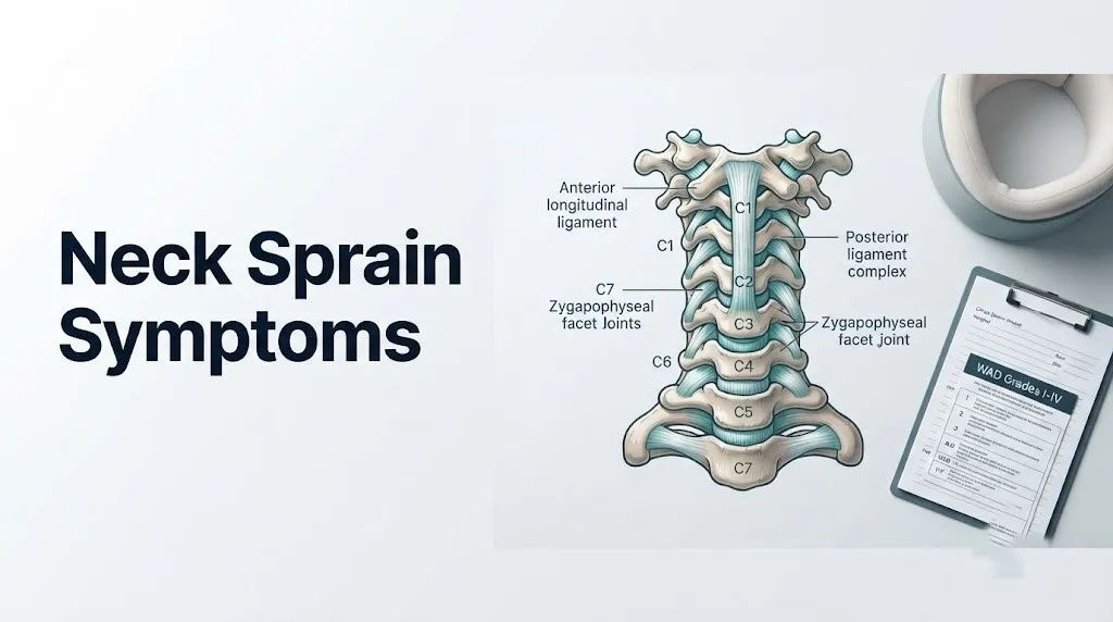
The cervical spine contains seven vertebrae (C1 through C7), and the ligament system holding them together is a layered, interlocking network. Understanding which specific ligaments are at risk explains why different injury mechanisms produce different symptom patterns. Think of these ligaments like the guy-wires holding a radio tower upright: each wire resists movement in a different direction, and which wire breaks determines which direction the tower becomes unstable.
The primary ligaments injured in a neck sprain include:
- Anterior longitudinal ligament (ALL): Runs along the front surfaces of the vertebral bodies from C1 to the sacrum. Resists hyperextension. Injured most commonly in rear-impact whiplash when the head snaps backward.
- Posterior longitudinal ligament (PLL): Runs along the posterior surfaces of the vertebral bodies inside the spinal canal. Resists hyperflexion. Injured in forward-impact mechanisms.
- Ligamentum nuchae: A thick posterior midline ligament running from the external occipital protuberance to the C7 spinous process. Resists forward flexion and provides attachment for posterior cervical muscles.
- Interspinous ligaments: Connect adjacent spinous processes. Resist flexion-induced separation. Tenderness directly over the posterior midline spinous processes often localizes to these.
- Capsular ligaments of the zygapophyseal (facet) joints: Among the richest sources of nociceptive fibers in the entire cervical spine. Injury to these capsular ligaments is responsible for a substantial proportion of post-sprain pain, particularly the referred headache pattern.
- Alar ligaments: Bilateral ligaments connecting the odontoid process (dens) of C2 to the occipital condyles. Resist excessive rotation and lateral flexion at the C1-C2 articulation.
According to the National Institute of Arthritis and Musculoskeletal and Skin Diseases (NIAMS), ligament injuries are graded by severity: Grade I involves microtearing of individual collagen fibers without structural failure, Grade II involves partial macroscopic tearing with some structural compromise, and Grade III involves complete ligament rupture.
What Does a Neck Sprain Feel Like
A neck sprain feels like a combination of local pain at the injury site, a deep aching stiffness that makes turning or tilting the head difficult, and often a sensation of soreness that extends into the base of the skull or upper shoulders.
The quality of the pain depends on which pain fibers are activated. Nociceptive A-delta fibers (thinly myelinated) in the injured ligament produce the initial sharp, acute pain felt at the moment of injury or within the first few hours. As the acute inflammatory response progresses over 12 to 24 hours, C-fibers (unmyelinated, slower conducting) carry a secondary wave of dull, deep, aching pain as bradykinin and prostaglandin E2 (produced via the COX-2 pathway in the injured connective tissue) accumulate in the peri-ligamentous space and sensitize peripheral nociceptors.
The stiffness component feels distinctly different from the pain itself. It is a mechanical restriction, a sense that the neck “won’t go there” rather than a pain so severe you are afraid to move. This is produced by the gamma motor neuron reflex arc: sensory spindle afferents in the injured tissue detect abnormal movement patterns and trigger reflexive contraction of the sternocleidomastoid, upper trapezius, and levator scapulae muscles as a protective guarding mechanism. The brain is essentially splinting the injured area by keeping the surrounding muscles in a state of sustained low-level contraction.
Quick Tip:
- Stiffness that is worse in the morning and loosens with gentle movement through the day suggests acute inflammatory-phase guarding, which is expected in the first 72 hours.
- Pain that is constant, identical in quality at rest and with movement, and does not change with position is a warning sign that warrants clinical evaluation rather than continued home monitoring.
- Patients who take opioid pain medications or have a high baseline pain tolerance may underreport early cervical sprain symptoms, which can delay recognition of a more serious injury.
Neck Pain and Stiffness: The Primary Symptoms of a Cervical Sprain
Neck pain and stiffness are the two most consistently reported primary symptoms of a cervical sprain, present in the vast majority of clinically confirmed cases and produced by distinct but overlapping physiological mechanisms.
Neck pain in a cervical sprain is primarily nociceptive in character, meaning it originates from tissue damage rather than from nerve injury. The injured ligament fibers release bradykinin and substance P from their torn ends. Substance P, released from the terminals of primary afferent C-fibers, promotes neurogenic inflammation: it causes vasodilation and plasma protein extravasation into the peri-ligamentous space, which amplifies local tenderness and creates the characteristic swelling and heat that accompany acute cervical ligament injury.
Pain localization provides a clinical clue to which structure is involved. Midline posterior cervical pain, directly over the spinous processes, suggests involvement of the interspinous ligaments or ligamentum nuchae. Lateral cervical pain, worse with rotation toward the painful side, suggests capsular ligament injury of the ipsilateral zygapophyseal joints. Anterior neck pain, reproduced by passive hyperextension, points toward anterior longitudinal ligament involvement.
The restricted range of motion of cervical stiffness has measurable clinical parameters. The American Academy of Family Physicians (AAFP) notes that normal cervical range of motion includes approximately 80 degrees of rotation in each direction, 45 to 50 degrees of lateral flexion, 60 degrees of flexion, and 75 degrees of extension. Acute cervical sprain typically reduces rotation and flexion-extension range of motion by 20 to 50 percent in moderate presentations.
| Movement | Normal Range | Typical Reduction in Moderate Sprain | Primary Limiting Structure |
|---|---|---|---|
| Rotation (each side) | 80 degrees | 30 to 50% reduction | Contralateral capsular ligaments, sternocleidomastoid |
| Flexion | 60 degrees | 20 to 40% reduction | Ligamentum nuchae, posterior musculature |
| Extension | 75 degrees | 20 to 40% reduction | Anterior longitudinal ligament, anterior muscles |
| Lateral flexion | 45 degrees | 15 to 30% reduction | Ipsilateral capsular ligaments, scalene muscles |
Children and adolescents with cervical sprains may show different range of motion restriction patterns than adults because their cervical ligaments are proportionally more elastic and the vertebral end plates are still developing. A pediatrician or pediatric orthopedic surgeon should evaluate cervical spine injuries in patients under 16 years of age.
Cervicogenic Headache and Referred Pain From a Neck Sprain
A cervicogenic headache is a headache caused by injury or dysfunction in the cervical spine, most commonly produced by injury to the C2-C3 zygapophyseal joint capsule, and it is one of the most frequently reported and least understood symptoms of a neck sprain.
The mechanism is specific. The trigemino-cervical nucleus is the caudal extension of the trigeminal nucleus caudalis, located in the brainstem and extending into the dorsal horn of the spinal cord at the C1-C3 levels. Nociceptive input from the upper cervical facet joint capsules (particularly C2-C3) converges onto this nucleus alongside input from the trigeminal nerve, which supplies sensation to the face, scalp, and head. Because these inputs share interneuron connections at the same spinal level, the brain cannot reliably distinguish whether a pain signal originated in the neck or in the head itself. The result is referred head pain that the patient experiences as a genuine headache, even though the actual injury is in the cervical ligament or joint capsule.
Research published in the Clinical Journal of Pain by Bogduk and colleagues established that C2-C3 facet joint injury produces a distinctive headache pattern: unilateral pain beginning at the suboccipital region (base of the skull on one side), radiating forward to the forehead, temple, or periorbital region on the same side. This is the classic cervicogenic headache of cervical sprain.
Distinguishing cervicogenic headache from other post-injury headache types matters clinically:
- Cervicogenic headache: Worsened by neck movement, relieved (partially) by maintaining a still head position, consistent unilateral distribution starting at the occiput.
- Post-concussion headache: Associated with a history of head impact, cognitive symptoms (difficulty concentrating, memory changes), light sensitivity, nausea. Requires neurological evaluation.
- Headache from vertebral artery dissection: Sudden onset, severe, described as the worst headache of life, may be accompanied by neck pain with neurological symptoms. Requires emergency evaluation.
Patients over 65 years of age who develop a new headache pattern after a neck injury warrant specific clinical attention: new headache in this age group carries a broader differential that includes temporal arteritis (giant cell arteritis), which requires prompt evaluation by a primary care physician or rheumatologist regardless of the neck injury context.
Key Takeaway: The headache after a neck sprain is not “just a headache.” It is a referred pain signal from injured C2-C3 facet joint capsules, transmitted through the trigemino-cervical nucleus. A headache that starts at the base of the skull and radiates forward on one side is a characteristic pattern of cervicogenic headache from a cervical sprain.
Neck Sprain Numbness, Tingling, and Nerve Symptoms
Numbness and tingling in the arm or hand after a neck injury indicate that a cervical nerve root is being irritated or compressed, which can occur in association with a sprain if the injury has also affected an adjacent intervertebral disc or if the inflammatory response within the spinal canal is irritating nearby neural structures.
The cervical nerve roots most commonly affected in neck sprain injuries are C5 through C8, because they exit the spinal canal through the intervertebral foramina at the most mechanically stressed segments of the mid and lower cervical spine. Each root produces a distinct pattern of numbness and tingling that follows the dermatome (the skin territory supplied by that specific nerve root):
- C5 nerve root (C4-C5 foramen): Numbness or tingling in the lateral upper arm and outer shoulder. Weakness of the deltoid muscle.
- C6 nerve root (C5-C6 foramen): Numbness or tingling in the thumb, index finger, and radial aspect of the forearm. Weakness of wrist extension.
- C7 nerve root (C6-C7 foramen): Numbness or tingling in the middle finger and dorsal forearm. Weakness of elbow extension (triceps).
- C8 nerve root (C7-T1 foramen): Numbness or tingling in the small finger and ulnar border of the hand. Weakness of intrinsic hand muscles.
A pure cervical sprain without disc involvement does not directly compress nerve roots. When numbness and tingling accompany a cervical sprain, it typically indicates one of three things: the injury has caused a secondary disc bulge or herniation that is pressing on an adjacent nerve root, the acute inflammatory swelling in the spinal canal is irritating a nerve root without physically compressing it, or the mechanism of injury was forceful enough to stretch a nerve root as well as the ligament.
According to the National Institute of Neurological Disorders and Stroke (NINDS), radicular arm symptoms after a neck injury that persist beyond 72 hours, or that are accompanied by arm weakness rather than just sensory symptoms, require clinical evaluation by a primary care physician within 24 to 48 hours to determine whether MRI of the cervical spine is indicated.
Patients taking anticoagulant medications such as warfarin, apixaban, or rivaroxaban who develop arm numbness or tingling after any cervical trauma require same-day emergency evaluation, because epidural hematoma (bleeding into the spinal canal compressing nerve roots or the spinal cord) is a rare but serious possibility in this population even with minor injury mechanisms.
Dizziness and Visual Symptoms With a Neck Sprain
Dizziness following a neck sprain is a recognized symptom that can arise from two distinct mechanisms: disruption of cervical proprioceptive joint receptors, or in more serious cases, involvement of the vertebral artery.
The cervical spine’s facet joint capsules contain a high density of mechanoreceptors (Ruffini endings, Pacinian corpuscles, and free nerve endings) that continuously transmit positional information to the brainstem’s balance centers. When these capsular mechanoreceptors are damaged or their input is distorted by ligament injury and muscle guarding, the vestibular system receives inconsistent positional data. The result is a persistent low-grade dizziness or unsteadiness called cervicogenic dizziness, which typically worsens with neck movement and improves when the head is held still.
Cervicogenic dizziness from a neck sprain is typically described as a sense of unsteadiness or “floating” rather than the room-spinning vertigo of a vestibular disorder like benign paroxysmal positional vertigo (BPPV). This distinction matters: if you feel the room spinning when you turn your head in a specific direction, that is more consistent with BPPV or an inner ear condition than with cervicogenic dizziness from a sprain.
Visual symptoms, including blurred vision or difficulty focusing, can occur in severe cervical sprains through sympathetic nervous system involvement. The cervical sympathetic chain runs anterior to the cervical vertebrae and can be irritated by significant anterior neck soft tissue injury, producing transient visual disturbances. However, visual symptoms after a neck injury should never be assumed to be benign without clinical evaluation.
The critical distinction that cannot be missed: dizziness with neck pain after trauma, accompanied by double vision, slurred speech, facial numbness, difficulty swallowing, or arm weakness, is a medical emergency. This symptom combination can indicate vertebral artery dissection, a tear in the wall of the vertebral artery that can cause stroke if not treated immediately.
Patients over 50 with a history of hypertension who develop dizziness after any neck trauma, even minor, should be evaluated the same day by a primary care physician or in an emergency room, because their baseline arterial fragility elevates the risk of vertebral artery injury with lower-force mechanisms.
Early Neck Sprain Symptoms in the First 24 to 72 Hours
Early neck sprain symptoms in the first 24 to 72 hours after injury are driven by the acute inflammatory phase, during which damaged ligament tissue releases inflammatory mediators that produce progressive pain amplification, increasing swelling, and escalating muscle guarding.
A clinically important feature of early cervical sprain is that symptoms often do not peak at the moment of injury. Instead, they intensify over the first 12 to 48 hours as the inflammatory cascade builds. Many people who sustain a neck sprain in a car accident feel relatively mild discomfort immediately afterward, then wake the following morning with marked pain, stiffness, and headache. This delayed-onset symptom intensification reflects the time course of prostaglandin E2 and substance P accumulation in the injured tissue and the progressive development of protective muscle guarding.
The Whiplash-Associated Disorder (WAD) grading scale, developed by the Quebec Task Force and published in Spine, classifies early symptom severity:
| WAD Grade | Early Symptoms | Clinical Action |
|---|---|---|
| Grade 0 | No neck pain or physical signs | Monitoring; no treatment needed |
| Grade I | Neck pain, stiffness, or tenderness only; no physical findings on exam | Home monitoring; primary care if no improvement in 72 hours |
| Grade II | Neck symptoms plus musculoskeletal signs (reduced range of motion, point tenderness) | Primary care evaluation within 24 to 48 hours |
| Grade III | Neck symptoms plus neurological signs (weakness, numbness, reflex changes) | Same-day primary care or urgent care; possible ER if rapid onset |
| Grade IV | Neck symptoms plus fracture or dislocation | Emergency room immediately |
Early symptoms that are expected and do not require immediate escalation include progressive soreness, difficulty looking fully left or right, mild headache at the base of the skull, and mild upper shoulder aching. None of these should prompt emergency evaluation on their own in an otherwise healthy adult with a low-mechanism injury.
Adolescent athletes who sustain a cervical sprain during contact sports should not return to play the same day, regardless of symptom severity. The American Academy of Family Physicians (AAFP) clinical guidance on sports-related cervical injuries recommends formal clearance by a primary care physician or sports medicine physician before any return to contact activity.
Key Takeaway: Neck sprain symptoms peaking at 24 to 48 hours after injury, not at the moment of impact, is a normal feature of the acute inflammatory phase. Worsening symptoms in the first two days do not automatically indicate a more serious injury, but any new neurological symptom appearing after initial onset requires same-day evaluation.
Chronic and Subacute Neck Sprain Symptoms
Chronic and subacute cervical sprain symptoms appear after the initial acute inflammatory phase resolves (typically after two to three weeks) and persist for six weeks or beyond, reflecting either incomplete ligament healing, the development of central pain sensitization, or a secondary structural issue that was not identified initially.
The subacute phase (two to six weeks after injury) is characterized by a shift in the dominant symptom driver. Acute peripheral inflammation from prostaglandins and bradykinin has partially resolved, but the dorsal horn of the cervical spinal cord may have undergone central sensitization: a process in which sustained nociceptive input from the injured tissue has lowered the activation threshold of dorsal horn neurons, causing them to fire in response to stimuli that would not normally produce pain. This is why some patients with subacute cervical sprains report persistent pain, hypersensitivity to touch over the posterior neck (allodynia), and a broader distribution of pain than the original injury would predict.
Symptoms specific to the subacute and chronic presentation include:
- Persistent neck pain that has not substantially improved from the peak acute phase
- Fatigue and sleep disruption from chronic pain signaling (well-documented in clinical observation studies of chronic whiplash-associated disorder published in Spine)
- Cognitive symptoms including difficulty concentrating, which the research literature consistently describes in patients with persistent whiplash-associated disorder, attributed to attentional resource diversion from ongoing pain processing
- Increased emotional irritability or low mood, which represent a biologically grounded response to persistent nociceptive input activating the HPA axis stress response and limbic system pathways, not a psychological “overreaction” to a minor injury
- Sensitization of the temporomandibular joint, producing jaw pain and clicking that was not present in the acute phase but develops from altered jaw positioning driven by neck posture changes
According to research published in Spine, approximately 10 to 15 percent of whiplash-associated disorder patients develop persistent symptoms lasting beyond one year. Predictors of prolonged recovery include high initial pain intensity, older age at time of injury, female biological sex (attributed in part to estrogen-modulated differences in central pain processing and inflammatory response), and the presence of psychological distress at the time of injury.
Patients with persistent cervical sprain symptoms beyond six weeks should be evaluated by a physiatrist (physical medicine and rehabilitation specialist) or a board-certified orthopedic spine surgeon, who can assess for undetected structural injury (disc herniation, facet joint arthrosis) and determine whether formal physical therapy with a cervical rehabilitation protocol is appropriate.
Neck Sprain Versus Neck Strain: How the Symptoms Differ
A neck sprain involves injury to the cervical ligaments, while a neck strain involves injury to the cervical muscles or the muscle-tendon units, and though the two conditions frequently co-occur, their primary symptoms have distinct anatomical signatures that help differentiate them.
The tissue difference is fundamental. Ligaments connect bone to bone and resist passive joint forces. Muscles generate active movement forces and are protected by active contraction and relaxation. When a sudden, unexpected force exceeds what the muscles can absorb through contraction (as in a rear-impact collision when the neck is relaxed), both the muscles and the ligaments sustain injury simultaneously. But in some injury scenarios, one tissue type bears the brunt more than the other.
| Feature | Neck Sprain (Ligament) | Neck Strain (Muscle/Tendon) |
|---|---|---|
| Primary pain location | Deeper, near the vertebral column; midline or joint-line | More superficial; lateral neck, upper shoulder, or paraspinal region |
| Pain character | Aching, positional, worsened by passive stretch | Aching or burning, worsened by active contraction against resistance |
| Tenderness | Over specific ligament attachment points or facet joints | Over the muscle belly or at the myotendinous junction |
| Spasm quality | Reflexive, involuntary; protective guarding | Fatigue-related or injury-related; may have palpable knots (trigger points) |
| Bruising | Absent (ligaments are avascular structures) | Possible if significant muscle fiber tearing (Grade II-III strain) |
| Response to rest | Some improvement; may feel worse after long immobility | Often better with rest, worse with activity that contracts the injured muscle |
Clinically, most acute cervical injuries involve both sprain and strain components. The distinction becomes more relevant for treatment planning than for initial symptom assessment: ligament-dominant injuries may benefit from stabilization-focused physical therapy, while muscle-dominant injuries respond well to targeted soft tissue techniques and progressive strengthening.
Sprained neck symptoms that are distinctly ligamentous, including deep midline pain, pain reproduced by passive joint motion rather than active muscle contraction, and referred headache from facet capsule injury, help the treating clinician distinguish the injury profile even without imaging.
Biological sex affects this distinction: female patients have statistically greater cervical ligament laxity compared to male patients, attributed to estrogen’s effects on collagen fiber organization. This means that with equivalent injury forces, female patients may sustain greater relative ligament involvement compared to muscle involvement than male patients.
Torn Ligament Neck Symptoms and How to Recognize Them
Torn ligament neck symptoms, corresponding to Grade II or Grade III cervical ligament tears, are more severe than simple sprain presentations and include marked instability, severe pain with minimal movement, and in the worst cases, neurological deficits from compromise of adjacent spinal cord or nerve root structures.
A Grade III cervical ligament tear (complete rupture) is a serious structural injury. When a ligament completely tears, the joint it was stabilizing loses passive restraint in that direction of movement. At the most severe end of this spectrum, complete rupture of the alar ligaments or the transverse ligament of the atlas (at the C1-C2 articulation) can allow the odontoid process (dens of C2) to move posteriorly into the spinal canal. This is a potential spinal cord emergency.
Recognizing torn ligament neck symptoms versus a simple sprain:
- Severe pain that is disproportionate to the apparent mechanism of injury
- A subjective sense that the head feels “heavy” or that the neck cannot support the head’s weight
- Pain with any attempt at passive range of motion testing, even very gentle movement
- Audible or palpable “clunking” with neck movement (may indicate facet joint subluxation)
- A dermatomal distribution of numbness or tingling consistent with a specific cervical nerve root (indicates nerve root compromise from structural instability)
- Loss of motor function in the arm or hand (weakness in grip, inability to raise arm) after neck trauma is a red flag for structural instability or disc herniation and requires emergency imaging
The AAOS notes that high-mechanism injury events (diving into shallow water, high-speed motor vehicle accidents, falls from height greater than 5 feet, direct blow to the head or neck during a collision sport) should be assumed to carry risk of a Grade III ligament tear or fracture until proven otherwise by clinical examination and appropriate imaging.
Patients with rheumatoid arthritis warrant particular attention in this context. Chronic synovitis of the transverse ligament of the atlas at the C1-C2 articulation is well-documented in rheumatoid arthritis, producing pre-existing ligamentous laxity that dramatically lowers the threshold for C1-C2 instability with even minor trauma. Any rheumatoid arthritis patient who sustains a neck injury should be evaluated emergently regardless of the apparent injury severity.
Key Takeaway: A sensation that the neck cannot support the head’s weight, or any arm weakness following neck trauma, are not symptoms of a simple sprain. They are red flags for Grade III ligament rupture or structural instability and require same-day emergency evaluation.
Risk Factors and Populations With More Severe Neck Sprain Symptoms
Several individual and situational factors predict more severe neck sprain symptoms, longer recovery times, and higher risk of progressing from acute to chronic presentation, all of which alter how the injury should be clinically managed.
Older adults with cervical spondylosis are the population at highest structural risk from cervical sprain. Age-related disc dehydration reduces disc height and alters load distribution across the cervical facet joints, producing osteophyte formation and narrowing of the intervertebral foramina. When a neck sprain occurs in this context, even moderate-grade ligament injury can produce nerve root irritation that would not occur in a young adult with the same injury severity. According to the Cleveland Clinic, older adults also have reduced collagen fiber turnover rates, which slows ligament healing and prolongs recovery compared to younger adults.
Additional risk factors for more severe or prolonged symptoms:
- Pre-existing cervical stenosis: Reduced spinal canal diameter means that any inflammatory swelling within the canal after injury brings the cord closer to compression. Even a Grade I sprain can produce myelopathic symptoms in a patient with severe pre-existing stenosis.
- Prior cervical surgery (fusion): Adjacent segment disease means that segments adjacent to a prior fusion bear increased mechanical load. Sprain at an adjacent level can be more severe than the same injury in a non-fused spine.
- Hypermobile connective tissue disorders (Ehlers-Danlos syndrome hypermobility type): Reduced baseline collagen mechanical integrity means that lower injury forces produce greater ligament fiber disruption.
- Osteoporosis: Reduces the mechanical strength of cervical vertebral bodies, increasing the risk that a force causing a sprain in a normal spine may cause a fracture in an osteoporotic spine. Imaging thresholds should be lower in this population.
- Female biological sex: Estrogen-related differences in cervical ligament laxity and in central pain processing pathways are associated with higher reported pain intensity and longer recovery times in studies of whiplash-associated disorders published in Spine.
- Psychological distress at time of injury: Research published in Spine identifies pre-injury anxiety and depression as independent predictors of chronic pain development after cervical sprain, not because the injury is “psychological,” but because pre-existing central sensitization lowers the threshold for persistent pain signal amplification.
Pregnant women who sustain a cervical sprain require careful evaluation because progesterone-mediated ligament laxity throughout the entire musculoskeletal system during pregnancy may amplify the extent of cervical ligament injury from a given force. Additionally, imaging decisions must account for fetal radiation exposure: plain cervical radiographs in pregnant women should be performed only when clinically necessary (NEXUS or Canadian C-Spine Rule criteria met), and MRI without gadolinium contrast is preferred over CT for further workup.
How a Neck Sprain Is Diagnosed
A neck sprain is diagnosed through clinical history taking and physical examination, with imaging studies ordered selectively based on validated clinical decision tools rather than reflexively for every neck injury.
The physical examination for a suspected cervical sprain includes several specific components:
- Assessment of mechanism of injury (force direction, speed, position of head at impact) to determine which ligament structures are most at risk
- Palpation of the posterior cervical midline spinous processes for bony tenderness (midline tenderness is a NEXUS criteria positive finding that mandates imaging)
- Assessment of active cervical range of motion in all six directions, with documentation of limitation and pain provocation
- Neurological examination including deep tendon reflexes (biceps C5, brachioradialis C6, triceps C7), sensation testing in each upper extremity dermatome, and grip strength testing bilaterally
- Spurling test: Lateral flexion and extension of the neck with downward axial pressure. Reproduction of ipsilateral arm pain or paresthesia is a positive test suggesting nerve root irritation.
- Assessment of level of consciousness and cognitive function (altered mental status is a NEXUS positive criterion)
The NEXUS low-risk criteria and the Canadian C-Spine Rule are two validated clinical decision tools that guide imaging decisions after cervical trauma. Both are well-established by prospective multicenter validation studies and are recommended by the American Academy of Family Physicians for emergency department and primary care use:
| Decision Tool | Imaging Required If: | Key Criteria |
|---|---|---|
| NEXUS | Any ONE criterion is positive | Midline tenderness, altered mental status, focal neurological deficit, intoxication, distracting injury |
| Canadian C-Spine Rule | Any HIGH-RISK factor present, OR low-risk factors absent | High-risk: age 65+, dangerous mechanism, extremity paresthesias. Low-risk factors allow safe range of motion testing. |
When imaging is indicated, plain cervical radiographs are the first-line study for fracture exclusion. CT of the cervical spine has higher sensitivity for bony injury and is used when plain film findings are equivocal or the mechanism is high-energy. MRI of the cervical spine is the preferred study for evaluating soft tissue injury (disc herniation, ligament disruption, spinal cord signal change) and is indicated when neurological symptoms are present or when clinical examination suggests structural instability.
When to See a Doctor for Neck Sprain Symptoms
A primary care physician or urgent care provider should evaluate any neck injury when clinical symptoms persist beyond 72 hours, worsen after the expected initial peak, or include any neurological component such as arm numbness, tingling, or weakness.
Symptom-based guidance for timing your evaluation:
Routine primary care appointment (within 5 to 7 days):
- Mild pain and stiffness with no neurological symptoms
- Gradual improvement since onset but not yet fully resolved
- No high-force mechanism (minor fender-bender, awkward pillow position, sleeping with neck twisted)
Urgent care or primary care within 24 to 48 hours:
- Moderate pain that has not begun to improve after 48 hours
- New headache pattern starting at the base of the skull after a neck injury
- Mild arm tingling without weakness that appeared after the injury
- WAD Grade II presentation (restricted range of motion plus musculoskeletal signs on self-examination)
Same-day evaluation by a primary care physician or emergency room:
- Any arm weakness, hand weakness, or grip reduction after neck trauma
- Persistent dizziness associated with neck movement
- Visual changes after a neck injury (blurred vision, double vision)
- Moderate to severe pain in a patient with rheumatoid arthritis, known cervical stenosis, osteoporosis, or prior cervical surgery
What to tell your provider at the appointment:
- The exact mechanism of injury: direction of force, speed involved, position of your head at impact
- When symptoms first appeared and how they have changed since
- The precise location of pain: front of neck, back of neck, sides, base of skull, shoulder
- Whether you have numbness, tingling, or weakness anywhere in your arms or hands
- Whether you have experienced dizziness, visual changes, or difficulty swallowing since the injury
- All current medications, including blood thinners, NSAIDs, and muscle relaxants
- Any prior neck surgery, rheumatoid arthritis diagnosis, cervical stenosis diagnosis, or prior neck injury
Emergency Symptoms: When to Call 911 or Go to the ER
Certain symptoms associated with neck injury require immediate emergency evaluation. Do not wait to see if these resolve on their own.
Call 911 or go to the nearest emergency room immediately if you experience:
- Numbness, tingling, or weakness in both arms, or in both legs, or in all four extremities (possible cervical spinal cord injury)
- Loss of bladder or bowel control after a neck injury (possible spinal cord compression)
- Inability to move your arms or legs after neck trauma (possible spinal cord injury requiring emergency immobilization)
- Severe neck pain after a high-force mechanism: motor vehicle accident at speed, a dive into shallow water, a fall from a height greater than 5 feet, a direct blow to the head in a collision sport
- Sudden severe headache described as the worst headache of your life, combined with neck pain (possible subarachnoid hemorrhage or vertebral artery dissection)
- Facial drooping, sudden slurred speech, or difficulty swallowing combined with neck pain (possible vertebral artery dissection causing posterior circulation stroke)
- Double vision, sudden loss of vision, or loss of balance combined with neck pain after trauma (possible posterior circulation ischemia from vertebral artery involvement)
- Horner syndrome: one-sided drooping eyelid, constricted pupil, and anhidrosis (absence of sweating on that side of the face) combined with neck pain (possible vertebral artery dissection)
- Any neck pain after trauma in a patient with known rheumatoid arthritis, regardless of symptom severity (C1-C2 instability risk from transverse ligament synovitis)
These presentations can indicate cervical fracture, traumatic spinal cord compression, or vertebral artery dissection and require emergency medical assessment, not an urgent care appointment or a phone call.
Frequently Asked Questions About Neck Sprain Symptoms
What are the first symptoms of a neck sprain?
The first symptoms of a neck sprain are typically neck pain and stiffness, which often appear within the first few hours after injury but may not peak until 24 to 48 hours later.
Protective muscle guarding of the sternocleidomastoid and upper trapezius muscles produces the characteristic stiffness that limits rotation and flexion, while bradykinin and prostaglandin E2 release from the injured ligament tissue drives the progressive pain amplification.
A headache at the base of the skull is also a common early symptom, produced by referred pain from injured C2-C3 facet joint capsular ligaments via the trigemino-cervical nucleus.
How do you know if your neck pain is a sprain or something more serious?
Neck pain that is accompanied by numbness, tingling, or weakness in the arm or hand, or by dizziness, visual changes, or difficulty swallowing, is not consistent with a simple sprain and requires same-day clinical evaluation.
Midline bony tenderness directly over the posterior cervical spinous processes, rather than in the paraspinal soft tissue, raises concern for a cervical fracture and meets NEXUS criteria for imaging.
A primary care physician or emergency medicine physician can assess these distinctions through physical examination and, when indicated, plain cervical radiography, CT, or MRI.
Can a neck sprain cause headaches and dizziness?
Yes, both headaches and dizziness are recognized symptoms of cervical sprain with specific anatomical mechanisms.
Headache is produced by referred pain from C2-C3 zygapophyseal joint capsule injury via convergence of cervical nociceptive input onto the trigemino-cervical nucleus.
Dizziness is produced by disruption of mechanoreceptors in the cervical facet joint capsules, which normally contribute continuous positional information to the brainstem’s balance centers.
How long do neck sprain symptoms last?
Most mild to moderate cervical sprains (WAD Grade I-II) resolve within two to four weeks with appropriate activity modification and physician-directed management.
According to research published in Spine, approximately 10 to 15 percent of patients with whiplash-associated disorder develop persistent symptoms lasting beyond one year, with higher initial pain intensity and older age as the strongest predictors of prolonged recovery.
Symptoms that show no improvement after six weeks, or that worsen after the expected initial peak, warrant re-evaluation by a primary care physician to assess for undetected structural injury or central sensitization.
What is the difference between a neck sprain and a neck strain?
A neck sprain involves injury to the cervical ligaments (connective tissue connecting bone to bone), while a neck strain involves injury to the cervical muscles or tendons (the contractile and tendinous tissue that generates movement).
Sprain pain tends to be deeper, located near the joint line or midline spinous processes, and worsened by passive joint movement rather than active muscle contraction.
Both conditions frequently occur simultaneously after acute cervical trauma, and the distinction is more relevant for treatment planning than for initial emergency decision-making.
When should I go to the emergency room for neck pain after an injury?
Go to the emergency room immediately for neck pain after any high-force mechanism (car accident, fall from height, diving injury, direct head impact in a contact sport), particularly if you also have arm weakness, numbness in both arms or legs, loss of bladder or bowel control, sudden severe headache, vision changes, or facial drooping.
The NEXUS criteria, a validated clinical decision tool, identify five findings that mandate emergency imaging: midline cervical tenderness, altered mental status, focal neurological deficit, intoxication, and distracting injury.
Patients with rheumatoid arthritis, known cervical stenosis, or osteoporosis should go to the emergency room for any neck injury rather than waiting for a routine appointment.
Closing
A neck sprain’s symptoms are not random. Every feature, from the stiffness that appears the morning after a car accident to the headache that starts at the base of your skull and radiates forward, has a specific anatomical explanation rooted in which cervical ligament was stretched, which inflammatory mediators were released, and which pain pathways were activated. Understanding that mechanism helps you recognize whether your symptom pattern is consistent with a self-limiting soft tissue injury or whether something in the clinical picture doesn’t fit.
The single most practical step is this: apply the triage framework from this article honestly. Mild pain and stiffness without neurological symptoms, improving over 48 to 72 hours, can be monitored. Arm numbness or weakness, dizziness combined with visual symptoms, or severe pain after a high-force mechanism cannot. Contact a primary care physician within 24 to 48 hours for any symptom that isn’t clearly improving. Go to an emergency room without delay for any of the red flag presentations listed above.
You now have the anatomical and clinical picture that the top five search results don’t provide. Use it