Cervical Instability Symptoms: What They Really Mean
Cervical instability symptoms can range from persistent neck pain and severe headaches to dizziness, neurological arm weakness, and autonomic problems that look nothing like a spine condition at first glance. The symptom picture depends heavily on which cervical structures have lost mechanical stability, which adjacent nerves and blood vessels are being irritated or compressed, and how far the condition has progressed before someone connects the dots.
According to the National Institute of Neurological Disorders and Stroke (NINDS), disorders of the cervical spine can produce a spectrum of symptoms from localized musculoskeletal pain to serious spinal cord dysfunction, depending on the degree and location of structural compromise. The cervical spine is the only section of the vertebral column where the spinal cord, the vertebral artery, multiple cranial nerve exit zones, and the autonomic sympathetic chain all occupy the same narrow anatomical corridor, which explains why instability here can produce symptoms in systems far removed from the neck itself.
This article covers the full clinical picture of cervical instability symptoms, organized by anatomical zone and physiological mechanism. It distinguishes early from advanced presentations, addresses the populations at highest risk, explains which symptoms require same-day evaluation, and identifies the red flags that constitute a neurological emergency.
Cervical Instability Symptoms
Cervical instability symptoms occur when the ligamentous and bony structures of the cervical spine fail to maintain normal vertebral alignment during movement, allowing abnormal motion that mechanically stresses the spinal cord, nerve roots, vertebral artery, and surrounding soft tissues.
The clinical picture divides into four main categories: musculoskeletal pain (neck pain, headache, muscle spasm), neurological symptoms (radiculopathy, myelopathy, sensory changes), vascular symptoms (cervicogenic dizziness, tinnitus, visual disturbance), and autonomic symptoms (heart rate irregularity, blood pressure changes, gastrointestinal dysfunction). Each category reflects a different anatomical structure being affected by the abnormal movement.
Symptoms are position-dependent in many cases. They worsen with specific neck positions, looking down for extended periods, or rapid head turns, and improve at rest or when the neck is supported. This positional pattern is a clinical clue that distinguishes cervical instability from conditions where pain is constant and posture-independent.
| Symptom Category | Primary Structure Affected | Position-Dependent? |
|---|---|---|
| Neck pain and stiffness | Cervical ligaments, facet joints, paraspinal muscles | Often yes |
| Headache | Suboccipital nerves, dural irritation, C2 to C3 nerve roots | Often yes |
| Dizziness and balance changes | Vertebral artery, cervical proprioceptors, vestibular nuclei | Often yes |
| Arm numbness or weakness | Cervical nerve roots C5 to C8 | Variable |
| Spinal cord dysfunction | Cervical spinal cord, corticospinal tract | Variable |
| Autonomic symptoms | Vagus nerve, sympathetic chain | Variable |
| Cognitive symptoms (brain fog) | Cerebral perfusion, brainstem arousal centers | Yes, often positional |
Women are diagnosed with cervical instability-related symptoms at higher rates than men, particularly in the hypermobility-associated forms. Research published in the European Spine Journal indicates that cervicogenic dizziness and cervicogenic headache show a female predominance of approximately 2:1 in clinical populations.
What Is Cervical Instability and How Does It Affect the Spine
Cervical instability is the failure of the cervical spine to maintain its normal positional relationships during physiological motion, resulting from disruption or insufficiency of the passive restraining structures including ligaments, the joint capsules, and the intervertebral discs.
Think of the cervical spine as a precision-engineered hinge system where seven vertebrae are stacked and held in alignment by a network of taut ligament cables, tensioned disc structures, and active muscle support. When the cables lose their tensile strength or are severed, the individual vertebrae can translate forward, backward, or rotate beyond their normal range during ordinary movement. That excessive motion is what produces the symptoms.
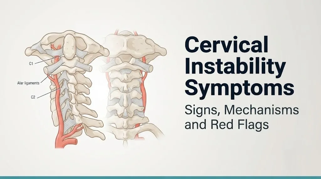
The alar ligament and the transverse atlantal ligament are the primary passive restraints of the upper cervical region. The alar ligament connects the odontoid process (dens) of C2 to the occipital condyles and limits rotation and lateral bending of C1 on C2. The transverse atlantal ligament holds the dens of C2 snugly against the anterior arch of C1, preventing the dens from being driven backward into the spinal cord. When either ligament is damaged or excessively lax, the atlantoaxial joint loses mechanical stability.
In the lower cervical spine, the anterior longitudinal ligament (ALL), the posterior longitudinal ligament (PLL), and the ligamentum flavum work together with the intervertebral discs to limit flexion-extension and translational movement between C3 and C7. Disc degeneration weakens this system progressively with age, which is why degenerative cervical instability becomes more common in adults over 50.
The American Academy of Orthopaedic Surgeons (AAOS) defines pathological instability in the cervical spine as intervertebral translation exceeding 3.5 mm or angulation exceeding 11 degrees between adjacent vertebrae on dynamic imaging, though clinical symptoms can appear before these thresholds are met when the spinal canal is narrow or when individual structural tolerance is reduced.
Upper Cervical Instability Symptoms
Upper cervical instability symptoms arise from pathological motion at the occipitoatlantal joint (C0 to C1) or the atlantoaxial joint (C1 to C2), two joints that are structurally and functionally distinct from every vertebral level below them.
The anatomical geography of the upper cervical spine makes it uniquely dangerous when instability develops. The brainstem sits at the level of the foramen magnum directly above C1. The vertebral arteries pass through the transverse foramina of C1 and C2 and make a distinctive lateral loop before entering the skull. The spinal cord at C1 occupies roughly 35 percent of the available spinal canal diameter. Any abnormal translational or rotational movement at this level can simultaneously compress the cord, kink the vertebral artery, and irritate the vagus nerve and upper cervical sympathetic ganglion.
Symptoms specific to upper cervical instability include:
- Suboccipital headache: burning or pressure-type pain at the base of the skull, produced by irritation of the C2 posterior ramus (greater occipital nerve) against abnormally mobile C1 to C2 joints
- Neck clunking or clicking: an audible or palpable shift during head rotation, representing the C1 ring translating on the C2 dens
- Cranial nerve symptoms: facial numbness (cranial nerve V), hoarseness or difficulty swallowing (cranial nerve IX, X, XII), produced by proximity of these nerve exit zones to the instability
- Visual disturbances: oscillopsia, blurred vision, or double vision with head movement, produced by vertebrobasilar vascular insufficiency or brainstem irritation
- Head-position intolerance: severe worsening of all symptoms with flexion, extension, or rotation, requiring frequent postural repositioning to manage symptom load
People with Down syndrome carry a well-documented elevated risk for atlantoaxial instability specifically, because of the genetic alteration in connective tissue that reduces transverse atlantal ligament tensile strength. The American Academy of Pediatrics recommends radiographic screening for atlantoaxial instability in children with Down syndrome before participation in high-impact sports.
Cervical Instability Neck Pain and Stiffness
Cervical instability produces neck pain through mechanical irritation of the cervical facet joints, abnormal tensile loading on the anterior and posterior longitudinal ligaments, sustained protective contraction of the paraspinal muscle group (particularly the semispinalis cervicis and multifidus), and direct irritation of the sinuvertebral nerves that innervate the disc, the ligaments, and the facet joint capsules.
The pain has a characteristic pattern that distinguishes it from simple muscular neck pain. It tends to be deep and aching, located in the midline or slightly lateral to the spinous processes. It worsens during position changes, particularly moving from lying to sitting or rotating the head while the neck is flexed. Patients often describe the need to support their head with their hands or to wear a soft cervical collar to reduce symptom intensity.
Stiffness in cervical instability is partly reflexive. When the ligamentous passive restraints are insufficient, the paraspinal muscles contract protectively to substitute for the lost ligamentous stability. This reflexive muscle guarding, which the semispinalis cervicis, the longus colli, and the rectus capitis posterior muscles all participate in, is physiologically appropriate in the short term but produces chronic muscular pain, reduced cervical range of motion, and eventually accelerated facet joint degeneration if the underlying instability is not addressed.
Quick Tip:
- Neck pain that worsens when you look down at a phone or desk for more than 10 to 15 minutes, then improves immediately when you lie flat, is a pattern consistent with positional cervical loading and warrants cervical spine evaluation.
- Pain that is constant and unchanged regardless of position is more likely to represent a non-mechanical cause and should be evaluated promptly by a primary care physician.
- Older adults over 65 with new-onset neck pain and any upper extremity symptom should be evaluated for degenerative cervical myelopathy, not assumed to have simple muscular pain.
Cervical Instability Headaches and Occipital Neuralgia
Cervical instability produces headaches through three distinct anatomical pathways, which is why the headache description varies so much between individuals.
The first pathway involves the C2 and C3 nerve roots. These posterior primary rami innervate the suboccipital region and communicate directly with the trigeminal nucleus caudalis in the brainstem via the trigeminocervical complex. When C2 to C3 nerve roots are irritated by abnormal movement or compression at the C1 to C2 or C2 to C3 joints, pain signals travel centrally and are perceived as headache extending from the occipital base forward to the frontal region and behind the eyes. This is cervicogenic headache, classified by the International Headache Society as a secondary headache disorder with specific diagnostic criteria including reproduction of the headache by cervical provocation maneuvers.
The second pathway involves occipital neuralgia, a distinct condition where the greater or lesser occipital nerves are mechanically compressed or irritated at the suboccipital region. Instability-related muscle hypercontraction in the semispinalis capitis and trapezius can entrap the greater occipital nerve as it pierces the semispinalis muscle belly. The pain is typically sharp, shooting, or electric, unilateral, and follows the dermatome of the occipital nerve from the base of the skull toward the vertex.
The third pathway involves dural irritation. The cervical dura is tethered to the posterior longitudinal ligament by ligamentous attachments at C1 to C2 and C2 to C3. Abnormal motion at these levels can produce traction on the dura itself, generating a diffuse, pressure-type headache that patients often describe as a sensation of fullness or pressure inside the skull.
According to research published in Cephalalgia (2022), cervicogenic headache accounts for approximately 15 to 20 percent of all chronic headache presentations in clinical headache centers. Women with hypermobility disorders represent a disproportionate subset of this population.
Cervicogenic Dizziness and Balance Problems From Cervical Instability
Cervicogenic dizziness from cervical instability arises when disrupted cervical proprioceptive signaling sends conflicting spatial orientation information to the vestibular nuclei in the brainstem, creating a mismatch between what the neck tells the brain about head position and what the inner ear and eyes simultaneously report.
Here is a way to understand the mechanism concretely. Imagine three navigation systems in your car: a GPS, a compass, and a backup camera. They normally agree. Now imagine the GPS starts giving readings that are slightly wrong because one of its sensors is malfunctioning. The compass and camera show you facing north, but the GPS says northwest. That conflict is disorienting and requires constant manual correction. Cervicogenic dizziness works similarly. The suboccipital muscles, the longus colli, and the sternocleidomastoid all contain dense concentrations of muscle spindle receptors that continuously feed proprioceptive data to the vestibular nuclei. When instability disrupts normal joint mechanics and muscle tension patterns in these muscles, that proprioceptive signal becomes inaccurate, creating a sensory mismatch that the brain experiences as dizziness, unsteadiness, or spatial disorientation.
Research published in the Journal of Vestibular Research found that cervical proprioceptive disturbances can produce measurable postural instability on computerized posturography testing in individuals with cervical spine dysfunction, independent of any inner ear pathology.
Dizziness from vertebral artery compromise is a distinct and more serious mechanism. When instability at C1 to C2 allows the atlas to rotate excessively, the vertebral artery can be mechanically kinked or stretched within its bony groove on the posterior arch of C1. This intermittent vertebrobasilar insufficiency produces dizziness that is abrupt, brief, and specifically triggered by particular head rotation positions rather than general movement, often accompanied by nausea, drop attacks, or visual disturbance.
Balance problems manifest as unsteadiness while walking, tendency to veer to one side, worsening in low-light environments (because the visual compensatory system is reduced), and difficulty with tandem walking. Older adults are at higher risk for fall-related injury from cervical instability-related balance dysfunction and should be evaluated by a neurologist if balance changes are present alongside neck symptoms.
Key Takeaway: Upper cervical instability can simultaneously irritate the vertebral artery, the greater occipital nerve, and the vagus nerve through proximity alone, which is why some patients experience headache, dizziness, and heart rate changes together as a cluster rather than as separate unrelated symptoms.
Neurological Symptoms of Cervical Instability
Neurological symptoms of cervical instability develop when the abnormal vertebral motion compresses or stretches one or more of the eight pairs of cervical nerve roots (C1 through C8) that exit the spinal canal through the intervertebral foramina, or when the cervical spinal cord itself is compressed.
Nerve root compression (radiculopathy) produces symptoms in a predictable dermatomal pattern. The specific nerve root involved determines exactly where the patient feels pain, numbness, tingling, or weakness.
| Compressed Nerve Root | Pain Location | Sensory Change | Motor Weakness |
|---|---|---|---|
| C4 | Shoulder and neck base | Reduced sensation at shoulder cap | Shoulder shrug (trapezius) |
| C5 | Outer upper arm | Outer shoulder numbness | Deltoid and biceps weakness |
| C6 | Outer forearm, thumb, index finger | Thumb and index finger numbness | Wrist extension weakness |
| C7 | Middle finger and posterior forearm | Middle finger numbness | Triceps weakness |
| C8 | Inner forearm and ring and little fingers | Ring and little finger numbness | Hand grip weakness |
The neurological symptoms of radiculopathy from cervical instability are intermittent initially, worsening with specific neck positions that narrow the intervertebral foramen and relieving when the neck is repositioned. This intermittency distinguishes early instability-related radiculopathy from disc herniation-related radiculopathy, which is often constant once established.
Reflexes change in a predictable pattern. The biceps reflex (C5 to C6) and the triceps reflex (C7) are the most commonly affected. Diminished reflexes at the level of compression and hyperreflexia below that level (in the legs) indicate spinal cord involvement rather than isolated nerve root compression.
According to the American Academy of Neurology (AAN), any patient presenting with new or progressive upper extremity neurological deficits (weakness, sensory loss, reflex changes) in the context of neck pain warrants cervical spine imaging to exclude cord compression as a cause.
Cervical Myelopathy Symptoms and When It Becomes an Emergency
Cervical myelopathy occurs when the spinal cord itself is compressed by abnormally translating vertebrae, hypertrophied ligamentum flavum, or protruding disc material in the setting of cervical instability, and it represents the most serious consequence of the condition.
The spinal cord does not have pain receptors. Myelopathy therefore does not typically present as neck pain worsening. Instead, it produces a characteristic pattern of symptoms that many patients initially attribute to aging, fatigue, or peripheral nerve problems, which leads to dangerous diagnostic delays.
Myelopathy symptoms include:
- Gait changes: a stiff, wide-based, unsteady walking pattern that worsens going down stairs or on uneven surfaces, produced by corticospinal tract dysfunction at the level of compression
- Hand clumsiness: dropping objects, difficulty buttoning shirts, deteriorating handwriting, or losing fine motor dexterity, produced by dorsal column dysfunction impairing fine sensory discrimination
- Lower extremity heaviness or stiffness: legs that feel heavy or that move slowly without clear weakness, produced by upper motor neuron dysfunction in the corticospinal tract
- Hyperreflexia: exaggerated deep tendon reflexes in the knees and ankles below the level of cord compression, a classical upper motor neuron sign
- Lhermitte’s sign: an electric shock or buzzing sensation that travels down the spine and into the limbs when the neck is flexed, produced by demyelination or mechanical deformation of the dorsal columns in the compressed cord segment
- Bowel or bladder dysfunction: urgency, incontinence, or retention, produced by disruption of the corticospinal control of the sacral micturition center
Research published in the Journal of Neurosurgery: Spine (2020) found that cervical myelopathy has an estimated prevalence of 1.6 per 1,000 adults and that spontaneous neurological deterioration occurs in approximately 20 to 60 percent of patients with moderate to severe myelopathy who are managed non-operatively, making the timing of intervention clinically important.
Emergency Symptoms: When to Call 911 or Go to the ER
Certain symptoms associated with cervical instability require immediate emergency evaluation. Do not wait to see if these resolve on their own.
Call 911 or go to the nearest emergency room immediately if you experience:
- Sudden inability to walk or rapidly worsening leg weakness after neck movement or trauma, which can indicate acute cord compression requiring emergency decompression
- Loss of bowel or bladder control in the setting of neck pain or neurological symptoms, a sign of spinal cord compression at the cervical or thoracic level
- Sudden quadriparesis (weakness in all four limbs) at any time, which represents a neurological emergency
- Acute traumatic neck injury with any neurological symptom, including numbness, tingling, arm or leg weakness, or loss of consciousness
- Drop attacks (sudden fall without loss of consciousness, triggered by head position change), which can indicate vertebrobasilar insufficiency from vertebral artery compromise at the C1 to C2 level
- Sudden severe headache described as the worst of your life combined with neck stiffness and neurological changes, which requires emergency evaluation to rule out subarachnoid hemorrhage
These presentations can indicate acute spinal cord compression, vertebrobasilar stroke, or catastrophic atlantoaxial instability, all of which require emergency medical assessment at a hospital, not an urgent care appointment or a phone call.
Key Takeaway: Cervical myelopathy does not hurt the way nerve root compression does. The first signs are often clumsy hands, a changed walking pattern, or unexplained difficulty with fine motor tasks, not neck pain. This is why myelopathy is dangerously easy to dismiss until it progresses.
Autonomic Dysfunction and Cervical Instability
Cervical instability can disrupt the autonomic nervous system when abnormal mechanical forces irritate, stretch, or compress the vagus nerve (cranial nerve X) and the superior cervical sympathetic ganglion, both of which run in close anatomical proximity to the upper cervical spine.
The vagus nerve exits the skull through the jugular foramen just lateral to the C1 vertebral body. It carries parasympathetic motor fibers to the heart, lungs, and gastrointestinal tract, and sensory afferents from these same visceral organs back to the brainstem. When instability at the occipitoatlantal or atlantoaxial joint creates mechanical tension or compression on the vagal trunk or its surrounding tissues, the downstream effects can include bradycardia (slow heart rate from excessive parasympathetic tone), nausea, early satiety, gastroparesis-like symptoms, and difficulty regulating breathing patterns.
The superior cervical sympathetic ganglion lies adjacent to the anterior bodies of C2 and C3. Irritation or mechanical disruption at this level can produce sympathetic dysregulation including temperature dysregulation in the head and face, unilateral facial sweating abnormalities (hyperhidrosis or anhidrosis), altered pupillary function, and contributions to chronic head pain through sympathetically maintained pain pathways.
The Ehlers-Danlos Society has noted in its clinical guidance that autonomic dysfunction in hEDS/HSD patients with upper cervical instability may present in ways that mimic primary dysautonomia, making careful structural evaluation necessary before attributing all autonomic symptoms to a centrally driven dysautonomia diagnosis.
Adults with autonomic symptoms (heart rate irregularity, blood pressure swings, temperature dysregulation) alongside neck pain and headache should request evaluation by a neurologist with experience in both spine and autonomic nervous system disorders, rather than being treated for each symptom independently.
Cervical Instability and POTS Symptoms
Postural orthostatic tachycardia syndrome (POTS) has been identified in a subset of patients with upper cervical instability as either a consequence of the structural instability or a condition that significantly overlaps with it in the hypermobility population.
POTS is characterized by a sustained heart rate increase of 30 beats per minute or more (40 bpm or more in individuals under 19) upon standing from a supine position, occurring within 10 minutes and in the absence of orthostatic hypotension. In the context of cervical instability, several proposed mechanisms may contribute. First, mechanical irritation of the superior cervical sympathetic ganglion can impair the normal sympathetic-mediated vasoconstriction that keeps blood from pooling in the lower extremities upon standing. Second, brainstem compression or irritation in upper cervical instability can disrupt the baroreflex arc, which relies on nucleus tractus solitarius function in the brainstem to regulate heart rate in response to blood pressure changes. Third, in the hEDS/HSD population, generalized vascular connective tissue laxity independently reduces venous tone throughout the body.
POTS symptoms in this context include:
- Rapid heart rate upon standing (palpitations described as pounding or racing)
- Lightheadedness or near-fainting when moving from sitting or lying to standing
- Fatigue disproportionate to activity level
- Cognitive impairment worsening when upright (brain fog)
- Nausea and gastrointestinal discomfort that worsens in the upright position
- Exercise intolerance with rapid heart rate escalation at low workloads
A 2021 study published in Frontiers in Neurology found measurable improvements in POTS symptom severity in a small cohort of patients with documented upper cervical instability following cervical stabilization treatment, suggesting a mechanistic link, though the authors noted the findings were preliminary and required replication in larger controlled trials.
Women between ages 15 and 50 are disproportionately affected by both POTS and cervical instability-related hypermobility disorders, and this demographic group should be evaluated for both conditions when presenting with unexplained tachycardia, presyncope, and neck symptoms together.
Cervical Instability in Ehlers-Danlos Syndrome and Hypermobility Disorders
Cervical instability is significantly more prevalent in individuals with hypermobile Ehlers-Danlos syndrome (hEDS) and hypermobility spectrum disorder (HSD) than in the general population, because the underlying collagen abnormality reduces the tensile strength of the ligaments that stabilize every joint, including the atlantoaxial and occipitoatlantal joints.
Collagen is the primary structural protein of ligaments. In hEDS, a genetic alteration in collagen processing (the precise molecular mechanism remains an active area of research) produces ligaments that stretch more than normal and return to resting length less efficiently. At the cervical spine, this means the alar ligament, the transverse atlantal ligament, and the capsular ligaments of the facet joints provide less positional stability than they would in a person with typical connective tissue. The instability in this population is often global across multiple cervical levels simultaneously, rather than focal at a single injured segment.
The clinical picture in hEDS-associated cervical instability has several distinguishing features:
- Symptoms often begin in adolescence or early adulthood, earlier than degenerative or post-traumatic instability
- Multiple body systems are affected concurrently (other hypermobile joints, GI dysfunction, POTS, chronic pain)
- Imaging in the standard supine MRI position may appear normal because instability is dynamic and position-dependent
- Standard cervical collars and physical therapy approaches may require modification for this population
- Surgical outcomes require specialist experience because connective tissue laxity affects the quality of the bone-hardware interface
The Ehlers-Danlos Society recommends that individuals with hEDS who have upper cervical symptoms be evaluated by a neurosurgeon or physiatrist with specific experience in hypermobility-related cervical instability before any management decisions are made, particularly before considering surgical stabilization, because the risk-benefit calculation differs substantially from post-traumatic or degenerative cervical instability.
Key Takeaway: In hEDS and hypermobility spectrum disorder, standard supine MRI may appear deceptively normal while significant instability is occurring dynamically. An upright or kinematic MRI is often needed to capture the functional instability that standard positioning misses.
Cervical Instability After Whiplash and Traumatic Injury
Whiplash injury produces cervical instability through a specific mechanism: the rapid, high-velocity flexion-extension movement of the neck during a collision or impact stretches the anterior and posterior cervical ligaments beyond their elastic limit, causing partial or complete tearing of ligamentous fibers without a fracture appearing on standard radiographs.
The Quebec Task Force on Whiplash-Associated Disorders classified whiplash injuries in a grading system from Grade 0 (no symptoms, no physical signs) to Grade IV (fracture or dislocation present). The majority of patients who develop chronic cervical instability symptoms after whiplash fall into the Grade II and III categories, where objective clinical signs (reduced range of motion, muscle tenderness, neurological signs) are present but standard imaging does not show fracture or dislocation. This classification gap is part of why post-whiplash instability is frequently underdiagnosed or dismissed.
At the ligamentous level, whiplash primarily injures the anterior longitudinal ligament, the alar ligament (particularly with rotational components of the mechanism), and the facet joint capsules. Research published in Spine (Panjabi et al., 2004) demonstrated measurable strain in the alar ligament at acceleration levels consistent with low-speed rear-end collisions, establishing that ligamentous injury can occur at impact speeds previously considered too minor to produce structural damage.
Symptoms following whiplash that suggest instability rather than simple muscular strain include:
- Persistent neck pain beyond six weeks despite conservative management
- Position-dependent neurological symptoms (arm numbness or tingling that varies with neck position)
- Severe headache beginning within days of the injury and persisting without improvement
- New-onset dizziness or balance changes not present before the injury
- Cognitive changes (difficulty concentrating, memory complaints) accompanying the above
Young adults aged 20 to 40 have the highest incidence of whiplash injury, and women in this age group show higher rates of persistent post-whiplash symptoms than men, possibly due to differences in cervical muscle mass and pre-existing cervical proprioceptive sensitivity.
Early Signs of Cervical Instability
The earliest signs of cervical instability often appear long before imaging abnormalities are detectable, because the initial failure of the passive restraint system produces functional changes before structural displacement becomes measurable on static imaging.
The first presentation is typically intermittent, position-dependent neck pain or headache. It occurs with specific activities, resolves with rest, and is often attributed to muscle strain, poor posture, or tension headache for months or years before the correct structural cause is identified. The distinguishing feature is the positional pattern: symptoms reliably appear with neck loading and reliably improve with neck rest or support.
Early signs to recognize:
- Neck pain or headache that begins during or immediately after sustained neck flexion (looking at a screen, reading, looking downward) and resolves when the neck is supported or returned to neutral
- A sensation of the neck “giving way” or needing to be held or supported during fatigue
- Subtle crepitus (grinding or clicking) with head movement at the C1 to C2 level (a midline posterior click)
- Mild paresthesias (tingling or buzzing) in the hands or scalp that come and go with neck movement
- Increased symptom frequency in the days after higher-impact activities (riding in a vehicle on rough roads, exercising with heavy neck loading, air travel with turbulence)
Research from the National Institute of Arthritis and Musculoskeletal and Skin Diseases (NIAMS) supports the concept that connective tissue injuries, including cervical ligamentous injuries, progress on a trajectory that can be slowed by early recognition and appropriate load management before structural displacement becomes established.
Children with Down syndrome represent a high-priority early detection group, with atlantoaxial instability present in approximately 10 to 20 percent of individuals with Down syndrome according to estimates cited in pediatric orthopedic literature, though most remain asymptomatic unless exposed to high-impact activities.
Chronic Cervical Instability Symptom Progression
Without appropriate management, cervical instability does not remain static. The symptom picture tends to broaden and worsen over time through two main mechanisms: structural progression and central sensitization.
Structural progression occurs because the unstable cervical segment creates abnormal mechanical loading on the adjacent facet joints, intervertebral discs, and uncovertebral joints with every head movement across years or decades. This accelerates degenerative changes at the unstable segment and at adjacent levels (adjacent segment disease), narrowing the spinal canal and the intervertebral foramina progressively. What begins as intermittent position-dependent pain may evolve into constant pain as the structural changes become fixed rather than dynamic.
Central sensitization adds a pain amplification layer that is not structural but neurological. Chronic nociceptive input from the unstable cervical structures upregulates substance P and N-methyl-D-aspartate (NMDA) receptor sensitivity in the dorsal horn of the cervical spinal cord. This process, well-documented in Spine journal research, reduces the threshold at which the nervous system generates pain signals, meaning that stimuli which would normally not produce pain (light touch, mild movement) begin to trigger significant pain responses. This is why chronic cervical instability patients often develop widespread pain sensitivity that exceeds what the structural picture alone would predict.
The progression pattern by timeframe:
| Timeframe | Typical Symptom Pattern |
|---|---|
| Acute (0 to 6 weeks) | Intermittent position-dependent neck pain, early headache, mild paresthesias |
| Subacute (6 weeks to 3 months) | Increasing frequency and duration of symptoms, early balance changes |
| Chronic (3 to 12 months) | Daily symptoms, established headache pattern, neurological signs emerging |
| Long-term (beyond 12 months untreated) | Fixed structural changes, possible myelopathy, central sensitization, widespread pain |
Older adults with chronic degenerative cervical instability are at particular risk for myelopathy development because the cervical canal narrows with age-related changes to the ligamentum flavum and disc height, leaving less reserve space before cord compression occurs.
Key Takeaway: Central sensitization in chronic cervical instability means the pain can become widespread and seem disproportionate to the structural finding on imaging, which does not mean the pain is not real or physically mediated. It means the nervous system has been sensitized by prolonged structural input and requires a treatment approach that addresses both the structural source and the central pain amplification.
How Cervical Instability Is Diagnosed
Cervical instability is diagnosed through a combination of clinical history, physical examination findings, and dynamic or positional imaging, because standard supine MRI often underestimates or misses functional instability that only becomes evident during movement.
The diagnostic process typically unfolds in this sequence:
- Clinical history: The physician documents the specific symptom pattern, onset, positional triggers, associated neurological symptoms, trauma history, and family history of connective tissue disorders. This history guides imaging selection.
- Physical examination: Includes cervical range of motion measurement, Spurling’s test (axial compression with lateral flexion to reproduce radicular symptoms), upper limb neurological examination (strength, sensation, reflexes), and lower limb examination for myelopathy signs (Babinski sign, hyperreflexia, gait assessment).
- Standard cervical MRI (supine): First-line imaging for soft tissue evaluation, cord compression assessment, and disc pathology. Standard in identifying cord compression, disc herniation, and ligamentous injury on T2 sequences. May miss dynamic instability.
- Flexion-extension radiography: Dynamic plain X-rays taken in maximum flexion and maximum extension. Identifies translational instability (greater than 3.5 mm between adjacent vertebrae) or angular instability (greater than 11 degrees). The most accessible and widely available dynamic imaging study.
- Upright or kinematic MRI: Performed in a weight-bearing position or through a range of motion. Captures cord compression or canal narrowing that may only occur during loading and movement, which supine MRI misses entirely. Availability varies by institution.
- CT myelogram: Used when MRI is contraindicated or when fine bony detail of the spinal canal is needed. Involves injection of contrast into the cerebrospinal fluid space. Provides excellent visualization of cord compression and nerve root compromise.
- Electromyography (EMG) and nerve conduction velocity (NCV) testing: Ordered when radiculopathy or peripheral nerve involvement needs to be confirmed or localized to a specific nerve root or plexus level.
When to See a Specialist for Cervical Instability Symptoms
The right specialist for cervical instability depends on the dominant symptom pattern, and choosing the appropriate provider type from the beginning prevents months of ineffective care.
Primary care physician: The appropriate first contact for new onset neck pain, headache, and early paresthesias. Can order initial imaging, rule out red flag causes, and provide appropriate referrals. Should not be bypassed in favor of direct specialist access without imaging.
Neurosurgeon (spine-specialized): Required when myelopathy signs are present, when imaging shows cord compression, when neurological deficits are progressive, or when surgical stabilization is being considered. Neurosurgeons with specific spine subspecialty training have the most direct authority to evaluate surgical candidacy.
Neurologist: Appropriate for confirmed or suspected myelopathy, peripheral neuropathy, autonomic dysfunction workup, or when the neurological picture is complex and requires clarification before surgical evaluation.
Physiatrist (Physical Medicine and Rehabilitation physician): Valuable for non-operative management optimization, guiding cervical stabilization exercise programs, coordinating interdisciplinary care, and evaluating functional impact. Physiatrists with spine subspecialty training can also manage advanced diagnostic workups in complex cases.
Rheumatologist: Required when inflammatory arthritis (particularly rheumatoid arthritis) is suspected as the cause, or when hEDS or HSD diagnosis has not yet been formally confirmed and the symptom picture suggests generalized connective tissue involvement.
When preparing for a specialist appointment, document the following to present to the provider:
- All imaging already performed, with dates and institutions
- Specific positional triggers: which positions worsen and which relieve symptoms
- Neurological symptoms: exact location of numbness, tingling, weakness, and whether they are constant or intermittent
- Functional impact: what activities you can no longer perform because of symptoms
- Any trauma history relevant to the neck, including minor events like heavy lifting or sports impact
- All current medications and any prior treatments tried
People with a known diagnosis of hEDS, HSD, or rheumatoid arthritis should specifically request referral to a specialist with documented experience in hypermobility-associated or inflammatory arthritis-associated cervical instability, because the management approach differs from post-traumatic or degenerative instability in ways that affect both conservative and surgical decision-making.
Frequently Asked Questions About Cervical Instability Symptoms
What does cervical instability feel like?
Cervical instability typically feels like deep, aching neck pain that worsens during movement and improves with rest or neck support.
Many people also describe a sensation of the neck “giving way,” frequent headaches at the base of the skull, and tingling or numbness in the arms or hands with specific neck positions.
The symptom experience depends heavily on which cervical structures are affected and whether the spinal cord, nerve roots, vertebral artery, or autonomic nerves are involved.
Can cervical instability cause dizziness and brain fog?
Cervical instability can cause dizziness through two mechanisms: disruption of cervical proprioceptive signals that feed into the vestibular nuclei in the brainstem, and intermittent compression of the vertebral artery at the C1 to C2 level during head rotation.
Brain fog in cervical instability is associated with reduced cerebral perfusion from vertebrobasilar insufficiency and, in the hypermobility population, from concurrent dysautonomia that impairs upright cerebral blood flow.
Research in the European Spine Journal supports a connection between cervical proprioceptive dysfunction and measurable postural instability, independent of inner ear disease.
What are the first signs of cervical instability?
The first signs of cervical instability are typically intermittent neck pain or headache that occurs with sustained neck loading and resolves with rest or neck support.
Subtle hand tingling with head turning, a clicking or grinding sensation at the upper neck, and increasing headache frequency after activities that load the cervical spine are common early presentations.
These early signs may precede measurable instability on standard imaging by months to years, which is why symptom history is as diagnostically important as imaging results.
Is cervical instability dangerous?
Cervical instability ranges from mildly symptomatic and manageable to potentially life-threatening, depending on the location, severity, and the structures being compressed.
Upper cervical instability at C1 to C2 carries the highest risk because the spinal cord at this level is adjacent to the brainstem, and the vertebral arteries are vulnerable to mechanical compromise during head rotation.
Cervical myelopathy, the most serious consequence, can produce progressive paralysis if cord compression is not addressed, which is why any myelopathy signs require urgent evaluation by a neurosurgeon.
Can cervical instability cause heart palpitations?
Cervical instability can produce heart palpitations through mechanical irritation of the vagus nerve or the superior cervical sympathetic ganglion in the upper cervical region.
Vagal irritation can produce bradycardia (slow, sometimes irregular heart rhythm), while sympathetic irritation or disruption of the baroreflex arc can produce tachycardia upon position change, a pattern overlapping with POTS.
A 2021 study published in Frontiers in Neurology found autonomic symptom improvement in a small cohort of upper cervical instability patients following cervical stabilization, suggesting structural origin for some palpitation presentations.
How do you know if you need surgery for cervical instability?
Surgery for cervical instability is considered when conservative management has failed to halt neurological progression, when myelopathy signs are present and cord compression is confirmed on imaging, or when the degree of structural instability places the spinal cord at immediate risk.
The decision requires evaluation by a spine-specialized neurosurgeon who can review dynamic imaging, assess the degree of cord compression, and weigh surgical risk against the natural history of untreated instability in the individual patient.
Not all cervical instability requires surgery; many patients with mild to moderate instability are managed effectively with cervical stabilization exercise, activity modification, and appropriate orthotic support under specialist supervision.
Putting the Clinical Picture Together
Cervical instability symptoms are rarely simple or isolated. The condition produces a web of overlapping musculoskeletal, neurological, vascular, and autonomic presentations because of the unique anatomical density of the upper and mid-cervical spine: the spinal cord, the vertebral arteries, the upper cervical nerve roots, the vagus nerve, and the sympathetic chain all occupy a remarkably small corridor.
The most important clinical step for anyone experiencing persistent neck pain with headaches, dizziness, arm symptoms, or autonomic changes is to connect these symptoms to a provider who can evaluate the cervical spine structurally, not treat each symptom in isolation. Standard supine MRI is the starting point, but dynamic or upright imaging is often the study that reveals what the standard scan misses. The right specialist depends on whether the dominant picture is structural, neurological, or autonomic.
If you are experiencing myelopathy signs, including hand clumsiness, gait changes, leg heaviness, or any bowel and bladder changes alongside neck symptoms, do not delay. These require same-week evaluation by a spine-specialized neurosurgeon. Your cervical spine is the only part of your body where ignoring a structural problem can produce consequences that progress faster than the appointment wait time