TMJ Symptoms Ear: Pain, Ringing, and Fullness (2026)
TMJ symptoms affecting the ear are among the most frequently misdiagnosed presentations in outpatient medicine, because ear pain, ringing, and a sensation of fullness in the ear can all originate from the temporomandibular joint without any primary problem in the ear itself. When the joint sitting directly in front of the ear canal becomes inflamed, displaced, or mechanically compromised, the shared nerve supply between that joint and your ear means the pain shows up in exactly the wrong place to get the right diagnosis.
According to the National Institute of Dental and Craniofacial Research (NIDCR), temporomandibular disorders affect an estimated 5 to 12 percent of the U.S. population, making them one of the most prevalent chronic orofacial pain conditions. Ear symptoms appear in a substantial proportion of those cases, with research published in the Journal of Oral Rehabilitation indicating that otalgia (ear pain) without primary ear pathology occurs in up to 42 percent of patients with confirmed temporomandibular disorder.
This guide covers every clinically recognized ear-related symptom of TMJ disorder, the specific anatomical and physiological reason each symptom occurs, how to distinguish a TMJ flare from a genuine ear infection, which symptoms require emergency evaluation, and exactly which specialist to see based on your specific presentation. The evidence here goes beyond what standard symptom checklists provide.
TMJ Symptoms Ear Overview: What’s Actually Happening
TMJ symptoms affecting the ear occur because the temporomandibular joint sits in direct anatomical contact with structures that serve the ear, meaning joint dysfunction produces symptoms that feel entirely auricular even when the ear itself is completely healthy.
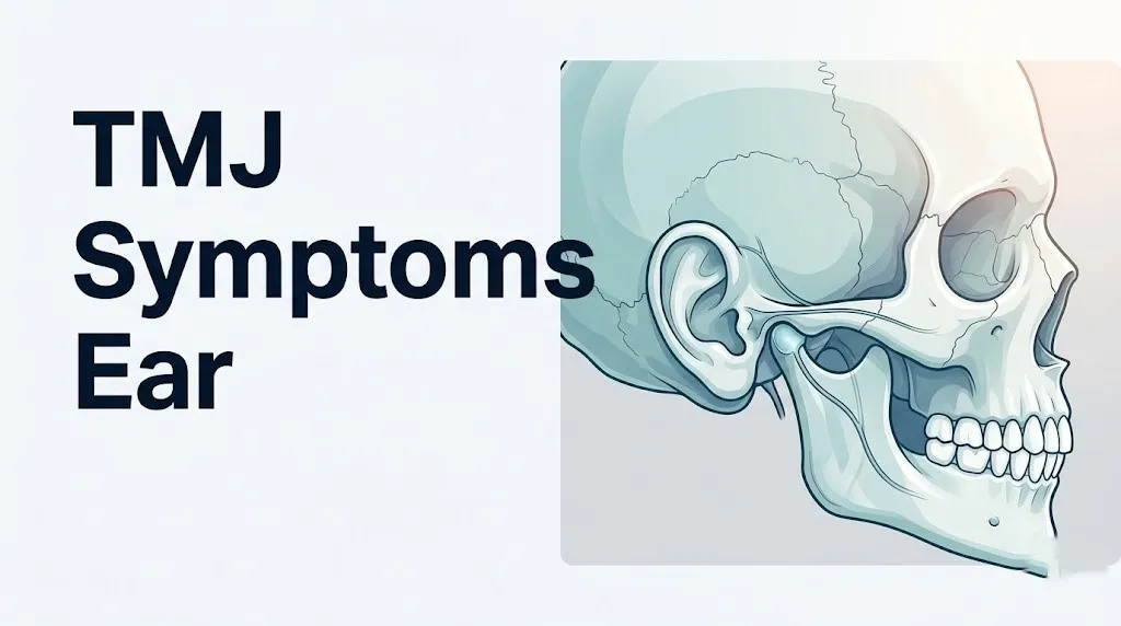
The temporomandibular joint is formed by the rounded head of the mandible, called the mandibular condyle, fitting into a shallow socket in the base of the skull called the glenoid fossa of the temporal bone. A fibrocartilaginous articular disc sits between these two bony surfaces, cushioning movement and distributing load during chewing, speaking, and swallowing. The entire joint capsule is lined with a synovial membrane that produces lubricating fluid.
The joint sits no more than one to two centimeters anterior to the opening of the external ear canal. That proximity is not coincidental; it’s the anatomical reason why any inflammation, disc displacement, or muscle dysfunction in the jaw complex immediately affects how the ear feels.
The National Institutes of Health OPPERA study (Orofacial Pain: Prospective Evaluation and Risk Assessment), the largest longitudinal study of TMD to date, tracked over 3,200 adults and found that individuals who developed temporomandibular disorder reported co-occurring ear symptoms in the majority of confirmed cases. The study established that ear symptoms in TMD are not secondary or rare; they are a core part of the clinical picture.
| TMJ Ear Symptom | Clinical Frequency | Primary Mechanism |
|---|---|---|
| Ear pain (otalgia) | Up to 42% of TMD patients | Auriculotemporal nerve referred pain |
| Ear fullness or pressure | Estimated 30 to 40% | Eustachian tube dysfunction via pterygoid tension |
| Tinnitus (ringing or buzzing) | Estimated 33% | Tensor tympani hyperactivity, central sensitization |
| Muffled or reduced hearing | Less common, estimated 10 to 15% | Eustachian tube pressure changes, tensor tympani reflex |
| Vertigo or dizziness | Less common, estimated 10 to 12% | Inner ear pressure effects, trigeminal-vestibular pathway |
Adolescents with TMD may present more prominently with muscle-related ear symptoms (myofascial otalgia) than with joint-specific clicking or locking, because their condylar cartilage is still developing and disc displacement is less common in younger patients. Adults over 50 are more likely to show degenerative joint changes alongside ear symptoms.
The Anatomical Reason TMJ Causes Ear Pain
TMJ causes ear pain through a specific nerve pathway: the auriculotemporal nerve, a branch of the mandibular division (V3) of the trigeminal nerve (cranial nerve V), supplies sensory fibers to both the TMJ capsule and the external ear canal, tragus, helix, and periauricular skin simultaneously.
Think of the auriculotemporal nerve like an electrical cable that powers two different rooms from one source. When inflammation at the TMJ generates pain signals through this nerve, your brain cannot reliably distinguish whether the signal is coming from the jaw joint or the ear. The result is genuine, physically real ear pain that has nothing to do with your ear drum, ear canal, or middle ear.
The nerve wraps around the neck of the mandible, passes through the substance of the parotid gland, and ascends anterior to the ear, branching to supply both the joint itself and the auricular structures. Research published in the Journal of Orofacial Pain has consistently documented that local anesthetic block of the auriculotemporal nerve eliminates TMD-related otalgia in the majority of test cases, confirming this nerve as the primary pain pathway.
This type of pain is called referred pain: pain perceived at a site anatomically distant from, but neurologically connected to, the actual source of the pathology. Referred pain is one of the most common reasons TMD-related ear pain is initially attributed to ear infection or otitis media when no infection exists.
Individual variation matters significantly here. People with pre-existing trigeminal neuralgia or a history of herpes zoster oticus (Ramsay Hunt syndrome) may find that TMD-related auriculotemporal nerve activation produces disproportionately severe ear pain, because the trigeminal nerve system is already in a sensitized state. For those individuals, even mild TMJ inflammation can trigger intense unilateral ear pain.
TMJ Tinnitus and Ear Ringing: Why Your Ears Ring
TMJ-related tinnitus is real, not imagined, and it arises through two distinct physiological pathways that most tinnitus workups fail to evaluate.
The first pathway involves the tensor tympani muscle, a small muscle in the middle ear that attaches to the handle of the malleus bone and dampens sound vibration. Critically, it is innervated by the medial pterygoid nerve, a branch of the same mandibular division (V3) of the trigeminal nerve that supplies the masticatory muscles. In patients with bruxism (teeth clenching and grinding) or active TMD myalgia, chronic overactivation of the masticatory muscles can produce reflex hyperactivity of the tensor tympani. That hyperactivity alters the normal oscillation mechanics of the middle ear ossicular chain, generating tinnitus perceived as ringing, buzzing, clicking, or rumbling.
The second pathway involves central sensitization of the trigeminal nucleus caudalis in the brainstem. Chronic TMD maintains a state of elevated neuronal excitability in this nucleus, which has known projections into the dorsal cochlear nucleus, an auditory brainstem structure involved in tinnitus generation. Research published in the Journal of Dental Research has found structural and functional overlap between trigeminal somatosensory inputs and cochlear processing, providing a mechanistic basis for somatic tinnitus in TMD patients.
A distinguishing clinical feature of TMJ-related tinnitus is that it often fluctuates with jaw position or jaw movement. Tinnitus that temporarily worsens when you clench your teeth, open your mouth wide, or press on the preauricular region (the skin just in front of the ear canal) strongly suggests a somatic, TMD-related origin.
- Tinnitus that changes with jaw position: strongly suggests TMD origin
- Tinnitus that is constant and unaffected by jaw movement: warrants ENT evaluation for cochlear or retrocochlear pathology
- Tinnitus accompanied by sudden unilateral hearing loss: requires same-day ENT evaluation regardless of TMD history
- Tinnitus with pulsing quality matching heartbeat (pulsatile tinnitus): requires prompt vascular evaluation, not a TMD workup
Women with TMD experience tinnitus more frequently than men with equivalent joint pathology, a pattern researchers attribute partly to estrogen’s modulatory effects on the central auditory pathway and partly to the higher prevalence of comorbid migraine, which further sensitizes brainstem auditory processing centers.
TMJ Ear Fullness and Pressure: The Eustachian Tube Connection
TMJ disorder produces a sensation of fullness or pressure in the ear by affecting the Eustachian tube (the pharyngotympanic tube), the narrow channel connecting the middle ear to the back of the throat that regulates air pressure on both sides of the eardrum.
The medial pole of the mandibular condyle sits in close anatomical proximity to the cartilaginous portion of the Eustachian tube. The tensor veli palatini muscle, which controls the opening of the Eustachian tube during swallowing and yawning to equalize middle ear pressure, shares its origin with the medial pterygoid muscle and both are modulated by branches of the mandibular nerve (V3). When TMD produces chronic tension in the medial pterygoid complex, that tension can mechanically impair the tensor veli palatini’s ability to open the Eustachian tube effectively.
The result is exactly the sensation you feel when an airplane descends and your ears won’t pop: a blocked, pressurized, cotton-wool-stuffed feeling that doesn’t respond to swallowing, yawning, or the Valsalva maneuver, because the problem isn’t pressure differential. It’s muscular tension restricting tube mechanics.
According to research published in the American Journal of Otolaryngology, a statistically meaningful proportion of patients referred to ENT clinics for Eustachian tube dysfunction with normal tympanometry and normal audiometry ultimately receive a diagnosis of temporomandibular disorder rather than primary Eustachian tube pathology. This is why normal tympanogram results in a patient reporting ear fullness should prompt clinical evaluation of the TMJ.
To differentiate TMJ-related ear fullness from primary Eustachian tube dysfunction:
- Assess whether the fullness changes with jaw position or jaw movement (TMD-related fullness often does).
- Note whether the fullness appeared alongside jaw pain, jaw clicking, or a period of increased bruxism.
- Determine whether tympanometry shows abnormal middle ear pressure (type B or type C tympanogram): if tympanogram is normal, primary Eustachian tube dysfunction is less likely.
- Ask whether the feeling worsens during stress: bruxism-driven pterygoid tension increases under psychological stress.
- Evaluate whether the fullness is bilateral or unilateral: unilateral ear fullness in an adult that persists beyond two weeks warrants nasopharyngoscopy to rule out nasopharyngeal pathology, regardless of TMJ findings.
Key Takeaway: TMJ ear symptoms, including pain, ringing, and fullness, are not imagined or secondary. They arise from direct anatomical proximity and shared nerve supply between the temporomandibular joint and the ear, specifically through the auriculotemporal nerve, tensor tympani hyperactivity, and Eustachian tube mechanical dysfunction via pterygoid muscle tension.
TMJ Flare Up Symptoms: What a Flare Feels Like
A TMJ flare up is an acute worsening of temporomandibular disorder symptoms driven by a specific inflammatory or mechanical trigger, overlaid on what may otherwise be a manageable baseline condition.
During a flare, the synovial membrane lining the TMJ capsule releases elevated concentrations of prostaglandin E2 (via cyclooxygenase-2 enzyme activation), interleukin-1 beta, and substance P into the joint space. These inflammatory mediators lower the firing threshold of auriculotemporal nerve afferents, which is why even light jaw movement, touch, or cold air can produce intense ear and jaw pain during a flare that would normally be only mildly uncomfortable.
What a TMJ flare actually feels like, symptom by symptom:
- Sudden or rapidly worsening ache or stabbing pain in front of or inside the ear, often on one side
- Jaw stiffness that makes it difficult to open the mouth to normal range (normal opening is approximately 40 to 50 mm between incisal edges)
- A noticeable increase in jaw clicking, popping, or grinding sounds, or a change in character of existing clicking
- Escalating sensation of ear fullness, as if flying and unable to clear the ears
- Intensified tinnitus: the ringing or buzzing gets louder or more intrusive during a flare
- Headache radiating from the temple into the ear or from behind the ear into the neck
- Jaw muscle fatigue and soreness, particularly in the masseter (the thick muscle felt when you clench at the angle of the jaw) and the temporalis (the muscle felt at the temple when biting down)
- Increased sensitivity to chewing: foods that were previously manageable become painful
Common triggers for TMJ flare-ups include: a period of increased stress with associated bruxism, eating hard or chewy foods, prolonged dental procedures requiring extended mouth opening, whiplash-type neck injury, hormonal fluctuation in women (particularly around menstruation, consistent with estrogen-modulated joint laxity), and cold or damp weather, which some patients consistently associate with increased joint stiffness through unclear physiological mechanisms.
People with fibromyalgia experience TMJ flares more frequently and more severely than the general TMD population, because the central sensitization that defines fibromyalgia amplifies all peripheral pain signals, including those from the TMJ and auriculotemporal nerve system.
TMJ Migraine Symptoms and Head Pain
TMJ migraine symptoms emerge through trigeminal nerve sensitization: the same nerve pathways carrying pain from the TMJ also carry pain signals that contribute to migraine generation and maintenance.
The trigeminal nerve (cranial nerve V) is the primary pain highway for both TMD and migraine. In migraine, inflammatory neuropeptides including calcitonin gene-related peptide (CGRP) are released from trigeminal nerve terminals in the meninges, producing throbbing headache, photophobia, and phonophobia. In TMD, the same trigeminal system is activated peripherally at the joint. Chronic peripheral TMJ pain can maintain a state of trigeminal sensitization that lowers the threshold for migraine attacks, meaning people with untreated TMD experience more frequent migraines and migraines that are harder to treat.
A 2021 study published in the Journal of Oral Rehabilitation found that individuals with confirmed temporomandibular disorder had a statistically higher prevalence of migraine compared to matched controls without TMD, and that the severity of TMD correlated with migraine attack frequency. The relationship appears bidirectional: migraine-related trigeminal activation can also trigger TMJ pain in people with underlying joint vulnerability.
TMJ-related head pain patterns the reader should know:
| Pain Location | Likely TMJ Structure Involved | Clinical Note |
|---|---|---|
| Temple (unilateral or bilateral) | Temporalis muscle referred pain | Often mistaken for tension headache |
| Ear and periauricular region | Auriculotemporal nerve | Classic TMD-otalgia pattern |
| Behind the eye (retro-orbital) | Trigeminal-ophthalmic division crossover | Can mimic cluster headache |
| Cheek and jaw | Masseter and medial pterygoid referral | Often most severe with chewing |
| Back of head and neck | Sternocleidomastoid, upper trapezius | Cervicogenic component of TMD |
| Forehead | Temporalis tendon referral | Often bilateral, dull, pressure-like |
People already diagnosed with chronic migraine (15 or more headache days per month) who also have jaw clicking or morning jaw stiffness should ask their neurologist about comorbid TMD evaluation. Treating the TMD component can reduce overall trigeminal sensitization and, in some patients, reduce migraine frequency without changes to migraine-specific medication.
TMJ Jaw Pain and Clicking Sounds
Jaw pain and clicking are the most recognized TMJ symptoms, and they occur when the fibrocartilaginous articular disc moves out of its normal position between the condyle and glenoid fossa.
In a healthy TMJ, the articular disc rides forward and back with condylar movement during jaw opening and closing, providing frictionless gliding. When the disc displaces anteriorly (forward) relative to the condyle, the condyle must click over the back edge of the disc to get into proper position during opening. That click, medically termed a reciprocal click, is the characteristic pop heard or felt when opening the mouth in disc displacement with reduction. A second click may occur on closing as the disc slips back. This is different from degenerative joint crepitus, which produces a grinding, grating sound rather than a single pop.
When the disc displaces and does not reduce (disc displacement without reduction), the jaw deflects toward the affected side on opening, mouth opening is limited (typically below 35 mm between upper and lower central incisors), and no click is heard because the disc never returns to position. This is the mechanism behind acute jaw locking.
According to the American Academy of Orofacial Pain (AAOP), not all TMJ clicking requires treatment. Asymptomatic clicking in the absence of pain, jaw limitation, or functional impairment is common in the general population and does not necessarily represent pathology requiring intervention.
Jaw pain associated with TMD most commonly originates from three sources:
- Synovial membrane inflammation (arthralgia): sharp, localized pain in the preauricular region worsened by jaw loading or palpation
- Masticatory muscle pain (myalgia or myofascial pain): dull, diffuse aching in the masseter, temporalis, or pterygoid muscles worsened by function
- Disc displacement pain: pressure or aching during movement, often associated with clicking or deviation
Key Takeaway: TMJ flare symptoms represent an acute inflammatory cascade in the joint’s synovial membrane, and TMJ-related headache arises from trigeminal nerve sensitization that directly overlaps with migraine pathophysiology. The disc displacement mechanism produces the characteristic clicking sound and, when the disc fails to reduce, can cause jaw locking.
TMJ Facial Pain and Referred Pain Patterns
TMJ referred pain spreads well beyond the jaw and ear: the masticatory muscle system produces predictable trigger point referral patterns that send pain to the cheek, forehead, temple, and even the teeth, making the source particularly difficult to identify without systematic clinical examination.
The masseter muscle, the primary jaw-closing muscle running from the zygomatic arch down to the angle and ramus of the mandible, is the most common source of referred facial pain in TMD. When the masseter develops myofascial trigger points (hyperirritable, taut bands within the muscle belly), it refers pain to the cheek, lower molars, jaw angle, and ear. Patients frequently present to their dentist reporting tooth pain that turns out to be referred masseter myalgia, not dental pathology.
The temporalis muscle, which fans out across the temporal bone from the temporal fossa to the coronoid process of the mandible, refers pain to the forehead, temple, upper teeth, and behind the eye when trigger points are active. This referral pattern is one of the primary reasons TMD-related facial pain is frequently misclassified as sinus headache: the pain over the forehead and cheek mirrors sinus pressure pain anatomically.
The medial pterygoid muscle, an internal jaw-closing muscle, refers pain to the back of the mouth, tongue, throat, and ear. When this muscle is the primary pain source, patients describe a sore-throat-like sensation combined with ear fullness, a combination that frequently leads to repeated negative ENT evaluations.
Central sensitization in chronic TMD expands these referral zones further. As the trigeminal nucleus caudalis becomes persistently sensitized, second-order neurons become hyperexcitable and respond to lower-intensity stimuli over a wider area. Patients with longstanding TMD may develop widespread facial allodynia (pain from normally non-painful stimuli, such as light touch to the face or cool air) that extends well beyond the original TMJ region.
People with post-traumatic stress disorder (PTSD) or anxiety disorders have measurably higher rates of TMD and broader referred pain patterns, likely because hyperactivation of the hypothalamic-pituitary-adrenal (HPA) axis amplifies central pain sensitization.
TMJ Pain Behind the Ear and Neck Pain
Pain behind the ear in TMD originates from two distinct anatomical sources that are often active simultaneously: the retrodiscal tissue (the highly vascularized and innervated connective tissue posterior to the articular disc), and the posterior digastric and stylohyoid muscles which connect the jaw to the upper neck and mastoid region.
When the articular disc displaces anteriorly, the condyle compresses the retrodiscal tissue (also called the bilaminar zone) during jaw loading and movement. This tissue contains a dense network of free nerve endings and is richly supplied by branches of the auriculotemporal and inferior alveolar nerves. Compression of this tissue generates a deep, aching pain behind the ear that worsens specifically with chewing and biting.
TMD neck pain develops through two mechanisms. First, postural compensation: people with chronic jaw pain unconsciously alter their head and neck posture to reduce jaw loading, which places sustained mechanical stress on the suboccipital muscle group (rectus capitis posterior major and minor, obliquus capitis superior and inferior) and upper cervical facet joints (C1-C2, C2-C3). Second, neurological convergence: cervical dorsal horn neurons and trigeminal nucleus caudalis neurons share convergent input, so chronic TMD sensitizes the cervical pain processing pathway and vice versa.
The National Institutes of Health has documented in OPPERA study data that neck pain and TMD co-occur at rates well above chance, with a substantial proportion of confirmed TMD patients reporting concurrent upper cervical pain as part of their symptom complex.
To describe TMJ-related neck and ear pain accurately to your provider:
- Note which ear or side of the neck is affected and whether it is consistently unilateral.
- Record whether the pain increases with chewing, yawning, or talking for extended periods.
- Note whether pressing gently in front of your ear (over the TMJ) reproduces or worsens the ear and neck pain.
- Describe your sleep position: side sleeping with the affected cheek on the pillow directly compresses the TMJ and often intensifies next-day neck and ear pain.
- Report any history of whiplash or cervical injury: the cervicogenic component of TMD is often more prominent after neck trauma.
Older adults (over 60) with TMD-related neck pain should have cervical spondylosis (degenerative changes in cervical vertebrae) evaluated before attributing all neck symptoms to the TMJ, because the two conditions frequently co-occur and each requires a different management approach.
Key Takeaway: TMJ facial pain follows predictable masseter, temporalis, and pterygoid trigger point referral patterns that mimic sinus headache and toothache, and TMJ pain behind the ear arises from condylar compression of the highly innervated retrodiscal tissue, not from ear pathology.
TMJ Dizziness and Vertigo Symptoms
TMJ-related dizziness is one of the most debated but clinically meaningful aspects of the disorder, and current evidence points to two plausible anatomical pathways, though the evidence for each is at different stages of confirmation.
The first pathway involves indirect pressure effects on the inner ear through Eustachian tube dysfunction and altered middle ear pressure. Sustained middle ear pressure imbalance can subtly affect the pressure differential across the oval window, potentially influencing the perilymph and endolymph fluid dynamics of the cochlea and vestibular labyrinth. This is the same mechanism by which severe Eustachian tube dysfunction can produce mild disequilibrium.
The second pathway is neurological: the trigeminal system has anatomical projections to the vestibular nuclei in the brainstem. Research published in the Journal of Orofacial Pain has identified trigeminal-vestibular convergence zones that may allow TMD-generated pain signals to produce vestibular disturbance in susceptible individuals.
Critically, not all dizziness in a TMD patient is TMD-related. Dizziness with a rotational, spinning quality (vertigo) that lasts seconds and is triggered by head position change is more consistent with benign paroxysmal positional vertigo (BPPV) than TMD. True BPPV is caused by dislodged otoconia (calcium carbonate crystals) in the semicircular canals and is treated with repositioning maneuvers, not jaw treatment.
Dizziness that accompanies TMD is typically described as a vague unsteadiness or light-headedness, worsened during jaw flares and improved as the flare resolves. It is rarely true spinning vertigo.
If dizziness is prominent, sudden, or accompanied by hearing loss, nausea, vomiting, or neurological symptoms (facial weakness, double vision, slurred speech), this is not TMD. Those presentations require urgent neurological or ENT evaluation, not a TMJ workup.
TMJ Symptoms in Women: Why Women Are More Affected
Women account for approximately 80 percent of patients seeking treatment for temporomandibular disorders, a sex disparity that goes beyond socialized health-seeking behavior and has a specific, researched biological basis.
The estrogen receptor hypothesis is the most evidence-supported explanation. Research documented in the Journal of Dental Research has confirmed the presence of estrogen receptors (specifically ERalpha and ERbeta subtypes) in the synovial tissue and posterior attachment (retrodiscal tissue) of the human TMJ. Estrogen fluctuations modulate both the mechanical properties of the joint (estrogen increases joint ligament laxity, making disc displacement more likely) and the sensitivity of joint afferent nerve fibers (higher estrogen correlates with lower pain threshold in joint tissue).
This is why TMD symptoms in many women are cyclical: they worsen in the premenstrual and menstrual phases (when estrogen falls sharply after midcycle), and some women report first-onset or dramatic worsening of TMJ ear symptoms during perimenopause, when estrogen fluctuation is most pronounced.
Oral contraceptive users show a different pattern: research from the OPPERA study found a statistically elevated risk of TMD in women using combined oral contraceptives containing synthetic estrogen compared to non-users of the same age, suggesting that supraphysiological or sustained estrogen exposure may sensitize joint tissue differently than naturally fluctuating estrogen.
Pregnant women with TMD deserve specific attention. Estrogen rises substantially throughout pregnancy, but the relaxin hormone, also elevated in pregnancy and responsible for ligamentous laxity, may increase TMJ disc displacement risk. Symptom management during pregnancy is limited because many anti-inflammatory medications are contraindicated; an orofacial pain specialist should guide management.
Adolescent girls and young women are the fastest-growing group presenting for first-time TMD evaluation, consistent with the post-pubertal onset of estrogen cycling. This group tends to have a higher proportion of myofascial pain relative to articular disc displacement compared to adult women.
TMJ Symptoms Worse at Night and During Sleep
Nocturnal bruxism is the single most powerful amplifier of TMJ ear symptoms, and it operates entirely outside conscious awareness, making it one of the most underrecognized contributors to morning-onset jaw and ear pain.
Bruxism is involuntary rhythmic or sustained contraction of the masseter, temporalis, and medial pterygoid muscles during sleep. The masticatory force generated during sleep bruxism can reach significantly higher levels than during voluntary chewing, because the conscious inhibitory pathways that prevent extreme jaw loading during waking are suppressed during sleep. Sustained pterygoid and masseter contraction compresses the TMJ directly, irritates the retrodiscal tissue, and maintains elevated prostaglandin E2 and interleukin-1 beta levels in the synovial fluid overnight.
The result is a predictable morning symptom pattern. People with nocturnal bruxism wake with:
- Jaw stiffness and difficulty opening the mouth fully, typically improving within 30 to 60 minutes after rising
- Ear pain or fullness that is worst in the first hour after waking
- Temple and jaw muscle aching, as if they have been exercising the jaw all night (which, functionally, they have)
- Heightened tinnitus in the morning that often lessens as the day progresses
- Tooth sensitivity from enamel wear, though this is a dental rather than ear symptom
According to Mayo Clinic, sleep bruxism is associated with sleep apnea, gastroesophageal reflux, and stress-related sleep disruption, all of which are common co-morbidities in the TMD population. Evaluating for obstructive sleep apnea (OSA) is clinically relevant in any TMD patient with prominent nocturnal symptoms, because OSA-related airway events are associated with arousals that trigger bruxism episodes.
Side sleepers who sleep with the affected cheek on the pillow add direct mechanical compression to the TMJ throughout the night. Sleeping in a supine position with a supportive cervical pillow can reduce positional TMJ loading, though this alone is insufficient to manage active TMD.
Key Takeaway: TMJ symptoms in women are driven by estrogen receptor expression in joint synovial tissue that modulates both joint laxity and pain threshold, and nocturnal bruxism is the primary mechanism behind morning-worst TMJ ear symptoms because sleeping masticatory forces exceed waking forces with no conscious inhibitory brake.
TMJ vs Ear Infection: How to Tell the Difference
TMJ vs ear infection is one of the most practically important distinctions in primary care, because the treatments are completely different and misdiagnosis leads to unnecessary antibiotic courses or missed TMJ treatment.
An ear infection (otitis media: infection of the middle ear; otitis externa: infection of the external ear canal) produces pain through bacterial or viral inflammation of the tympanic membrane, middle ear mucosa, or external ear canal epithelium. A TMJ disorder produces pain through auriculotemporal nerve referred pain from a joint and muscle complex that sits adjacent to but entirely outside the ear.
| Feature | TMJ-Related Ear Pain | Ear Infection (Otitis Media) | Ear Infection (Otitis Externa) |
|---|---|---|---|
| Location of pain | In front of or around the ear | Deep inside the ear | In the ear canal, worsened by pulling the outer ear |
| Discharge from ear | None | Possible (if TM rupture occurs) | Possible (watery or purulent) |
| Fever | Not a TMJ symptom | Common in acute bacterial otitis | Possible in severe cases |
| Worse with jaw movement | Yes: chewing, yawning worsen it | No: jaw movement does not affect it | Not typically worsened by jaw movement |
| Ear canal appearance on exam | Normal | Red, bulging, or fluid behind TM | Swollen, red canal; white exudate possible |
| Tympanogram | Normal | Abnormal (type B flat curve) | Normal or slightly abnormal |
| Responds to antibiotics | No | Often yes (bacterial type) | Yes (topical antibiotic drops) |
| Associated jaw symptoms | Yes: clicking, stiffness, limited opening | No | No |
| Recent upper respiratory infection | Not required | Common precipitant | Less common |
The clinical test that most reliably distinguishes the two is tympanometry. A normal tympanogram (type A curve) in a patient with ear pain strongly suggests the pain is not arising from middle ear pathology and directs the workup toward TMD or other referred pain sources.
Children are more susceptible to genuine otitis media than adults, so ear pain in a child with fever, cold symptoms, or fluid behind the eardrum should always be evaluated by a pediatrician or family physician before attributing symptoms to TMD, even if the child grinds their teeth.
TMJ Red Flag Symptoms
TMJ red flag symptoms are specific presentations that indicate either a more serious underlying condition masquerading as TMD, or a complication of TMD severe enough to require prompt specialist evaluation rather than watchful waiting.
Certain symptoms appearing in the context of apparent TMJ disorder should trigger immediate clinical re-evaluation:
- Unilateral sudden-onset hearing loss: a decrease in hearing that appears rapidly (over hours to days) in one ear is a medical urgency. It may represent sudden sensorineural hearing loss, which has a time-sensitive treatment window (corticosteroid therapy is most effective within 72 hours of onset). This is not TMD, and treating it as TMD causes harmful delay.
- Jaw locked in the closed position: acute closed lock (inability to open the mouth beyond approximately 25 to 30 mm) indicates disc displacement without reduction and warrants evaluation by an oral and maxillofacial surgeon within 24 hours. Delay allows fibrous adhesions to form in the joint space, reducing the success of non-surgical management.
- Facial swelling with fever: preauricular or facial swelling combined with fever and trismus (difficulty opening the mouth) may represent septic arthritis of the TMJ or a spreading odontogenic infection. This requires emergency evaluation.
- Facial numbness or weakness: numbness of the cheek, lip, or chin, or weakness of facial muscles, is not a TMD symptom. These findings suggest involvement of the facial nerve (cranial nerve VII) or infraorbital nerve and may indicate a parotid gland tumor, acoustic neuroma, or other serious pathology.
- Progressive or painless trismus: gradual, painless limitation of jaw opening without a history of clicking or joint pain warrants imaging to rule out fibrosis, tumor, or radiation-related tissue changes.
- Night sweats, unexplained weight loss, or lymphadenopathy alongside jaw or ear symptoms: this constellation of systemic symptoms alongside regional pain is a red flag for malignancy or serious systemic disease, not TMD.
- Ear pain in an adult over 50 who smokes: persistent unilateral ear pain in an older adult with tobacco use history requires nasopharyngoscopy to evaluate the nasopharynx and pyriform sinuses before a TMD diagnosis is accepted.
A single red flag symptom is a reason to accelerate evaluation, not to wait and see.
Emergency Symptoms: When to Call 911 or Go to the ER
Certain symptoms that can occur in the setting of TMJ disorder or ear pain require immediate emergency evaluation. Do not wait to see if these resolve on their own.
Call 911 or go to the nearest emergency room immediately if you experience:
- Sudden facial weakness, drooping on one side of the face, or inability to close one eye: this may indicate Bell’s palsy, stroke, or a brainstem event and is a neurological emergency
- Sudden severe headache described as the worst headache of your life, especially if accompanied by neck stiffness or visual changes: this may indicate subarachnoid hemorrhage and requires immediate CT evaluation
- Double vision, slurred speech, or sudden difficulty swallowing alongside jaw or ear pain: these are stroke warning signs and require immediate emergency evaluation
- Complete and sudden loss of hearing in one ear with rapid onset: this represents sudden sensorineural hearing loss, which has a 72-hour treatment window, and requires same-day emergency evaluation at minimum
- Jaw pain with fever above 38.5 degrees Celsius (101.3 degrees Fahrenheit) and significant facial swelling: this may represent septic arthritis of the TMJ or a spreading deep space infection of the head and neck, which can compromise the airway
- Inability to open the mouth at all (acute closed lock) combined with fever or chills: warrants emergency evaluation to rule out infection
These presentations can indicate a serious complication or an entirely different emergency condition and require emergency medical assessment, not an urgent care appointment or a scheduled dentist visit.
How TMJ Is Diagnosed and Which Specialist to See
TMJ disorder is diagnosed clinically through a structured physical examination combined with targeted imaging, using the Diagnostic Criteria for Temporomandibular Disorders (DC/TMD), the internationally validated research and clinical diagnostic framework developed through collaboration of TMD researchers and endorsed by the American Academy of Orofacial Pain (AAOP).
The diagnostic examination includes: measurement of maximum mouth opening (normal: 40 to 50 mm), assessment of jaw deviation or deflection on opening, palpation of the TMJ capsule for tenderness and joint sounds, palpation of the masticatory muscles for tenderness, assessment of occlusion (bite relationship), and a pain interview using standardized validated tools including the Graded Chronic Pain Scale and PHQ-4 (for comorbid anxiety and depression screening, which is clinically relevant because psychological distress amplifies TMD severity and treatment response).
Imaging is not required for all TMD presentations but is indicated when joint sounds suggest internal derangement, when jaw opening is limited, or when the clinical picture suggests degenerative joint disease:
| Imaging Modality | What It Shows | When It’s Ordered |
|---|---|---|
| Panoramic radiograph | Bony condylar morphology, gross degenerative changes | Initial screening for bony pathology |
| Cone-beam CT (CBCT) | Detailed condylar bone detail, erosion, subchondral changes | When degenerative joint disease is suspected |
| MRI (magnetic resonance imaging) | Articular disc position, disc shape, joint effusion, soft tissue | When disc displacement or closed lock is suspected |
| Ultrasound | Joint effusion, synovial thickening, guided injections | Increasingly used in orofacial pain clinics |
Which specialist to see, by symptom pattern:
- Jaw pain with clicking, limited opening, or ear symptoms present for more than 2 to 4 weeks: see a dentist trained in TMD management or an orofacial pain specialist (a dentist with postgraduate training in chronic orofacial pain, recognized by the American Academy of Orofacial Pain)
- Ear symptoms with normal ENT examination and normal tympanometry: request a referral to an orofacial pain specialist and bring all ENT records to that appointment
- Jaw locked closed: see an oral and maxillofacial surgeon within 24 hours
- Prominent tinnitus with normal audiogram: see an orofacial pain specialist and consider concurrent evaluation by an audiologist experienced in somatic tinnitus
- Headache as primary complaint alongside jaw symptoms: the workup may require both an orofacial pain specialist and a neurologist, as migraine-TMD comorbidity requires coordinated management
- Any red flag symptoms listed above: emergency medicine physician immediately, not a TMD specialist first
Key Takeaway: The DC/TMD diagnostic criteria provide a validated structured framework for diagnosing TMD, and the appropriate specialist depends on your specific symptom pattern. Ear symptoms with a normal ENT workup are the clearest indication to seek evaluation from an orofacial pain specialist with experience in temporomandibular disorders.
Frequently Asked Questions About TMJ Symptoms and Ear Pain
Can TMJ disorder cause ear pain without any jaw pain?
TMJ disorder can absolutely cause ear pain without noticeable jaw pain, because the auriculotemporal nerve supplies sensory fibers to both the joint capsule and the external ear canal simultaneously.
When the disc or synovial tissue is mildly inflamed, the referred pain from the joint may be perceived entirely in the ear while the joint itself produces no strong local pain signal.
This presentation is one of the primary reasons TMD-related ear pain is frequently misdiagnosed as recurrent ear infection, particularly when the jaw is not clinically examined.
What does TMJ ear pain feel like compared to an ear infection?
TMJ ear pain typically presents as a dull or aching discomfort in front of or around the ear that worsens with jaw movement, chewing, yawning, or wide mouth opening.
Ear infection pain (particularly otitis media) presents as a deep, pressurized ache inside the ear that does not change with jaw movement and is frequently accompanied by fever, recent upper respiratory infection, or fluid drainage.
A normal tympanogram in the setting of ear pain is the single most useful clinical indicator that the pain source is not the middle ear.
Why does TMJ cause ringing in the ears?
TMJ causes tinnitus through two mechanisms: hyperactivity of the tensor tympani muscle (a middle ear muscle innervated by the same trigeminal nerve branch that controls masticatory muscles) and central sensitization of the trigeminal nucleus caudalis, which has projections into the dorsal cochlear nucleus.
A key distinguishing feature is that TMJ-related tinnitus typically changes in intensity with jaw position or jaw clenching, while tinnitus from cochlear pathology does not.
A dentist or orofacial pain specialist trained in TMD can perform a manual examination that reproduces or modifies the tinnitus through jaw muscle palpation, helping confirm the somatic origin.
How do I know if my ear fullness is from TMJ or something else?
Ear fullness from TMJ is caused by tension in the pterygoid muscles impairing the tensor veli palatini’s ability to equalize Eustachian tube pressure, and it typically fluctuates with jaw position, stress levels, and bruxism activity.
The most practical distinguishing test is tympanometry: a flat or type B tympanogram indicates fluid or pressure behind the eardrum from primary Eustachian tube or middle ear disease, while a normal tympanogram in the setting of ear fullness strongly points to a non-middle-ear source like TMD.
Unilateral ear fullness persisting beyond two weeks in an adult, regardless of suspected cause, warrants examination by both an ENT and an orofacial pain specialist.
Can TMJ cause hearing loss?
TMJ disorder does not typically cause permanent hearing loss, but it can produce a temporary, reversible decrease in hearing perception through Eustachian tube dysfunction causing middle ear pressure changes.
Sudden, persistent, or complete unilateral hearing loss is not a symptom of TMD and requires same-day evaluation by an otolaryngologist (ENT) to assess for sudden sensorineural hearing loss, which has a time-sensitive treatment window.
If your audiogram shows measurable hearing change alongside TMD symptoms, the two findings need to be evaluated independently before attributing the hearing change to the jaw disorder.
Which doctor should I see for TMJ ear symptoms?
For ear symptoms combined with jaw pain, clicking, or jaw stiffness lasting beyond two to four weeks, the most appropriate initial specialist is a dentist trained in TMD or a board-certified orofacial pain specialist.
If your primary care physician or ENT has evaluated your ear and found no ear pathology, ask for a referral specifically to an orofacial pain specialist, and bring all ENT records including tympanometry and audiogram results to that appointment.
For any red flag symptoms, including sudden hearing loss, jaw lock with fever, facial weakness, or neurological symptoms, go to the emergency room or call 911 immediately.
Closing
TMJ symptoms affecting the ear are not mysterious or psychosomatic. They arise from specific, well-mapped anatomical pathways: the auriculotemporal nerve carrying referred pain from the joint to the ear canal, the pterygoid muscles impairing Eustachian tube function, and the tensor tympani generating tinnitus through trigeminal nerve activation. Understanding these mechanisms tells you why a normal ear exam does not rule out the diagnosis.
If you have had unexplained ear pain, ear ringing, or ear fullness for more than two to four weeks, and your ENT evaluation was normal, ask your primary care physician specifically for a referral to an orofacial pain specialist trained in DC/TMD diagnostic criteria. Bring your tympanometry results, your audiogram, and a written list of when your symptoms are worst. That specificity makes the appointment more productive.
Any sudden change in hearing, jaw lock with fever, facial numbness, or neurological symptoms requires emergency evaluation the same day. Every other presentation of TMJ ear symptoms has time for a structured clinical evaluation, and getting the right specialist involved is the step that changes the clinical trajectory from chronic mismanagement to accurate diagnosis and effective treatment.