Broken Wrist Symptoms: Pain, Swelling and Emergency Signs
Broken wrist symptoms include immediate pain, swelling, tenderness, bruising, deformity, and reduced range of motion, all produced by fracture of one or more bones in the wrist region from a sudden high-force impact or fall. The clinical presentation ranges from obvious and severe, with visible bone displacement, to surprisingly subtle, as in certain scaphoid fractures that produce only mild aching and a small area of tenderness.
According to the American Academy of Orthopaedic Surgeons, distal radius fractures are among the most frequently occurring fractures in the human body, representing roughly one-sixth of all fractures seen in emergency departments in the United States. The injury mechanism in the vast majority of cases is a fall on an outstretched hand (FOOSH), in which the wrist instinctively extends to break the fall and absorbs the full impact force through the distal forearm.
This guide covers every recognized broken wrist symptom with the anatomical and physiological reason behind each one. It also addresses fracture-specific symptom patterns, how presentations differ by age and bone health, the critical distinction between a fracture and a sprain, and the specific emergency thresholds that require immediate action rather than watchful waiting.
Broken Wrist Symptoms: What They Are and Why They Happen
Broken wrist symptoms are produced by a combination of periosteal nerve activation, fracture hematoma formation, and the acute inflammatory cascade that follows any cortical bone disruption in the wrist region.
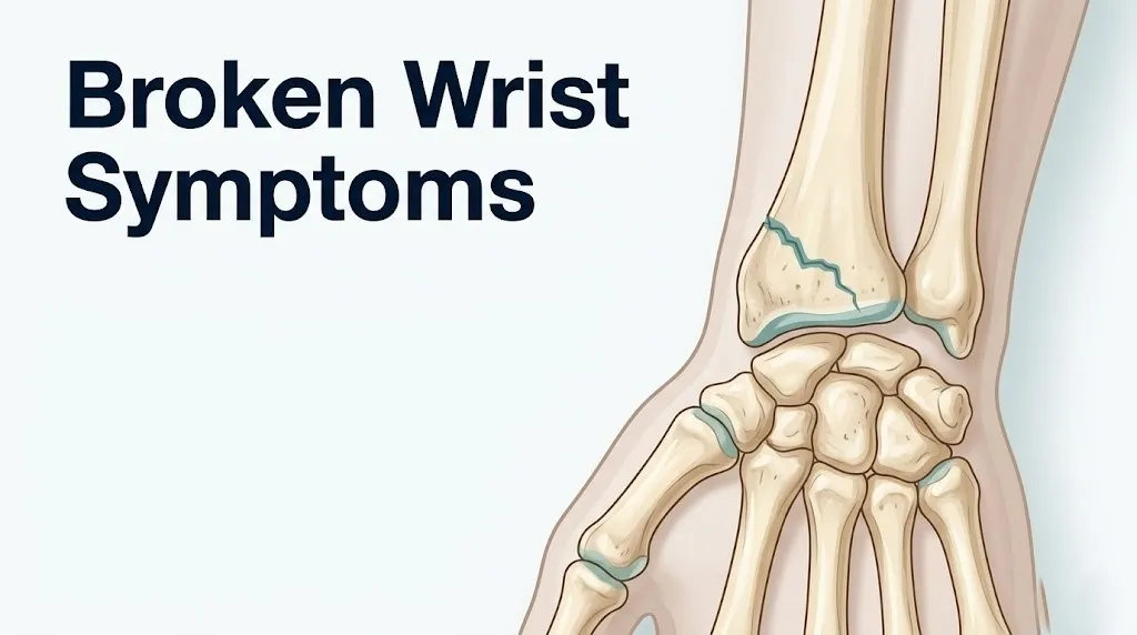
The wrist is not a single bone. The anatomical wrist includes the distal radius, the distal ulna, and eight carpal bones: the scaphoid, lunate, triquetrum, pisiform, hamate, capitate, trapezoid, and trapezium. The distal radius is by far the most commonly fractured, accounting for the majority of what clinicians and patients refer to as a “broken wrist.” The scaphoid ranks second in fracture frequency.
When bone cortex breaks, the dense periosteum, the fibrous outer sheath covering bone that is richly supplied with sensory nerve fibers, is disrupted or stretched. Those nerve fibers release substance P and bradykinin, two nociceptive signaling molecules that immediately generate intense pain signals. Simultaneously, the periosteal vascular plexus ruptures, causing a fracture hematoma that begins within minutes and expands over the first few hours as the inflammatory response amplifies.
The inflammatory response then triggers release of prostaglandin E2 (PGE2) via the cyclooxygenase pathway in injured periosteal cells, which sensitizes surrounding nociceptors and causes local vasodilation. Increased capillary permeability allows plasma proteins and fluid to leave the intravascular space and enter the surrounding tissue, producing visible swelling.
Summary of broken wrist symptoms and their mechanisms:
| Symptom | Mechanism | Onset Timing |
|---|---|---|
| Immediate pain | Periosteal nociceptor activation, substance P and bradykinin release | Within seconds of injury |
| Swelling (edema) | PGE2-driven vasodilation and capillary permeability, fracture hematoma expansion | Minutes to hours |
| Tenderness on palpation | Ongoing nociceptor sensitization from inflammatory mediators | Immediately after injury |
| Bruising (ecchymosis) | Hemoglobin degradation to biliverdin and bilirubin as hematoma spreads subcutaneously | Hours to days |
| Deformity | Displacement of fracture fragments from force of injury | Immediate in displaced fractures |
| Numbness or tingling | Median or ulnar nerve compression from edema or fracture fragment displacement | Variable; minutes to hours |
People on blood thinners, including warfarin, rivaroxaban, or apixaban, tend to experience faster and more extensive hematoma formation after a wrist fracture. This can accelerate the development of significant swelling and increases the theoretical risk of compartment pressure elevation.
What Does a Broken Wrist Feel Like
A broken wrist typically feels like an immediate, sharp, severe pain at the moment of impact, followed rapidly by a deep, throbbing ache that intensifies as swelling develops over the first hour after injury.
The initial pain is periosteal in character. It is sharp, intense, and localized. Many people describe a “crack” sensation at the moment of the break, though this is not universally present and its absence does not rule out a fracture. Within the first ten to thirty minutes, the pain often transitions from sharp to a deep, constant pressure-ache as the fracture hematoma expands and inflammatory edema begins filling the local tissue compartment.
Touching the wrist at any point near the fracture site produces a specific, point-tender response called point tenderness, which is one of the most clinically useful physical examination findings for distinguishing a fracture from a soft tissue injury. The area directly over the fracture hurts distinctly more than adjacent areas, and the pain is often described as a sharp “electric” sensation with firm palpation.
The overall sensation is typically described by patients as feeling “different” from the pain of a soft tissue bruise. A sprain aches diffusely. A fracture produces a localized, constant, and often worsening pain that does not ease with rest the way a muscular injury typically does within the first thirty to sixty minutes.
According to the Mayo Clinic, most people with a displaced wrist fracture find it impossible to use the injured hand for any gripping or weight-bearing function immediately after injury, in contrast to many sprains where some limited function persists despite pain.
How a broken wrist feels through the first 24 hours:
- First 0 to 5 minutes: sharp, severe localized pain at point of impact; possible audible or felt “crack”
- First 5 to 30 minutes: pain deepens from sharp to constant throbbing; visible swelling begins at fracture site
- First 1 to 4 hours: swelling spreads beyond immediate fracture site; gripping or wrist movement becomes impossible without severe pain; bruising may not yet be visible
- First 4 to 12 hours: bruising becomes visible as hematoma tracks subcutaneously; swelling reaches maximum early peak; numbness or tingling may develop if nerve compression is present
- First 12 to 24 hours: pain may feel slightly less sharp as acute inflammatory peak begins stabilizing, but swelling and tenderness remain; any reduction in pain at this stage does not indicate the fracture is less serious
Children often report the sensation more dramatically than adults, partly because their periosteum is thicker and more vascular, producing a more intense early hematoma and more immediate swelling.
Signs of a Broken Wrist You Should Not Ignore
The recognizable signs of a broken wrist include visible swelling, tenderness at a specific wrist location, bruising, visible deformity, warmth over the injury site, inability to grip, and in some cases numbness in the fingers.
Not every sign is always present. A displaced fracture with obvious deformity is impossible to miss. An undisplaced scaphoid fracture may produce only mild swelling and a single area of localized tenderness in the anatomical snuffbox, the small depression at the base of the thumb between the extensor pollicis longus and extensor pollicis brevis tendons.
The clinical rule in emergency and orthopedic medicine is straightforward: any wrist injury with localized bone tenderness following a significant impact should be treated as a fracture until imaging confirms otherwise. Waiting to see if the pain “goes away” is not clinically appropriate when a fracture is possible.
Red flag signs that indicate immediate evaluation is needed:
- Visible bone displacement or a wrist that appears bent or angled unnaturally
- Skin broken at the injury site with bone visible or suspected beneath (open fracture)
- Fingers that are pale, cold, or bluish after the injury (possible arterial compromise)
- Numbness or complete loss of sensation in one or more fingers immediately after impact
- Severe pain in the forearm that intensifies with passive finger extension (possible compartment syndrome)
- Inability to feel sensation in the palm or thumb-side of the hand (possible median nerve injury)
The American Academy of Orthopaedic Surgeons states that any wrist fracture with associated nerve symptoms, including tingling, numbness, or weakness in the fingers, warrants same-day evaluation at an emergency department rather than urgent care, because these symptoms may indicate nerve compression requiring prompt surgical decompression.
Older adults taking corticosteroids for conditions including rheumatoid arthritis or chronic obstructive pulmonary disease have steroid-induced bone fragility that can produce wrist fractures from forces that would not fracture a younger person’s bone, such as stumbling at home or pushing off a chair.
Key Takeaway: A broken wrist produces a localized, worsening, constant pain that is distinctly different from a bruise or sprain; any wrist injury with point tenderness directly over bone after a significant impact needs same-day imaging, not watchful waiting.
Broken Wrist Swelling: Why It Happens and What It Means
Broken wrist swelling is produced by two overlapping processes: the acute fracture hematoma that forms as the periosteal vascular plexus ruptures, and the inflammatory vasodilation and increased capillary permeability driven by prostaglandin E2 and histamine release at the fracture site.
The swelling typically begins within minutes of injury. Initially it is localized to the fracture area, but within one to two hours it spreads through the loose connective tissue of the wrist and hand dorsum. The dorsal wrist tends to swell more visibly than the palmar surface because the dorsal subcutaneous tissue is thinner and more compliant, allowing fluid to accumulate and become visible earlier.
Think of the fracture hematoma as a water balloon inflating under the skin at the injury site. At first it stays local. But as inflammatory mediators signal neighboring blood vessels to dilate and leak fluid, that balloon effect spreads outward through the surrounding tissue planes, producing the diffuse, puffy, warm swelling visible within one to two hours of a serious wrist fracture.
Swelling that continues to increase significantly beyond six to twelve hours, particularly if accompanied by worsening pain in the forearm rather than just the wrist, is a warning sign of compartment syndrome and warrants emergency evaluation.
According to the National Institute of Arthritis and Musculoskeletal and Skin Diseases (NIAMS), controlling early swelling in wrist fractures is a clinical priority because excessive soft tissue swelling can delay the application of a definitive cast and complicate the accuracy of fracture reduction.
Practical guidance for managing swelling before medical evaluation:
- Apply a bag of ice or a cold pack wrapped in a thin cloth to the wrist for 20 minutes, then remove for 20 minutes; repeat until medical evaluation
- Keep the injured wrist elevated above the level of the heart, resting on pillows, to reduce hydrostatic pressure driving fluid into the wrist tissue
- Do not apply a tight bandage or wrap that compresses the entire wrist circumferentially, as this can convert manageable swelling into compartment pressure elevation
- Do not apply heat within the first 48 hours; heat increases vasodilation and worsens acute swelling
- Do not attempt to move the wrist to test range of motion; movement at a fracture site displaces fragments and worsens hematoma
Patients with poorly controlled diabetes may experience more substantial swelling due to baseline microvascular disease and impaired inflammatory regulation, and should not delay evaluation even if the swelling appears moderate.
Broken Wrist Pain Location: Where It Hurts and Why
The location of wrist fracture pain is determined by which bone or bones are broken, because each bone in the wrist has a distinct anatomical position and a distinct periosteal innervation territory.
The most frequently fractured bone, the distal radius, produces pain and tenderness on the radial (thumb) side of the wrist and just proximal to the wrist crease along the front and back of the forearm. Pressing directly on the distal radial metaphysis, the flared section of the radius just above the wrist joint, reproduces intense, localized pain in virtually all cases of distal radius fracture.
The scaphoid fracture produces pain and tenderness in a very specific location: the anatomical snuffbox, the small hollow depression on the thumb side of the wrist between the tendons of the extensor pollicis longus and extensor pollicis brevis. Pain there, particularly when accompanied by pain on axial loading of the thumb (pressing firmly along the line of the thumb into the scaphoid), is highly suggestive of scaphoid involvement.
The ulnar styloid, the bony prominence on the little-finger side of the wrist, produces pain and tenderness on the ulnar side when fractured. Ulnar styloid fractures often accompany distal radius fractures. Isolated ulnar styloid pain with intact wrist motion raises the possibility of triangular fibrocartilage complex (TFCC) injury rather than a fracture, though both can coexist.
Pain location map for wrist fractures:
| Pain Location | Most Likely Structure | Clinical Feature |
|---|---|---|
| Dorsal-radial wrist, above wrist crease | Distal radius (dorsal cortex) | Colles fracture pattern; visible “dinner fork” deformity if displaced |
| Palmar wrist, proximal to wrist crease | Distal radius (volar cortex) | Smith fracture pattern; wrist flexed deformity |
| Anatomical snuffbox (thumb-side hollow) | Scaphoid | Tenderness here with negative X-ray still warrants MRI |
| Ulnar wrist, bony knob | Ulnar styloid | Often accompanies distal radius fracture |
| Central base of palm / hypothenar eminence | Hamate (hook of hamate) | Common in golfers and racquet sport athletes from grip-impact |
Athletes who participate in sports involving racquet grips, golf clubs, or baseball bats are at elevated risk for hook of hamate fractures, which produce pain in the heel of the palm on the little-finger side rather than at the dorsal wrist, a location that often delays diagnosis because patients describe it as “hand pain” rather than wrist pain.
Broken Wrist Bruising and Numbness
Bruising after a broken wrist appears as discoloration ranging from red-purple initially to yellow-green over days, produced by the subcutaneous tracking of blood from the fracture hematoma; numbness reflects compression or contusion of the median nerve, ulnar nerve, or radial sensory nerve.
Bruising does not appear immediately. It typically becomes visible six to twenty-four hours after injury as the fracture hematoma, initially contained near the periosteum, dissects through subcutaneous tissue planes toward the skin surface. The hemoglobin within the hematoma is broken down sequentially into biliverdin (green) and bilirubin (yellow) by local tissue macrophages over five to ten days, producing the characteristic color progression from deep purple through green to yellow as healing progresses.
The distribution of bruising gives clinical information. Bruising isolated to the dorsal wrist suggests a dorsal cortex fracture. Bruising that tracks down into the palm or fingers within the first twelve to twenty-four hours indicates a larger hematoma with more extensive soft tissue dissection, which correlates with higher-energy fractures and greater fracture displacement.
Numbness in the thumb, index finger, middle finger, and the radial (thumb) half of the ring finger suggests compression or contusion of the median nerve, which travels through the carpal tunnel directly beneath the flexor retinaculum. Fracture fragment displacement or acute swelling within the carpal tunnel can compress the median nerve at this location, producing the sensory distribution of acute carpal tunnel syndrome.
A 2019 study published in the Journal of Hand Surgery found that acute median nerve symptoms following distal radius fracture occur in approximately 10 to 17 percent of cases, and that early fracture reduction typically resolves the nerve compression in the majority of affected patients without additional surgical intervention.
Numbness on the little-finger and ring-finger ulnar side suggests ulnar nerve involvement, which is less common but occurs with specific fracture patterns affecting the medial wrist. Any finger numbness after a wrist fracture should be specifically reported to the evaluating emergency medicine physician or orthopedic surgeon, as persistent nerve compression after fracture reduction may require urgent carpal tunnel release.
Older adults are at higher risk for persistent post-fracture median nerve symptoms because age-related narrowing of the carpal tunnel from degenerative arthritis leaves less room for the additional swelling that accompanies a fracture.
Key Takeaway: Numbness in the thumb, index, or middle fingers after a wrist injury is not just a sensation to monitor at home; it indicates median nerve compression that must be evaluated by an emergency physician or orthopedic surgeon the same day.
Distal Radius Fracture Symptoms
Distal radius fracture symptoms are the most commonly encountered broken wrist presentation, typically including immediate severe pain at the radial wrist, rapid swelling, visible deformity in displaced cases, and tenderness directly over the distal radial metaphysis.
The distal radius is the wider, flared end of the radius bone that forms the primary weight-bearing surface of the wrist joint, articulating with the proximal row of carpal bones at the radiocarpal joint. When a person falls onto an outstretched hand, the impact force travels from the heel of the hand through the carpal bones into the distal radius, which absorbs the majority of the load. When that force exceeds the structural capacity of the radial cortex, the bone fractures, typically at a point one to two centimeters proximal to the radiocarpal joint surface.
The most recognized distal radius fracture type is the Colles fracture, in which the fracture fragment is displaced dorsally (toward the back of the wrist), producing the classic “dinner fork” or “bayonet” deformity visible as a step-off on the dorsal wrist when viewed from the side. The Smith fracture is the reverse pattern, with volar (palm-side) displacement of the fracture fragment, producing a visible wrist flexion deformity.
Colles fracture vs. Smith fracture symptom comparison:
| Feature | Colles Fracture | Smith Fracture |
|---|---|---|
| Direction of displacement | Dorsal (backward) | Volar (forward, toward palm) |
| Mechanism | Fall on extended (outstretched) hand | Fall on flexed wrist, or direct blow |
| Visible deformity | “Dinner fork” dorsal step-off | Wrist appears flexed or angled toward palm |
| Who it affects most | Postmenopausal women, older adults | Younger adults, high-energy injuries |
| Median nerve risk | Moderate | Lower |
According to the American Academy of Orthopaedic Surgeons, distal radius fractures are the most frequently occurring fractures in people under age 75 and the second most frequent fracture overall in people over 75, after hip fractures. The age distribution reflects the central role of bone density in fracture susceptibility.
Postmenopausal women experience substantially higher rates of distal radius fracture than men of the same age because estrogen deficiency following menopause reduces osteoclast inhibition, accelerating trabecular bone resorption in the distal radius metaphysis, where trabecular bone is particularly dense and therefore particularly vulnerable to estrogen-deficiency-driven structural weakening.
Scaphoid Fracture Symptoms
Scaphoid fracture symptoms are often deceptively mild, typically producing only localized tenderness in the anatomical snuffbox, mild wrist swelling, and a dull aching pain with gripping, without the dramatic deformity seen in displaced distal radius fractures.
The scaphoid is the small, boat-shaped bone sitting at the thumb-base side of the proximal carpal row. It is the most frequently fractured carpal bone, representing approximately 70 percent of all isolated carpal bone fractures, according to research published in the Journal of Bone and Joint Surgery. Scaphoid fractures typically result from a FOOSH mechanism at a slightly different wrist position than distal radius fractures, with the wrist in radial deviation and extension at the moment of impact.
The diagnostic challenge of scaphoid fractures is their subtle clinical presentation and the unreliability of initial plain film X-rays. Standard wrist X-rays miss up to 20 percent of scaphoid fractures at initial presentation, according to a systematic review published in Radiology. The fracture line may not be visible until five to ten days after injury when bone resorption at the fracture site makes the gap radiographically apparent.
This missed diagnosis is clinically serious because the scaphoid has a retrograde blood supply through the dorsal scaphoid branch of the radial artery, which enters the bone distally and flows toward the proximal pole. A fracture through the waist or proximal pole of the scaphoid interrupts this blood supply to the proximal fragment. Without revascularization from proper treatment and immobilization, the proximal pole undergoes avascular necrosis, a condition in which the bone tissue dies from ischemia, leading to wrist arthritis and chronic pain.
Key clinical signs of a scaphoid fracture:
- Point tenderness specifically in the anatomical snuffbox when the examiner’s thumb presses into that hollow with the wrist held in neutral position
- Pain with axial loading of the thumb: pressing firmly along the thumb axis toward the wrist reproduces scaphoid pain
- Pain with resisted pronation or supination of the forearm
- Mild swelling over the radial wrist without dramatic deformity
- Grip strength reduced, with pain localized to the radial wrist rather than diffusely across the joint
Young athletic men aged 15 to 40 are disproportionately affected by scaphoid fractures, likely because their bone density protects the distal radius from fracturing under similar forces, directing the impact energy further into the carpal bones instead. Any athlete in this demographic presenting with radial wrist pain after a fall or contact injury should have scaphoid-specific evaluation even if initial X-rays are negative.
Broken Wrist Without Deformity: When There Is No Obvious Sign
A broken wrist without deformity occurs when the fracture is non-displaced, meaning the bone has cracked or fractured but the fragments have not shifted position, leaving the wrist appearing externally normal.
This is one of the most clinically important points about wrist fractures: the absence of visible deformity does not rule out a fracture. Non-displaced distal radius fractures, all scaphoid fractures presenting without significant carpal malalignment, and stress fractures of the carpal bones can all produce a completely normal-looking wrist on visual inspection.
The critical mistake people make in this scenario is attributing their wrist pain to a sprain and continuing activities without seeking imaging. A non-displaced scaphoid fracture treated as a sprain and allowed to bear load and continue normal function risks fracture displacement, collapse of the proximal fragment from avascular necrosis, and permanent wrist dysfunction.
The clinical standard, stated by the American Academy of Orthopaedic Surgeons, is that a wrist injury with anatomical snuffbox tenderness, radial wrist pain, and a mechanism consistent with a fall on an outstretched hand should be treated as a presumed scaphoid fracture and immobilized in a thumb spica splint until advanced imaging confirms or excludes the diagnosis, even if initial X-rays appear normal.
Situations where a broken wrist without deformity is especially likely:
- Young adult athlete with radial wrist pain after a fall, with anatomical snuffbox tenderness and negative initial X-ray
- Older adult with osteoporosis who reports wrist pain after a relatively low-energy fall, even with minimal visible swelling
- Child with wrist pain and tenderness after a fall, particularly if tenderness is at the growth plate level rather than the joint space
- Any patient with persistent wrist pain beyond two weeks after a “sprain” that has not improved with rest and anti-inflammatory measures
Patients with rheumatoid arthritis have structurally weakened wrist bones from chronic synovial inflammation and may fracture with minimal trauma, presenting with what appears to be a routine flare rather than an acute injury.
Key Takeaway: A normal-looking wrist after a fall does not mean it is not broken; scaphoid and non-displaced distal radius fractures require imaging to diagnose, and treating them as sprains risks permanent wrist damage.
Can You Move a Broken Wrist
Some people can move a broken wrist to a limited degree, particularly with non-displaced fractures, but movement is almost always painful and always inadvisable before imaging confirms the nature of the injury.
This is the question that sends the most people astray. The belief that “if I can move it, it isn’t broken” is clinically incorrect. Movement capacity after a wrist fracture depends entirely on the fracture type, displacement, and the individual’s pain tolerance. A person with a high pain threshold and a non-displaced distal radius fracture may be able to flex and extend their wrist partially before pain stops them. A person with a severely displaced Colles fracture will be unable to attempt any movement without extreme pain and may have visible loss of normal anatomy.
The Mayo Clinic states clearly that limited ability to move the wrist does not exclude a fracture and that all suspected wrist fractures require imaging for definitive evaluation regardless of whether movement is possible.
What typically distinguishes fracture movement from sprain movement is quality rather than quantity. With a sprain, movement is limited by pain that is spread across a range of motion. With a fracture, movement typically hits a specific, sharp pain barrier at a certain point, and produces a deeply unpleasant sensation described as “wrong” or “bone-level” rather than muscular.
What to avoid doing with a suspected broken wrist before imaging:
- Gripping or squeezing anything with the injured hand
- Rotating the forearm (pronation and supination movements), which stresses the distal radioulnar joint
- Bearing weight on the wrist, as when pushing up from a chair or the floor
- Attempting to straighten a visibly deformed wrist; this can damage nerves and blood vessels
- Continuing sport or work activity because movement “seems okay”
Children often continue using an injured wrist briefly after a fracture because their acute pain response may be temporarily masked by adrenaline and distraction, particularly in playground and sport injuries. A child who was crying immediately after a fall but then “seemed fine” should still be evaluated if localized wrist tenderness is present on gentle examination.
Broken Wrist vs Sprained Wrist Symptoms
Distinguishing broken wrist vs sprained wrist symptoms requires assessment of pain character, tenderness location, swelling pattern, mechanism severity, and specific physical examination findings, because the two conditions can initially feel similar but have distinctly different physiological origins.
A wrist sprain involves stretching or tearing of the ligaments connecting the carpal bones to each other or to the distal radius. The most commonly sprained wrist ligaments are the scapholunate ligament and the components of the triangular fibrocartilage complex (TFCC) on the ulnar side. Sprains produce pain, swelling, and tenderness, but the pain is diffuse rather than focused on a specific bony point, and the swelling typically develops more slowly than in a fracture.
A fracture produces point tenderness directly over the bone itself, not over the ligament. This distinction is the most clinically useful physical examination finding for initial triage before imaging.
Broken wrist vs. sprained wrist: key distinguishing features:
| Feature | Broken Wrist (Fracture) | Sprained Wrist (Ligament Injury) |
|---|---|---|
| Pain onset | Immediate, severe at moment of injury | Immediate or delayed; often builds over minutes |
| Point tenderness | Directly over bone surface | Over ligament or joint space, not bone |
| Deformity | Possible in displaced fractures | Absent (no structural bone displacement) |
| Swelling speed | Rapid; within minutes to 1 hour | Typically within 1 to 4 hours |
| Bruising extent | Can be extensive if hematoma tracks widely | Usually limited and diffuse |
| Grip function | Severely impaired or impossible | Reduced but often partially retained |
| X-ray finding | May show fracture line (not always visible) | Normal; diagnosis by clinical exam and MRI |
| Hearing/feeling a crack | Possible | Uncommon; pop possible with scapholunate tear |
The honest clinical reality is that distinguishing a significant wrist sprain from a non-displaced fracture on physical examination alone is often impossible, even for experienced emergency physicians. Research published in Injury has found that clinical examination without imaging has insufficient sensitivity to reliably exclude wrist fractures in the acute setting.
Postmenopausal women should not rely on the sprain versus fracture distinction to avoid imaging. Their trabecular bone architecture is weakened enough that fractures can occur with the same low-energy mechanisms that produce sprains in younger adults.
Broken Wrist in Children: How Symptoms Differ
Broken wrist symptoms in children differ from adult presentations because the pediatric skeleton contains growth plates (physes), cartilaginous zones of active bone growth located at the ends of long bones that are mechanically weaker than the surrounding cortical bone and fracture more easily.
The most frequently occurring pediatric wrist fracture involves the distal radial physis, the growth plate at the lower end of the radius. This is classified using the Salter-Harris classification system, which describes fractures by their relationship to the growth plate. A Salter-Harris Type II fracture, in which the fracture passes through the physis and exits through the metaphysis (the wider section of bone just above the growth plate), is the most common growth plate fracture pattern in the distal radius.
In children, swelling at the distal forearm and wrist after a fall tends to be rapid and more pronounced than in adults because the pediatric periosteum is thicker, more vascular, and produces a larger and faster inflammatory response. Bruising may appear sooner and spread more widely. Children are more likely to cry immediately after a growth plate fracture and may be unwilling to allow anyone to touch the injured area.
One important diagnostic nuance: torus fractures (also called buckle fractures) are incomplete cortical fractures common in young children aged 6 to 12, in which only one side of the cortex buckles without a complete fracture line. These fractures may produce only mild, localized tenderness at the distal radius with minimal swelling, and the child may continue to use the wrist with only mild guarding, leading parents to underestimate the injury severity.
According to the American Academy of Pediatrics, any child with tenderness over the distal forearm and wrist after a fall should have wrist radiographs performed at a minimum of two views, because growth plate injuries that are inadequately treated can result in growth disturbance and limb length discrepancy.
Any parent whose child sustained a wrist injury during sport or play and is guarding the arm, refusing to grip, or crying with any wrist movement should take the child for same-day imaging at an urgent care facility equipped with pediatric X-ray capability or a pediatric emergency department, and ask the treating physician specifically whether a growth plate injury is present and whether pediatric orthopedic consultation is warranted.
Key Takeaway: Children with wrist pain after a fall may have a buckle or growth plate fracture that looks minor on the surface; they need same-day X-rays because undertreated physeal injuries can permanently affect bone growth.
Broken Wrist in Elderly Patients: Higher Risk, Different Presentation
Broken wrist presentations in older adults, particularly those over age 65, differ from younger adult presentations because age-related bone density loss means fractures occur with lower-energy mechanisms, fracture patterns are more likely to be comminuted, and healing takes longer.
Osteoporosis is the primary structural risk factor for wrist fractures in older adults. Osteoporosis is a condition of reduced bone mineral density and degraded trabecular microarchitecture, classified by the World Health Organization as a bone mineral density T-score of negative 2.5 or lower on dual-energy X-ray absorptiometry (DEXA) scan. In osteoporotic bone, the trabecular struts within the distal radial metaphysis are thinner, fewer, and more disconnected, reducing the bone’s ability to absorb and distribute impact forces.
An older adult with osteoporosis may sustain a distal radius fracture from simply tripping on a curb, stumbling in a bathroom, or catching themselves on a counter edge. The force threshold for fracture is far lower than in younger adults. This means the dramatic, high-velocity fall mechanism of younger patients may be completely absent, leading some older adults to underestimate the likelihood that they have a true fracture.
Clinically, fractures in osteoporotic bone are more often comminuted, meaning the bone breaks into multiple fragments rather than two clean pieces. This makes surgical fixation more technically demanding and increases the likelihood that open reduction internal fixation (ORIF) will be required rather than simple casting.
Older adults taking chronic corticosteroids for conditions including polymyalgia rheumatica, inflammatory bowel disease, or asthma face compounded risk because corticosteroids reduce bone formation by inhibiting osteoblast activity and accelerate bone resorption by increasing osteoclast lifespan, producing steroid-induced osteoporosis that can cause fragility fractures after trivially low-force impacts.
Risk factors for wrist fracture in older adults:
- Female sex and postmenopausal status (reduced estrogen, accelerated trabecular resorption)
- Chronic corticosteroid use exceeding three months
- Vitamin D deficiency (reduced calcium absorption, impaired bone mineralization)
- History of prior low-energy fracture (the strongest predictor of future fracture)
- Reduced muscle mass and balance impairment increasing fall frequency
- Chronic kidney disease (disrupted calcium-phosphate regulation and secondary hyperparathyroidism)
Any older adult who sustains a wrist injury should be evaluated at an emergency department or urgent care facility the same day. A board-certified emergency medicine physician or orthopedic surgeon should assess both the acute fracture and perform or refer for a falls risk assessment, because a first distal radius fracture in an older adult is a strong clinical indicator of underlying osteoporosis that warrants a formal bone density evaluation.
Complications of Wrist Fracture
Complications of wrist fracture range from the common and manageable, including post-fracture stiffness and reduced grip strength, to the rare but limb-threatening, including compartment syndrome and avascular necrosis.
The most feared acute complication is compartment syndrome of the forearm. Compartment syndrome occurs when interstitial fluid pressure within the closed myofascial compartments of the forearm (the anterior and posterior compartments, enclosed by the antebrachial fascia) rises above the perfusion pressure threshold for the muscles and nerves within those compartments. Ischemia of the forearm muscles and nerves begins within hours of compartment pressure elevation, and permanent muscle and nerve damage can occur within four to six hours of the onset of critical pressure elevation.
The characteristic symptom pattern of early compartment syndrome includes pain that is out of proportion to the injury (far more severe than expected for a simple fracture), pain with passive extension of the fingers (stretching the ischemic flexor muscles of the anterior compartment), forearm tightness or a woody firmness on palpation, and numbness or tingling in the fingers.
Malunion occurs when a fracture heals in a suboptimal position, producing wrist deformity, reduced range of motion, and chronic pain. This is more common in fractures treated with casting alone when the fracture was significantly displaced at the time of reduction.
Nonunion is less common in distal radius fractures but clinically important in scaphoid fractures, where the retrograde blood supply to the proximal pole can be interrupted by the fracture itself, leading to failure of fracture healing. Scaphoid nonunion can progress to avascular necrosis and scaphoid nonunion advanced collapse (SNAC), a pattern of progressive wrist arthritis.
Complications summary:
| Complication | Mechanism | Time Frame | Urgency |
|---|---|---|---|
| Compartment syndrome | Forearm fascial compartment pressure exceeds perfusion pressure | Hours after injury | 911 emergency |
| Acute carpal tunnel syndrome | Median nerve compressed by fracture fragment or edema | Hours to days | Same-day evaluation |
| Malunion | Fracture heals in displaced position | Weeks to months | Outpatient orthopedic follow-up |
| Scaphoid nonunion | Interrupted retrograde blood supply prevents healing | Months to years | Orthopedic hand surgery evaluation |
| Avascular necrosis | Bone death from ischemia in scaphoid proximal pole | Months | Hand surgeon evaluation |
| Post-traumatic arthritis | Articular surface damage leads to joint degeneration | Years | Rheumatology and hand surgery co-management |
People on immunosuppressive medications, including those used for organ transplantation, have impaired fracture healing because immunosuppressive agents reduce the osteoblast activity and inflammatory signaling that drive callus formation.
How a Broken Wrist Is Diagnosed
A broken wrist is diagnosed through a combination of clinical history, physical examination findings, and imaging studies, with plain film radiography as the first-line imaging tool and computed tomography or magnetic resonance imaging used for fractures not visible on initial X-rays.
The standard initial imaging protocol for a suspected wrist fracture includes at minimum three views of the wrist: posteroanterior (PA), lateral, and oblique projections. These three views together give the treating physician the ability to identify fracture lines, assess the degree of displacement, evaluate articular surface involvement, and identify associated carpal fractures.
For scaphoid fractures specifically, a dedicated scaphoid series adds two additional angled views that increase the sensitivity of plain film imaging for this bone. Despite optimized X-ray technique, research published in Radiology confirms that plain film imaging still misses a clinically meaningful proportion of scaphoid fractures at initial presentation. When clinical suspicion is high (anatomical snuffbox tenderness, positive axial load test), the preferred next-step imaging is magnetic resonance imaging (MRI) of the wrist, which can identify scaphoid fractures within 24 hours of injury with a sensitivity approaching 100 percent in experienced radiological hands.
Computed tomography (CT) scanning of the wrist is used to characterize complex or comminuted fracture patterns, assess articular surface disruption, and plan surgical approaches when ORIF is being considered.
Diagnostic process for a suspected wrist fracture:
- Clinical history: mechanism of injury, location of pain, timing of symptom onset, prior wrist injuries, medications (anticoagulants, corticosteroids), and medical history relevant to bone density
- Physical examination: visual inspection for deformity, skin integrity, palpation for point tenderness over specific bones (distal radius, anatomical snuffbox, ulnar styloid), neurovascular assessment (capillary refill, median and ulnar nerve sensation testing), compartment assessment (forearm firmness)
- Plain film X-rays (minimum three views): assess for fracture line, displacement, articular surface involvement
- MRI or CT if X-rays are negative but clinical suspicion remains high: MRI preferred for soft tissue and occult bone injury; CT preferred for complex fracture geometry
- Orthopedic surgery consultation if fracture is displaced, articular, associated with neurovascular symptoms, or if compartment syndrome is suspected
Children require age-appropriate radiation dose protocols and physeal-specific evaluation by a radiologist or orthopedic surgeon familiar with pediatric growth plate anatomy, because the normal physis can be mistaken for a fracture by an inexperienced reader.
Key Takeaway: A negative wrist X-ray does not rule out a scaphoid fracture; if you have anatomical snuffbox tenderness after a fall, ask specifically whether MRI is indicated before accepting a diagnosis of “sprain.”
Broken Wrist Treatment and Recovery
Broken wrist treatment depends on fracture type, degree of displacement, patient age and bone health, and functional demands, ranging from cast immobilization alone for non-displaced fractures to open reduction internal fixation with metal plate and screws for displaced or articular fractures.
Non-displaced or minimally displaced distal radius fractures are typically managed with a short-arm cast (below elbow) or long-arm cast (above elbow) for four to six weeks, depending on fracture stability and location. During this period, the fracture heals through a sequence of physiological events: the fracture hematoma organizes into a fibrous callus, which is then progressively mineralized into a hard callus, and finally remodeled into normal bone architecture over months.
Displaced fractures requiring reduction are initially managed in the emergency setting with a closed reduction procedure, in which the treating physician manipulates the fracture fragments back into alignment under local or regional anesthesia. A post-reduction X-ray confirms the quality of alignment. If adequate reduction is achieved and maintained, casting proceeds. If reduction cannot be maintained or the fracture pattern is inherently unstable, open reduction internal fixation (ORIF) with a volar locking plate is the most commonly used surgical approach for distal radius fractures, with extensive evidence supporting its use in active patients and in fractures with significant intra-articular involvement.
Scaphoid fractures are treated with a thumb spica cast that immobilizes the wrist and thumb joint for six to twelve weeks for non-displaced waist fractures. Proximal pole fractures or any scaphoid fracture with displacement typically require surgical fixation with a headless compression screw to restore the bone’s structural integrity and protect the retrograde blood supply.
General recovery timeline by fracture type:
| Fracture Type | Immobilization Period | Return to Light Activity | Full Recovery |
|---|---|---|---|
| Non-displaced distal radius | 4 to 6 weeks cast | 6 to 8 weeks | 3 to 6 months |
| Displaced distal radius (ORIF) | 4 to 6 weeks post-surgery | 8 to 12 weeks | 6 to 12 months |
| Non-displaced scaphoid waist | 6 to 12 weeks cast | 12 to 16 weeks | 4 to 6 months |
| Proximal pole scaphoid (surgical) | 8 to 12 weeks post-surgery | 12 to 16 weeks | 6 to 12+ months |
| Torus fracture in child | 3 to 4 weeks splint | 4 to 6 weeks | 6 to 8 weeks |
A board-certified occupational therapist specializing in hand therapy plays a central role in wrist fracture rehabilitation, guiding progressive range-of-motion exercises, grip strengthening, and functional activity retraining after cast removal or surgical healing. Research in the Journal of Bone and Joint Surgery supports early hand therapy involvement in improving functional outcomes after distal radius fracture.
Emergency Symptoms: When to Call 911 or Go to the ER
Certain symptoms associated with a broken wrist and its complications require immediate emergency evaluation. Do not wait to see if these resolve on their own.
Call 911 or go to the nearest emergency room immediately if you or someone you are with experiences:
- Visible bone protruding through the skin at the wrist or forearm: this is an open fracture requiring emergency surgical irrigation and stabilization to prevent bone infection (osteomyelitis)
- Fingers that are pale, white, or bluish and cool to the touch after a wrist injury: this indicates possible arterial compromise to the hand and is a vascular emergency
- Severe forearm pain that worsens continuously and is out of proportion to the injury, particularly pain with passive straightening of the fingers: these are the cardinal early signs of compartment syndrome, a limb-threatening emergency
- Forearm that feels abnormally tight, hard, or “wooden” to the touch: indicates elevated compartment pressure requiring emergency fasciotomy
- Complete loss of sensation in the palm or all fingers after a wrist injury: indicates possible median or ulnar nerve injury requiring emergency evaluation
- Wrist deformity with the hand hanging at an abnormal angle: indicates significant displacement requiring emergency reduction and possible surgery
- A child who refuses to move the arm at all and is crying inconsolably after a wrist injury: warrants emergency pediatric evaluation for growth plate fracture or vascular injury
These presentations can indicate open fracture with infection risk, vascular compromise of the hand, acute compartment syndrome, or displaced fracture with nerve and vessel injury, all of which require emergency department evaluation and immediate orthopedic surgery consultation, not an urgent care appointment scheduled for the following day.
Frequently Asked Questions About Broken Wrist Symptoms
How do I know if my wrist is broken or just sprained?
A broken wrist typically produces point tenderness directly over the bone surface, rapid swelling within the first hour, and severe pain that worsens with any attempted movement.
A sprain produces more diffuse tenderness over the ligament or joint space rather than over the bone itself, and swelling is usually slower to develop.
Because clinical examination alone cannot reliably distinguish a non-displaced fracture from a sprain, an X-ray is the appropriate step for any wrist injury with significant mechanism of injury, localized bone tenderness, or persistent pain after 48 hours.
Can you move your wrist if it’s broken?
Some people can move a broken wrist to a limited degree, particularly when the fracture is non-displaced and pain tolerance is high.
Limited movement does not rule out a fracture; the ability to flex or extend the wrist partially is not a reliable indicator of whether bone is intact.
Any wrist with localized bone tenderness, swelling, or a traumatic mechanism of injury needs imaging before movement capability is used to decide whether medical care is needed.
What does a broken wrist feel like right after injury?
A broken wrist typically produces an immediate sharp, severe pain at the moment of impact, often accompanied by a feeling or sound of a crack.
Within the first thirty minutes, the pain usually transitions from sharp to a deep, constant throbbing ache as the fracture hematoma forms and swelling begins.
The sensation is generally described as distinctly more localized and severe than a bruise, and most people find it impossible to grip or use the hand normally within the first hour.
How long does a broken wrist take to heal?
A non-displaced distal radius fracture typically heals within six to eight weeks of immobilization, though full strength and range of motion recovery takes three to six months.
Scaphoid fractures take longer, ranging from six weeks for non-displaced waist fractures in cast to six to twelve months for proximal pole fractures requiring surgical fixation.
Healing times are longer in older adults with osteoporosis, in patients taking corticosteroids, and in smokers, because nicotine impairs periosteal blood flow and slows the callus formation process.
Do all broken wrists show up on X-ray?
Not all wrist fractures are visible on initial X-ray; scaphoid fractures are missed on plain film in up to 20 percent of cases at initial presentation, according to research published in Radiology.
Torus (buckle) fractures in children may also be subtle and require experienced radiological review of properly positioned pediatric X-ray views.
When clinical suspicion is high and initial X-rays are negative, magnetic resonance imaging (MRI) is the most sensitive imaging method for detecting occult wrist fractures, including scaphoid injuries.
What are the signs a broken wrist needs surgery?
Signs that a broken wrist needs surgery include significant fracture displacement, intra-articular fracture extension (the fracture line entering the joint surface), inability to maintain reduction after closed manipulation, and associated neurovascular injury.
Open fractures (bone visible through skin), irreducible dislocations, and scaphoid fractures of the proximal pole also typically require surgical fixation.
A board-certified orthopedic surgeon will assess these factors through physical examination and post-reduction imaging; any patient with a displaced distal radius fracture or a scaphoid fracture should ask at initial evaluation whether they need an orthopedic surgery referral.
Putting It All Together
The single most important takeaway from this guide is that a broken wrist does not always look broken. The absence of visible deformity, the ability to move the fingers, or a mechanism that feels too minor to cause a fracture are not reliable reasons to avoid imaging. Scaphoid fractures in particular can destroy wrist function over months if dismissed as sprains.
If you or someone with you has wrist pain after a fall, impact, or twisting injury, seek same-day evaluation at an urgent care facility with X-ray capability or an emergency department. Bring information about any blood thinners, corticosteroids, or osteoporosis medications being taken, and specifically ask the treating physician whether a scaphoid fracture is possible if your pain and tenderness are on the thumb side of the wrist.
Symptoms that worsen progressively, fingers that change color or lose sensation, or forearm pain that feels wildly out of proportion to the injury require immediate emergency room evaluation. Those are the signs the clock is running.