Pelvic Congestion Syndrome Symptoms: Full 2026 Guide
Pelvic congestion syndrome symptoms include chronic pelvic pain, a persistent dragging pressure in the lower abdomen, pain that intensifies after intercourse, and discomfort that reliably worsens with prolonged standing. These symptoms arise from dilated, dysfunctional pelvic veins that trap blood in the pelvis rather than returning it efficiently to the systemic circulation.
According to the American College of Obstetricians and Gynecologists, chronic pelvic pain affects approximately 15 percent of women of reproductive age in the United States, and pelvic congestion syndrome is estimated to account for 30 percent or more of those cases. Despite this, the condition remains one of the most underdiagnosed causes of persistent pelvic pain, partly because standard pelvic ultrasound performed with a woman lying down often fails to reveal the engorged varicosities that become most visible in the upright posture.
This guide covers the full symptom spectrum of pelvic congestion syndrome with detailed physiological explanation for each symptom, the clinical features that distinguish it from endometriosis and other look-alikes, the populations at greatest risk, how the condition is diagnosed, and when symptoms require urgent evaluation rather than watchful waiting.
Pelvic Congestion Syndrome Symptoms: What They Are and Why They Happen
Pelvic congestion syndrome symptoms are caused by chronically elevated venous pressure within dilated, incompetent veins of the pelvic venous plexus, producing a pattern of pain and pressure that is distinctively position-dependent, cyclically variable, and often postcoitally prolonged.
The core symptom cluster recognized by vascular and gynecological specialists includes:
- Dull, aching pelvic pain lasting longer than six months, typically bilateral but sometimes lateralized to the left side
- Pelvic heaviness or dragging sensation that worsens as the day progresses
- Pain that is significantly worse after intercourse and may persist for 24 hours or more
- Discomfort that intensifies with prolonged standing, walking, or physical activity
- Pain relief when lying down, particularly with the hips elevated
- Visible or palpable varicosities around the vulva, inner thighs, or buttocks
- Dysmenorrhea (painful menstruation) that exceeds what was previously normal for that individual
- Urinary urgency or frequency without infection
What distinguishes this symptom pattern clinically is its postural behavior. Pain that reliably worsens when vertical and improves when horizontal is a highly specific feature pointing toward a venous pressure-dependent mechanism rather than endometrial, musculoskeletal, or infectious causes.
| Symptom | Clinical Characteristic | Physiological Basis |
|---|---|---|
| Chronic pelvic pain | Dull, aching, bilateral or left-sided, greater than 6 months | Visceral afferent stimulation from dilated pelvic varicosities |
| Postcoital aching | Pain persisting 12 to 24 hours after intercourse | Compression of engorged paravaginal venous plexus during intercourse |
| Postural worsening | Pain increases with standing, improves lying down | Increased hydrostatic venous pressure in the upright posture |
| Vulvar varicosities | Visible tortuous veins at the vulva or inner thigh | Retrograde venous reflux extending into perineal tributaries |
| Bladder urgency | Urinary frequency without infection | Varicosities adjacent to the bladder neck |
| Dysmenorrhea | Worsening menstrual pain relative to prior cycles | Prostaglandin amplification in congested pelvic tissue |
Women with polycystic ovary syndrome (PCOS) may experience a more pronounced symptom pattern due to higher circulating estrogen levels and altered pelvic hemodynamics associated with enlarged ovarian volume.
What Is Pelvic Venous Insufficiency
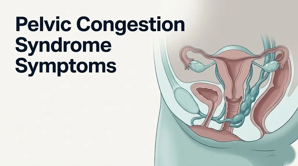
Pelvic venous insufficiency refers to the functional failure of the venous drainage system of the pelvis, specifically the inability of the ovarian veins and their tributaries to return blood from the pelvic organs to the central circulation against gravitational forces without retrograde leakage.
The left ovarian vein is involved in most cases. It drains at a near-perpendicular angle into the left renal vein rather than directly into the inferior vena cava, creating a higher resistance drainage pathway. Functional venous valves are absent in approximately 13 to 15 percent of women in the left ovarian vein, according to anatomical studies cited in the Journal of Vascular and Interventional Radiology. When those valves fail or are absent, blood refluxes back down the ovarian vein toward the pelvis under the force of gravity.
The right ovarian vein drains directly into the inferior vena cava at an oblique angle, which produces lower venous resistance and explains why right-sided involvement is less common but not rare.
Pelvic venous insufficiency is distinct from varicose veins of the lower legs in its location and clinical presentation, but the underlying mechanism of valve incompetence and venous reflux is physiologically identical. Think of it like a water pipe with a broken check valve: instead of fluid moving in one direction and stopping, it flows backward, building pressure in structures that were never designed to handle sustained high-pressure filling.
The European Society of Vascular Medicine has endorsed a formal classification system called the Pelvic Venous Disorder Classification (PVDCS) that recognizes pelvic venous insufficiency as a distinct clinical entity requiring targeted vascular assessment rather than routine gynecological workup alone.
Younger nulliparous women are not exempt. While multiparity is the most commonly cited risk factor, pelvic venous insufficiency has been documented in adolescents and women who have never been pregnant, typically in association with primary venous valve deficiency or compression syndromes such as nutcracker syndrome.
How Pelvic Vein Reflux Causes Pelvic Pain
Pelvic vein reflux causes pelvic pain by producing sustained venous hypertension within the parametrial, paravaginal, and uterine arcuate venous networks, which stimulates visceral afferent nociceptive nerve fibers that carry pain signals to the spinal cord and brain.
When venous valves fail and reflux occurs, blood pools chronically within the parametrial venous plexus (the venous network running through the broad ligament alongside the uterus) and the paravaginal venous plexus (the venous network surrounding the vaginal walls). These plexuses become progressively dilated, reaching diameters greater than 5 mm, which is the threshold above which they are classified as varicosities by the Society of Interventional Radiology diagnostic criteria.
Chronically distended veins stretch their vessel walls. The stretched venous walls contain visceral afferent nerve endings that are exquisitely sensitive to mechanical stretch. When these fibers are repeatedly or continuously activated, they release substance P and calcitonin gene-related peptide (CGRP), nociceptive mediators that sensitize surrounding tissue and contribute to a state of central sensitization. This is why, in longstanding PCS, even gentle palpation of the pelvic floor or mild physical activity can provoke pain that seems disproportionate to the stimulus.
A research review published in Phlebology described this process as a form of visceral hyperalgesia driven by chronic venous distension, analogous in mechanism to the gut hypersensitivity seen in irritable bowel syndrome but located within the pelvic venous bed.
Women with pre-existing pelvic floor dysfunction experience an amplified pain response. The pelvic floor muscles may reflexively contract in response to chronic pelvic pain, creating a secondary myofascial pain layer that persists even after the venous pressure is addressed, which is why multidisciplinary treatment that includes pelvic floor physiotherapy is often necessary.
Quick Tip:
- Tracking pain intensity on a simple 0 to 10 scale at different times of day (morning, midday, evening) and in different postures (sitting, standing, lying) for two weeks creates a posture-pain diary that provides a vascular and interventional radiologist or gynecologist with immediate clinical insight
- Note whether pain worsens in the week before menstruation and improves after menstruation begins, as this cyclical pattern directly supports a hormonal-venous mechanism
- Women on oral contraceptives may have a suppressed or altered symptom cycle because exogenous hormones change ovarian blood flow patterns
Chronic Pelvic Pain and Heaviness in Pelvic Congestion Syndrome
Chronic pelvic pain and a sensation of pelvic heaviness are the cardinal symptoms of pelvic congestion syndrome, affecting the vast majority of women with the condition and typically described as a dull, aching pressure concentrated in the lower abdomen and deep pelvis.
The National Institute of Child Health and Human Development (NICHD) categorizes chronic pelvic pain as pain below the umbilicus lasting at least six months and severe enough to cause functional disability or require medical care. In PCS, this pain is not sharp, stabbing, or colicky. It is characteristically dull, deep, and often described by patients as a feeling of fullness or dragging, as though the pelvis is too heavy to carry comfortably.
The heaviness sensation is mechanically accurate. The pelvic varicosities in moderate-to-severe PCS can hold a substantial volume of pooled venous blood, adding genuine mass to the pelvic floor structures. This engorgement is most pronounced at the end of the day after prolonged upright activity and typically resolves significantly after a full night’s sleep in a supine position.
Pain intensity in PCS correlates imperfectly with the anatomical severity of venous dilation on imaging. Some women with extensively dilated pelvic varicosities report relatively tolerable discomfort, while others with moderate imaging findings report severe functional impairment. Research published in the Journal of Vascular and Interventional Radiology has confirmed this disconnect, noting that symptom severity depends partly on individual nociceptive threshold, degree of central sensitization, and concurrent pelvic floor muscle involvement rather than varicosity diameter alone.
Key considerations for specific populations:
- Women with a history of cesarean section may have additional scar tissue affecting pelvic venous drainage patterns
- Adolescents presenting with severe dysmenorrhea and chronic pelvic pain warrant evaluation for both endometriosis and pelvic venous insufficiency, as both can be present simultaneously
- Women approaching perimenopause may notice symptom escalation as hormonal fluctuations destabilize venous tone before the stabilizing effect of the postmenopausal hormonal environment
Key Takeaway: Chronic pelvic pain in PCS is characteristically dull, pressure-like, and worsens throughout the day with upright activity, a pattern driven by gravity-dependent pooling of blood in incompetent pelvic veins that improves when lying flat.
Dyspareunia and Pain After Intercourse in Pelvic Congestion Syndrome
Dyspareunia in pelvic congestion syndrome refers specifically to deep pelvic pain during or after intercourse, produced by the direct mechanical compression of engorged paravaginal and parametrial venous plexuses during sexual activity.
Deep dyspareunia is one of the most diagnostically distinctive symptoms of PCS. Unlike the superficial vulvar pain seen in conditions such as vulvodynia or vaginismus, PCS-related dyspareunia is felt deep in the pelvis. It may occur during penetration but it is most characteristic when it persists as a dull aching pain for hours or even through the following day, a presentation termed postcoital aching or post-orgasmic pelvic pain.
The mechanism is direct. During intercourse, physical pressure is applied to the vaginal walls, which directly overlies the paravaginal venous plexus. In a woman with normal venous drainage, this temporary compression has no significant effect because blood drains efficiently. In PCS, those plexuses are already engorged with pooled blood that has no efficient outlet due to reflux-compromised ovarian vein drainage. Compression of already-distended vessels produces acute pain and initiates a post-activity inflammatory response that sustains the aching for many hours afterward.
According to a study published in Cardiovascular and Interventional Radiology, postcoital aching lasting more than 24 hours was reported by approximately 65 percent of women subsequently confirmed to have pelvic congestion syndrome on catheter-directed venography.
This symptom is clinically significant because it is frequently misattributed to vaginismus, pelvic inflammatory disease, or psychological causes by providers who are not specifically looking for a vascular etiology. Women who report this symptom to their gynecologist should ask specifically whether pelvic venous insufficiency has been considered.
Women who have recently given birth may find that postcoital pain is newly severe or has returned. This is consistent with the venous dilation that occurs during pregnancy and may not fully resolve in the postpartum period, particularly after multiple pregnancies.
Why Pelvic Congestion Symptoms Worsen When Standing
Pelvic congestion symptoms worsen when standing because the upright posture increases hydrostatic venous pressure within the pelvic venous plexuses, forcing additional blood volume into already-dilated and incompetent pelvic varicosities.
Hydrostatic pressure in any fluid-filled tube increases proportionally with the height of the fluid column above it. In upright posture, the ovarian veins and pelvic venous plexuses lie below the incompetent venous valves, meaning the weight of the refluxing blood column above presses downward into the pelvic varicosities. The longer a woman stands or walks, the greater the cumulative engorgement.
This is one of the most clinically distinctive features of PCS and a critical differentiator from endometriosis, which typically does not produce the same posture-dependent pain behavior. A woman with PCS often reports that she feels fine when she wakes up, develops discomfort mid-morning after an hour of standing, and experiences significant pelvic pressure and pain by the end of a working day spent largely on her feet.
The relief obtained by lying down reflects the reversal of this hydrostatic mechanism. In the supine position, the pelvic veins are at the same level as the heart and venous return improves. Blood drains from the engorged plexuses, pressure falls, and pain diminishes, often within 20 to 30 minutes.
| Posture / Activity | Effect on Pelvic Venous Pressure | Typical Symptom Outcome |
|---|---|---|
| Lying flat (supine) | Reduced hydrostatic pressure, improved drainage | Pain relief within 15 to 30 minutes |
| Seated | Moderate hydrostatic pressure | Partial relief; less effective than lying flat |
| Standing stationary | Elevated hydrostatic pressure from pooling | Progressive pain and heaviness worsening |
| Walking or physical activity | Elevated pressure plus muscle movement compressing varicosities | Variable; moderate activity may temporarily help, prolonged activity worsens symptoms |
| Bearing down or straining | Acute increase in intra-abdominal and venous pressure | Immediate symptom worsening |
Women who work in occupations requiring prolonged standing, including nurses, teachers, and retail workers, often report the most severe functional impairment from PCS. Scheduled rest breaks in a supine or semi-recumbent position can provide partial symptom management while awaiting definitive treatment.
Pelvic Congestion Syndrome Back Pain and Bladder Symptoms
Pelvic congestion syndrome back pain arises from referred pain transmitted through visceral afferent nerve fibers of the sacral plexus and lumbar somatic nerves, while bladder symptoms occur when engorged pelvic varicosities impinge on the bladder neck and lower urinary tract.
Low back pain is present in a substantial proportion of women with PCS, estimated at 50 to 60 percent in clinical case series published in Phlebology. It is characteristically located in the sacral region and the lower lumbar spine, follows the same posture-dependent pattern as the pelvic pain itself, and is not associated with neurological deficit, sciatica-pattern radiation to the foot, or the mechanical reproduction found in disc herniation.
The mechanism involves convergence of visceral afferent signals from the pelvic venous plexuses onto the same dorsal horn neurons that receive input from the lower back musculature and sacroiliac joint structures. This convergence causes the brain to mislocalize the pain source, producing what clinicians describe as referred pain.
Bladder symptoms, including urinary urgency, increased daytime frequency, and sometimes discomfort with bladder filling, occur when pelvic varicosities develop in close proximity to the bladder neck and the vesical venous plexus. These symptoms are often misdiagnosed as recurrent urinary tract infections, and women with PCS frequently undergo multiple courses of antibiotics for negative urine cultures before the pelvic vascular etiology is considered.
According to data from the Cleveland Clinic’s pelvic pain program, bladder symptoms in PCS may improve significantly following ovarian vein embolization, confirming the venous origin of the lower urinary tract dysfunction in affected women.
Women with interstitial cystitis (bladder pain syndrome) should be assessed for co-occurring PCS, as the two conditions can amplify each other and share a clinical presentation of pelvic pressure and urinary urgency without infection.
Key Takeaway: Low back pain and urinary urgency in PCS are referred or pressure effects of engorged pelvic varicosities, not primary spine or bladder conditions, and they typically respond to vascular rather than orthopedic or urological treatment.
Menstrual Changes and Pelvic Congestion Syndrome
Menstrual changes in pelvic congestion syndrome include worsening dysmenorrhea, heavier menstrual flow, and premenstrual pelvic pain that is more intense than prior menstrual cycles, all driven by increased prostaglandin activity in congested pelvic tissue and altered uterine venous drainage.
Dysmenorrhea in PCS differs in quality from the cramping dysmenorrhea of primary dysmenorrhea (which is prostaglandin-driven uterine contraction). PCS-related menstrual pain has a deeper, more aching character. It often begins several days before menstruation, peaks during the first day or two of flow, and may persist for longer than expected after bleeding stops.
The uterine arcuate veins, which drain the myometrium, connect directly to the parametrial plexus. When that plexus is chronically hypertensive from reflux, the arcuate venous drainage is impaired and the uterine wall itself becomes relatively congested. This impairs normal prostaglandin clearance from the uterine tissue, allowing prostaglandins to accumulate and amplify contractile pain signals during menstruation.
Increased menstrual flow may occur because venous congestion of the uterine lining slows resorption of shed endometrial tissue and blood. Some women with PCS report that their periods became significantly heavier after their second or third pregnancy, correlating with the progressive venous dilation each pregnancy produces.
Tracking menstrual changes:
- Record cycle length, flow volume (number of pads or tampons per day), and pain intensity on a 0 to 10 scale for at least two full cycles before a gynecology appointment
- Note specifically whether pain begins before menstruation starts (premenstrual onset points toward a venous or endometrial mechanism)
- Document whether pain persists after menstruation ends, as this persistence is more characteristic of PCS than primary dysmenorrhea
- Note any correlation between the severity of menstrual pain and the severity of the month’s pelvic heaviness or postural symptoms
Adolescents with severe dysmenorrhea from their first menstrual cycles warrant gynecological evaluation regardless of age. PCS has been documented in teenage patients, particularly those with connective tissue laxity or a family history of venous insufficiency.
Atypical Symptoms: Leg Varicosities, Vulvar Varicosities, and Referred Pain
Atypical pelvic congestion syndrome symptoms include varicose veins of the vulva, inner thighs, and buttocks; pain radiating to the groin and posterior thigh; and in some cases, nausea and abdominal bloating, all arising from the same retrograde venous reflux that extends beyond the pelvis proper into adjacent venous territories.
Vulvar varicosities affect an estimated 18 to 22 percent of women with PCS, according to data reviewed in Phlebology, and they represent the most visible external sign of the underlying pelvic venous disorder. These tortuous, dilated veins appear at the labia majora and minora, and can extend along the inner thigh, the perineum, and the buttocks following the course of the internal pudendal and inferior gluteal venous tributaries.
Women often notice these veins worsen during pregnancy and expect them to resolve postpartum. When they persist or worsen beyond six months after delivery, this is a strong clinical indicator that the underlying pelvic venous reflux has not resolved and is likely to become progressively symptomatic.
Leg varicosities in an atypical distribution, specifically veins arising from the upper inner thigh, the posterior thigh, or the buttock rather than the medial calf and ankle, suggest a pelvic source of reflux rather than the saphenous system origin seen in typical lower extremity venous insufficiency. Vascular surgeons and phlebologists call these “escape points” from the pelvic venous bed.
Nausea and bloating occur in some women with PCS due to compression of retroperitoneal structures and altered motility of the sigmoid colon and rectum from adjacent varicosities. These symptoms are among the most frequently misattributed, often leading to a diagnosis of irritable bowel syndrome before the vascular etiology is considered.
- Vulvar varicosities that appear or worsen during pregnancy and fail to resolve by 6 months postpartum warrant pelvic venous assessment
- Inner thigh or buttock varicose veins without medial calf involvement suggest pelvic rather than saphenous origin
- Unexplained left-sided abdominal fullness or bloating with pelvic pain may reflect sigmoid or descending colon displacement from left-sided pelvic varicosities
Hormonal Influence on Pelvic Congestion Syndrome Severity
Hormones significantly influence pelvic congestion syndrome severity because estrogen and progesterone act directly on venous smooth muscle cells, modulating venous wall tone and the degree of venous dilation across the menstrual cycle and during pregnancy.
Estrogen binds to estrogen receptors expressed on the smooth muscle cells of pelvic vein walls. At the cellular level, estrogen promotes the synthesis of nitric oxide within endothelial cells, which relaxes vascular smooth muscle and increases venous compliance. This means pelvic veins become more distensible under estrogen influence, allowing greater engorgement at the same venous pressure. The estrogen-dominant follicular phase of the menstrual cycle often produces relatively tolerable symptoms, while the later luteal phase, when progesterone rises alongside estrogen, produces the greatest symptom severity.
Progesterone also relaxes venous smooth muscle and promotes venous wall laxity. This is the same mechanism that causes the dramatic venous dilation of pregnancy, during which pelvic blood flow increases by 40 to 60 percent. Women who develop PCS after pregnancy often recall that their first severe symptoms began in the second or third trimester and never fully resolved postpartum.
Research published in the American Journal of Obstetrics and Gynecology has demonstrated that treatment with progestins (synthetic progesterone analogs) can reduce PCS symptom scores in some women, likely by inducing a controlled hormonal suppression of the ovarian cycle and reducing estrogen-driven venous laxity. This pharmacological response also provides indirect mechanistic evidence for the hormonal contribution to symptom severity.
Women using systemic hormone replacement therapy (HRT) in the postmenopausal period may experience persistent or re-emergent PCS symptoms because exogenous estrogen continues to act on venous smooth muscle receptors. Postmenopausal women on HRT who develop new-onset chronic pelvic pain warrant pelvic venous assessment before attributing symptoms to other causes.
Women using combined oral contraceptives may have a partially suppressed PCS symptom pattern during use, which can make diagnosis more difficult and may create the false impression that symptoms have resolved until the contraceptive is discontinued.
Key Takeaway: Symptoms of pelvic congestion syndrome reliably worsen during the luteal phase of the menstrual cycle and during pregnancy because estrogen and progesterone both increase venous wall laxity, allowing greater blood pooling in already-incompetent pelvic varicosities.
How Pelvic Congestion Syndrome Progresses Without Treatment
Without treatment, pelvic congestion syndrome tends to progress in most women because the underlying venous valve incompetence is a structural defect that does not self-correct, and the venous dilation caused by repeated reflux cycles is cumulative over time.
In the early stages, symptoms may be intermittent. A woman might notice discomfort only after an unusually demanding physical day or in the late luteal phase before menstruation. This intermittent pattern can lead to delayed diagnosis because symptoms seem manageable and non-urgent.
Over months to years, the varicosities enlarge progressively as the vessel walls are repeatedly stretched by high-pressure reflux. Larger varicosities sustain higher internal pressure, stimulate more visceral afferent nerve fibers, and produce more severe and persistent pain. The pain transition from intermittent to constant, dull background discomfort represents the crossing of a threshold at which the venous bed is so chronically engorged that no normal daily activity allows adequate decompression.
Central sensitization develops in some women with longstanding PCS. Once the spinal cord dorsal horn neurons that process pelvic visceral pain have been repeatedly activated over months or years, they can become hypersensitized, meaning they fire pain signals at lower stimulus thresholds and produce pain in response to inputs that would not normally be painful. This explains why some women with chronic PCS develop pain with pelvic floor muscle palpation, gentle pressure over the lower abdomen, or even emotional stress.
The natural history data, drawn primarily from observational studies in Phlebology and Clinical Radiology, suggests that without vascular intervention, the majority of affected women do not experience spontaneous improvement after the initial diagnosis. A minority with mild initial symptoms and favourable hormonal changes entering menopause may notice modest natural reduction in symptom burden, but this is not a reliable expectation.
Each additional pregnancy in a woman with untreated PCS carries a substantial risk of worsening the underlying venous dilation, because the hormonal and hemodynamic demands of pregnancy are the most potent drivers of pelvic venous engorgement known.
Pelvic Congestion Syndrome vs Endometriosis and Other Look-Alikes
Distinguishing pelvic congestion syndrome from endometriosis requires attention to specific symptom characteristics, because both conditions cause chronic pelvic pain in reproductive-age women but differ in their pain pattern, postural behavior, and relationship to menstruation.
Endometriosis produces pain that is most intense during menstruation (cyclic pattern), during specific phases of bowel movements in rectovaginal endometriosis, or during deep penetration in posterior cul-de-sac disease. The pain of endometriosis is typically not postural and does not improve when the patient lies down during a non-menstrual day.
PCS pain, by contrast, is chronic and daily, worsens progressively throughout the upright day, and reliably improves with recumbency. Both conditions produce deep dyspareunia, which is one of the most frequent causes of misclassification between the two.
| Feature | Pelvic Congestion Syndrome | Endometriosis | May-Thurner Syndrome | Nutcracker Syndrome |
|---|---|---|---|---|
| Pain pattern | Chronic, daily, position-dependent | Cyclical, worst at menstruation | Left leg swelling plus pelvic pain | Left flank pain, hematuria |
| Postural worsening | Consistent, worsens upright | Not characteristic | Worsens with left leg dependency | Not characteristic |
| Postcoital pain | Prolonged aching, 12 to 24 hours | Present, often immediate | Not typical | Rare |
| Visible varicosities | Vulvar, thigh, buttock common | Not associated | Lower extremity left-sided possible | Vulvar varicosities possible |
| Imaging finding | Pelvic varicosities, ovarian vein reflux on Doppler | Endometrial implants, endometrioma on MRI | Left iliac vein compression on CT | Left renal vein compression on CT |
| Gold standard diagnosis | Catheter-directed pelvic venography | Diagnostic laparoscopy | CT venography or IVUS | CT angiography |
May-Thurner syndrome (iliac vein compression syndrome) involves compression of the left common iliac vein between the right common iliac artery and the lumbar spine. It produces predominantly left leg swelling, left leg deep vein thrombosis risk, and left-sided pelvic pain. It can co-occur with PCS.
Nutcracker syndrome involves compression of the left renal vein between the superior mesenteric artery and the aorta, which impairs drainage of the left ovarian vein into the left renal vein and produces left flank pain, microscopic or gross hematuria, and pelvic varicosities.
Only a vascular and interventional radiologist performing catheter-directed venography can definitively distinguish these overlapping conditions in complex presentations. A gynecologist can initiate the diagnostic workup, but vascular specialist involvement is necessary for definitive classification.
Who Is Most at Risk for Pelvic Congestion Syndrome
The women most at risk for pelvic congestion syndrome are multiparous women of reproductive age, though the condition is documented across a wide range of ages and parity levels, including nulliparous women and adolescents.
Multiparity is the most consistently identified risk factor. Each pregnancy exposes the pelvic veins to sustained elevations of progesterone, increased pelvic blood volume (by up to 40 percent at term), and the mechanical pressure of an enlarging uterus compressing pelvic venous outflow. These combined forces progressively stretch and damage venous valve leaflets. By the third or fourth pregnancy, the structural integrity of the ovarian vein valves is frequently compromised sufficiently to permit sustained reflux.
Additional risk factors include:
- Polycystic ovary syndrome (PCOS): Associated with elevated estrogen levels, enlarged ovarian volume, and increased pelvic blood flow, all of which increase venous loading on the ovarian drainage system
- Retroversion of the uterus: A retroverted uterus may compress the ovarian vein drainage pathway and impair venous outflow from the parametrial plexus
- Family history of venous insufficiency: Primary venous valve deficiency has a heritable component, and women with first-degree relatives affected by lower extremity venous insufficiency or varicose veins are at elevated risk
- Tall stature and low body weight: Associated with longer, higher-pressure venous columns in the upright posture
- Connective tissue disorders: Including hypermobile Ehlers-Danlos syndrome (hEDS), in which generalized connective tissue laxity affects venous wall structural integrity
- Occupations requiring prolonged standing: Nurses, teachers, retail workers, and similar occupations increase cumulative daily venous hydrostatic loading
According to the National Institutes of Health, PCS is estimated to affect between 10 and 15 percent of women during their reproductive years when pelvic imaging is specifically performed in symptomatic populations, though prevalence in the general population is lower and the condition remains significantly underdiagnosed.
Postmenopausal women not on hormone replacement therapy typically experience natural symptom reduction as estrogen levels fall and venous tone partially improves. However, women who begin HRT postmenopausally may experience re-emergence of symptoms as exogenous estrogen restores venous wall laxity.
Key Takeaway: Multiparity is the dominant risk factor for pelvic congestion syndrome, but polycystic ovary syndrome, connective tissue disorders, and occupational prolonged standing also increase risk independently, and the condition is not limited to women who have been pregnant.
How Pelvic Congestion Syndrome Is Diagnosed
Pelvic congestion syndrome is diagnosed through a combination of clinical history assessment, pelvic imaging studies to identify varicosities and reflux, and, when definitively required, catheter-directed pelvic venography, which remains the gold standard procedure.
The diagnostic process typically begins with a gynecology evaluation. A thorough clinical history identifying the posture-dependent pain pattern, postcoital aching, vulvar varicosities, and the relationship of symptoms to menstrual cycle phase provides the essential clinical context. Pelvic examination may reveal a tender, slightly enlarged uterus and bilateral adnexal tenderness, but a normal pelvic exam does not exclude the diagnosis.
Transvaginal Doppler ultrasound is the first-line imaging study. It can detect pelvic varicosities when performed with the patient in a standing position or during a Valsalva maneuver, which temporarily increases intra-abdominal pressure and forces reflux into the pelvic varicosities. In supine imaging, varicosities may compress and become nearly invisible, which is a primary reason PCS is missed on routine pelvic ultrasound.
Pelvic MRI provides detailed anatomical imaging of varicosities throughout the parametrium and paravaginal space, confirms the relationship of varicosities to pelvic organs, and can identify co-occurring conditions including endometriosis, adenomyosis, and nutcracker syndrome anatomy. MR venography with contrast enhancement further characterizes the reflux pattern.
Diagnostic evaluation steps:
- Document full symptom history with posture and cycle correlation for at least two menstrual cycles
- Undergo transvaginal Doppler ultrasound performed in standing or Valsalva position, not supine only
- Proceed to pelvic MRI or CT venography if Doppler findings are inconclusive but clinical suspicion remains high
- Referral to a vascular and interventional radiologist for catheter-directed pelvic venography if treatment planning requires definitive anatomical mapping
- Simultaneous evaluation for May-Thurner syndrome and nutcracker syndrome if left-sided symptoms predominate or if left leg swelling is present
The Society of Interventional Radiology defines diagnostic criteria for PCS on venography as ovarian vein diameter greater than 6 mm, retrograde flow on contrast injection, and the presence of pelvic varicosities opacifying on venography with associated patient symptoms.
Women who have repeatedly received a “diagnosis of exclusion” label for their pelvic pain should specifically request that their gynecologist order a standing-position pelvic Doppler ultrasound and consider referral to a vascular and interventional radiologist for formal pelvic venous assessment.
Emergency Symptoms: When to Call 911 or Go to the ER
Certain symptoms occurring in the context of known or suspected pelvic congestion syndrome require immediate emergency evaluation. Do not wait to see if these resolve on their own.
Call 911 or go to the nearest emergency room immediately if you experience:
- Sudden, severe pelvic pain that is dramatically different in character or intensity from your usual PCS pain, particularly if it reaches peak intensity within minutes
- Severe left or right lower abdominal pain with nausea and vomiting that does not respond to position change, as this presentation may indicate ovarian torsion, a surgical emergency requiring time-sensitive intervention
- Pelvic or abdominal pain with hemodynamic symptoms including lightheadedness, sudden pallor, rapid heart rate, or fainting, which may indicate hemorrhagic rupture of a pelvic varicosity or ovarian cyst
- Fever above 38.5 degrees Celsius (101.3 degrees Fahrenheit) with severe pelvic pain, as this combination may indicate thrombophlebitis of pelvic varicosities or pelvic inflammatory disease
- Sudden onset severe unilateral leg swelling with pelvic pain, as this combination may indicate a deep vein thrombosis, which is a potentially life-threatening clotting complication that requires immediate anticoagulation assessment
- Loss of consciousness or near-syncope associated with pelvic pain
- Sudden onset of extremely severe lower back and flank pain with visible blood in urine, which may indicate an acute complication related to nutcracker syndrome or renal vein involvement
These presentations can indicate acute ovarian torsion, variceal hemorrhage, pelvic sepsis, or deep vein thrombosis, all of which require emergency medical assessment in an emergency room setting, not an urgent care appointment or a next-day call to your gynecologist.
Pelvic Congestion Syndrome Treatment Options Overview
Pelvic congestion syndrome treatment ranges from conservative hormonal management to minimally invasive vascular procedures, with the selection of approach guided by symptom severity, reproductive plans, imaging findings, and patient preference.
Conservative and hormonal treatments are typically the first step. Progestins, including medroxyprogesterone acetate (MPA), have been studied in several clinical trials and can reduce PCS pain scores by suppressing ovarian activity and reducing estrogen-driven venous laxity. Gonadotropin-releasing hormone (GnRH) agonists, which induce a temporary medically menopausal state, have also been used for symptom control, but their side effects and bone density impact limit long-term use. These treatments manage symptoms but do not correct the underlying venous reflux.
Ovarian vein embolization (OVE) is currently the most effective definitive treatment for PCS supported by the available clinical evidence. Performed by a vascular and interventional radiologist under fluoroscopic guidance, OVE involves threading a catheter through the femoral vein to the ovarian vein and deploying coils or sclerosant agents to occlude the incompetent vessel. Research published in the Journal of Vascular and Interventional Radiology reported significant pain reduction in 75 to 85 percent of treated patients at 12-month follow-up.
A 2023 systematic review published in Phlebology confirmed that embolization produces more durable pain relief than hormonal therapy alone, though both approaches may be used sequentially or in combination.
Surgical ligation of the ovarian veins was historically performed but has been largely replaced by embolization due to lower procedural risk and equivalent or superior efficacy. It remains an option in specific anatomical situations.
Pelvic floor physiotherapy addresses the secondary myofascial pain component that develops in many women with longstanding PCS due to reflex pelvic floor muscle guarding. It does not treat the venous pathology but significantly improves functional outcomes when combined with vascular treatment.
Women who wish to preserve fertility should discuss treatment timing and sequencing with both their gynecologist and their vascular and interventional radiologist before proceeding, since reproductive plans may influence the choice and timing of interventional procedures.
Frequently Asked Questions About Pelvic Congestion Syndrome
What does pelvic congestion syndrome feel like?
Pelvic congestion syndrome most commonly feels like a dull, aching heaviness deep in the lower pelvis that builds throughout the day with upright activity and partially relieves when lying down.
Many women describe it as a dragging or fullness below the navel, often accompanied by a prolonged deep aching sensation for 12 to 24 hours after intercourse.
The pain is typically chronic and daily rather than episodic, and it is often most intense in the week before menstruation.
Can pelvic congestion syndrome cause leg pain?
Pelvic congestion syndrome can cause pain in the inner thigh, buttock, and groin when retrograde venous reflux extends from the pelvic varicosities into the perineal and pudendal venous tributaries.
Leg varicose veins arising from the upper inner thigh or buttock rather than the medial calf are a visible sign that the reflux is originating from the pelvic venous bed rather than the leg’s saphenous system.
If leg pain is accompanied by significant leg swelling, a vascular specialist should assess for concurrent May-Thurner syndrome (iliac vein compression), which is a distinct but related condition.
How is pelvic congestion syndrome different from endometriosis?
Pelvic congestion syndrome pain is chronic, daily, and reliably worsens with upright posture and improves when lying down, a posture-dependent pattern not seen in endometriosis.
Endometriosis pain is characteristically cyclical, peaking during menstruation, and is not relieved by changes in posture during non-menstrual days.
Both conditions cause deep dyspareunia, which is why they are frequently confused, but catheter-directed pelvic venography differentiates PCS while diagnostic laparoscopy is required to confirm endometriosis.
Does pelvic congestion syndrome get worse over time?
Pelvic congestion syndrome tends to worsen gradually over time without treatment because venous valve incompetence is a structural defect that does not self-correct, and each episode of reflux progressively dilates the affected pelvic varicosities.
Central sensitization can develop in women with longstanding untreated PCS, causing the nervous system to amplify pain signals at progressively lower stimulus thresholds.
Each subsequent pregnancy in an affected woman carries a high risk of further worsening venous dilation, due to the dramatic increase in pelvic blood volume and progesterone levels during gestation.
What type of doctor diagnoses pelvic congestion syndrome?
Pelvic congestion syndrome is typically first evaluated by a gynecologist, who can perform an initial clinical assessment and order pelvic Doppler ultrasound.
Definitive diagnosis and treatment are carried out by a vascular and interventional radiologist, who performs catheter-directed pelvic venography and, when appropriate, ovarian vein embolization.
Women whose symptoms strongly suggest PCS but who have received only routine gynecological assessment should specifically request referral to a vascular and interventional radiologist for formal pelvic venous evaluation.
Can pelvic congestion syndrome occur in postmenopausal women?
Pelvic congestion syndrome can persist in postmenopausal women, particularly those using systemic hormone replacement therapy containing estrogen, because exogenous estrogen continues to act on venous smooth muscle receptors and maintain venous wall laxity.
Women who experience new-onset or worsening chronic pelvic pain after menopause while on HRT should be evaluated for pelvic venous insufficiency before the pain is attributed solely to musculoskeletal or gastrointestinal causes.
Natural menopause without HRT is associated with partial reduction in symptom severity for many women, as falling estrogen levels allow some improvement in venous wall tone, but this is not universal.
Closing
Pelvic congestion syndrome is a real, anatomically specific condition with a clear physiological explanation for every symptom in its clinical picture. The pain is not psychosomatic, not unexplained, and not simply the price of having a uterus. It is the predictable consequence of blood pooling in incompetent pelvic veins under hydrostatic pressure, distending nerve-rich vessel walls, and triggering a chain of referred and sensitized pain responses.
The most productive next step for any woman who recognizes this symptom pattern is a structured conversation with her gynecologist: bring a two-cycle pain and posture diary, ask specifically about pelvic venous insufficiency, and request a standing-position transvaginal Doppler ultrasound rather than a routine supine scan. If imaging is inconclusive but symptoms are strongly suggestive, ask for a referral to a vascular and interventional radiologist for formal venographic assessment.
You now have the mechanistic understanding of this condition that most diagnostic paths take years to reach. Use that understanding to ask the right questions and advocate for the right evaluation.