Symptoms of Pinched Nerve in Neck: Full 2026 Guide
The symptoms of a pinched nerve in the neck include radiating pain, numbness, tingling, and weakness that travel from the cervical spine into the shoulder, arm, forearm, and hand, following a predictable path determined by which specific nerve root is compressed. These symptoms are caused by a condition clinicians call cervical radiculopathy, a term covering the full range of sensory and motor changes that occur when a cervical nerve root is mechanically compressed or chemically irritated at or near the spinal column.
According to the National Institute of Neurological Disorders and Stroke (NINDS), cervical radiculopathy affects an estimated 83 people per 100,000 annually in the United States, with peak incidence occurring between the ages of 50 and 54. The condition accounts for a substantial portion of neck-related disability in working-age adults, and when symptoms are left unrecognized or attributed to unrelated causes, the delay in appropriate management can allow motor deficits to become permanent.
This guide covers every cervical nerve root level from C3-C4 through C8-T1, explains the physiological reason each level produces its specific symptom pattern, identifies the signs that distinguish a manageable nerve root irritation from a spinal cord emergency, and tells you precisely when to see which type of provider and how urgently.
Symptoms of Pinched Nerve in Neck: What You Need to Know First
The symptoms of a pinched nerve in the neck depend almost entirely on which cervical nerve root is compressed, because each root supplies a specific band of skin, a specific set of muscles, and a specific deep tendon reflex.
This is not a condition with one universal symptom pattern. A person with compression at C5-C6 will have very different symptoms from a person with compression at C8-T1, even though both have a pinched nerve in the neck. Pain that radiates into the thumb and index finger points to a different nerve level than weakness in the intrinsic muscles of the hand. Knowing this distinction determines which diagnostic tests are ordered and which surgical level, if surgery is needed, is addressed.
The two most common structural causes of cervical nerve root compression are intervertebral disc herniation and foraminal stenosis from osteophyte formation. In younger patients under 45, acute disc herniation accounts for the majority of presentations. In adults over 55, observational data from the journal Spine consistently shows that foraminal narrowing from cervical spondylosis becomes the predominant mechanism.
Key facts to understand before reading the level-specific sections below:
- Symptoms are almost always unilateral (affecting one side) in true cervical radiculopathy.
- Bilateral arm symptoms, leg symptoms, or gait changes suggest spinal cord involvement, not a simple nerve root pinch.
- Pain is frequently the earliest symptom. Numbness, weakness, and reflex loss tend to develop as compression continues or intensifies.
- Position matters. Many people find that symptoms worsen when they extend the neck or rotate it toward the affected side, and improve when they raise the arm above the head (a clinical observation called the shoulder abduction relief sign).
What Is a Pinched Nerve in the Neck?
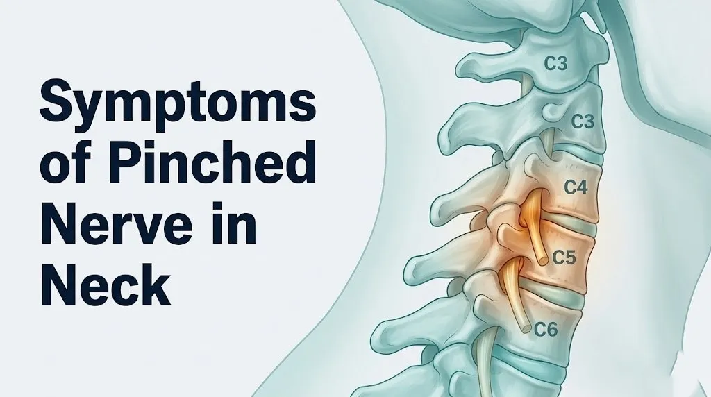
A pinched nerve in the neck occurs when one of the eight cervical nerve roots is compressed, stretched, or chemically irritated as it exits the spinal column through a bony opening called the intervertebral foramen.
The cervical spine consists of seven vertebrae labeled C1 through C7. Between each pair of vertebrae sits an intervertebral disc, a structure that functions like a hydraulic shock absorber. The disc has a tough outer ring called the annulus fibrosus and a gel-like center called the nucleus pulposus. When the annulus develops a tear, the nucleus can push outward and press against the nearby nerve root. That physical pressure, combined with inflammatory chemicals released from the nucleus pulposus material (primarily tumor necrosis factor-alpha and prostaglandins, as identified in research published in Spine), irritates the nerve root’s electrical signaling and produces the pain, numbness, and weakness pattern that most people call a pinched nerve.
Think of the nerve root like an electrical cable running from a server (the spinal cord) to a peripheral device (your hand or arm). When someone pinches that cable at its exit point, the signal to the entire cable is disrupted. It does not matter how far the device is from the server: the disruption travels the full length of the cable.
Foraminal stenosis works differently. Here, bony spurs (osteophytes) that form during normal aging of the cervical spine gradually narrow the foramen through which the nerve root passes. The nerve is not acutely herniated upon, but the progressive narrowing produces a slowly worsening radiculopathy, often without a clear precipitating event.
The American Academy of Orthopaedic Surgeons reports that C6 and C7 nerve root compression are the two most frequently affected levels, together accounting for approximately 70% of all cervical radiculopathy presentations in clinical practice.
Pinched Nerve Neck Symptoms: The Core Clinical Picture
Pinched nerve neck symptoms fall into three categories: sensory symptoms (pain, numbness, tingling), motor symptoms (weakness, loss of coordination), and reflex changes (diminished or absent deep tendon reflexes in the arm).
Radicular pain is typically the dominant early symptom. This is not the same as simple neck stiffness or muscle soreness. Radicular pain from a cervical nerve root has a burning, sharp, or electric quality, and it travels from the neck into the shoulder, arm, forearm, or hand following the anatomical path of the affected nerve. Many people describe it as a hot wire or electric shock sensation running down the arm. This quality is explained by the fact that inflammatory mediators, particularly substance P and prostaglandins, sensitize the dorsal root ganglion (the cluster of sensory nerve cell bodies just outside the spinal cord), lowering the threshold for pain signaling across the entire nerve distribution.
Paresthesia (tingling or pins-and-needles sensation) occurs because compression disrupts the transmission of sensory signals along the nerve’s large-diameter, myelinated A-beta fibers, which normally transmit light touch and proprioceptive information. When these fibers are compressed, they generate spontaneous electrical activity, which the brain interprets as tingling in the skin territory those fibers serve.
Numbness follows a different mechanism. When compression is severe enough to reduce axonal blood flow through the nerve’s internal vascular supply (the vasa nervorum), the nerve fibers begin to lose their ability to conduct signals altogether, producing a numb or dead feeling in the same skin territory.
| Symptom Category | Clinical Symptom | Mechanism | Onset Timing |
|---|---|---|---|
| Sensory | Burning or electric radicular pain | TNF-alpha and prostaglandin sensitization of dorsal root ganglion | Early, often first symptom |
| Sensory | Paresthesia (tingling, pins-and-needles) | Spontaneous discharge of compressed A-beta sensory fibers | Early to middle |
| Sensory | Numbness, reduced sensation | Reduced axonal blood flow, blocked signal conduction | Middle to later |
| Motor | Arm or hand weakness | Compression of alpha motor neuron axons in the ventral root | Middle to later |
| Reflex | Diminished deep tendon reflex | Interruption of monosynaptic reflex arc at compressed level | Persists as long as compression continues |
What Does a Pinched Nerve in the Neck Feel Like?
A pinched nerve in the neck most commonly feels like a sharp, burning, or electric pain that starts in the neck or shoulder and travels down the arm, often reaching the forearm, hand, or specific fingers depending on which nerve root is involved.
The pain is different from the dull, diffuse ache of a sore muscle. Most people with cervical radiculopathy describe the pain as having a distinct pathway, as if the pain is being injected along a line rather than spreading from a single point. Neck movement often makes it worse, particularly tilting or rotating the head toward the affected side. This is the clinical basis of the Spurling test, in which a clinician applies gentle downward pressure on the skull with the neck slightly extended and rotated toward the symptomatic side; reproduction of the radiating arm symptoms is considered a positive finding.
Beyond pain, a pinched nerve in the neck often produces a feeling that something is wrong with the arm or hand without obvious physical weakness. People describe their hand feeling clumsy, their grip feeling unreliable, or their fingers feeling thick and unresponsive. This happens because compression affects both sensory and motor fibers simultaneously, degrading both the sensory feedback the brain uses to calibrate fine movement and the motor commands that drive finger and hand function.
People also report that symptoms are position-dependent in a highly specific way. Raising the arm above the head or placing the hand on top of the head (the shoulder abduction sign) often reduces symptoms, because this movement reduces tension on the compressed nerve root. Conversely, reaching downward or carrying heavy loads often worsens symptoms.
For people with rheumatoid arthritis involving the cervical spine, the symptom experience may be more variable and unpredictable due to inflammatory joint changes at multiple levels, and the clinical picture can change quickly with disease flares. These individuals warrant closer monitoring by a rheumatologist and a spine specialist working in coordination.
Pinched Nerve C3-C4 Symptoms
Pinched nerve C3-C4 symptoms are distinct from lower cervical levels because the C3 and C4 nerve roots do not enter the brachial plexus and therefore do not produce the classic arm radiating pain that most people associate with a pinched nerve in the neck.
The C3 nerve root supplies sensation to the back of the scalp, behind the ear, and the side of the neck. C4 supplies the lower neck, the top of the shoulder (the cape area), and contributes to diaphragm innervation through the phrenic nerve. Compression at C3-C4 typically produces pain in the neck and the top of the shoulder, sometimes extending to the upper periscapular region (the area around the shoulder blade), without producing the forearm or hand symptoms that characterize C5 through C8 compression.
This symptom pattern is frequently misattributed to a shoulder problem, cervicogenic headache, or muscle tension, especially because the pain stays proximal (close to the spine and shoulder) rather than radiating down the arm. A 2019 review in the journal Spine noted that upper cervical levels are the most diagnostically delayed subset of cervical radiculopathy precisely because their symptoms do not fit the classic arm-pain pattern clinicians expect.
C3-C4 compression may also produce:
- A sensation of neck stiffness that is out of proportion to visible range-of-motion limitation
- Pain with head rotation toward the symptomatic side
- Referred pain to the ear or jaw that can mimic temporomandibular joint (TMJ) disorder or atypical facial pain
- Occasional shortness of breath with significant C4 compression, because the C4 nerve root contributes to phrenic nerve function and diaphragm innervation
Older adults with multilevel degenerative disease often have concurrent C3-C4 and C5-C6 involvement, producing a mixed symptom picture that is harder to localize without MRI confirmation.
C5-C6 Pinched Nerve Symptoms
C5-C6 is the most frequently affected level in cervical radiculopathy, and its symptoms affect the outer shoulder, upper arm, forearm, thumb, and index finger.
The C5 nerve root exits between the C4 and C5 vertebrae. It supplies the deltoid muscle (responsible for lifting the arm away from the body), the biceps brachii, and the skin over the outer shoulder and upper arm. Compression at C5 produces pain and numbness in the outer shoulder and upper arm, weakness in shoulder abduction and elbow flexion, and a diminished biceps reflex. People with C5 radiculopathy often report difficulty lifting the arm, particularly raising it out to the side to reach a shelf.
The C6 nerve root exits between the C5 and C6 vertebrae, the single most commonly herniated disc level in the cervical spine. C6 supplies the brachioradialis muscle, contributes to the biceps, and provides sensation to the radial forearm, the thumb, and the index finger. Compression at C6 produces:
- Burning or electric pain running from the neck through the outer forearm to the thumb and index finger
- Numbness or tingling specifically in the thumb and index finger (the diagnostic finger pattern for C6)
- Weakness in wrist extension and elbow flexion
- A diminished brachioradialis reflex (tested at the outer forearm)
| C5 vs C6 Compression: Key Distinctions |
|—|—|—|—|
| Feature | C5 Radiculopathy | C6 Radiculopathy |
| Pain location | Outer shoulder, upper arm | Outer forearm, thumb, index finger |
| Numb finger(s) | Outer upper arm | Thumb and index finger |
| Weak muscle(s) | Deltoid, biceps (arm lifting) | Brachioradialis, wrist extensors |
| Reflex affected | Biceps reflex | Brachioradialis reflex |
For women, C5-C6 pinched nerve symptoms may initially present as shoulder girdle aching without a clear arm radiation pattern, leading to an initial diagnosis of rotator cuff pathology. This is one reason women with cervical radiculopathy receive an accurate diagnosis on average three months later than men, according to observational data published in Spine.
Pinched Nerve C6-C7 Symptoms
C6-C7 disc herniation is the second most common level of cervical radiculopathy, and its symptoms are distinctive because they affect the middle finger and the triceps muscle, a pattern that most other conditions do not replicate.
The C7 nerve root exits between the C6 and C7 vertebrae. It is the dominant root of the upper extremity in terms of motor function, supplying the triceps brachii (the primary elbow extensor), the wrist flexors, and the finger extensors. Its sensory territory runs down the back of the upper arm and forearm and into the middle finger, which is the specific fingertip most characteristically numb in C7 radiculopathy.
The reflex affected is the triceps reflex, tested by tapping the triceps tendon at the elbow with the arm hanging loosely. A diminished triceps reflex in the context of middle finger numbness and outer triceps-distribution arm pain is a highly specific clinical marker for C7 nerve root compression.
People with C7 radiculopathy often describe:
- Difficulty with pushing movements (pushing open a heavy door, triceps-heavy exercises like pushups or overhead pressing)
- Weakness in writing or typing due to combined finger extensor and wrist flexor involvement
- Pain running down the back of the arm and forearm to the middle finger
- A sensation that the arm fatigues faster than normal during any pushing or extending activity
Quick Tip:
- The simplest at-home functional test for C7 motor involvement is attempting a wall push-up with both arms. If the affected-side elbow collapses or fatigues noticeably faster, triceps weakness from C7 involvement should be discussed with a physician.
- People with diabetes may have an altered or absent triceps reflex from diabetic peripheral neuropathy independent of any cervical radiculopathy, making clinical localization by reflex testing unreliable without electrodiagnostic confirmation.
- Among athletes who perform heavy overhead pressing or throwing, C6-C7 disc herniation can present acutely without a preceding trauma event, triggered by repetitive compressive loading during exercise.
Key Takeaway: The nerve root level of a pinched nerve in the neck determines which finger goes numb, which muscle weakens, and which reflex diminishes. C5 affects the outer shoulder and upper arm, C6 affects the thumb and index finger, and C7 affects the middle finger and the triceps.
C8-T1 Nerve Damage Symptoms
C8-T1 nerve damage produces a symptom pattern affecting the ring and little fingers and the intrinsic muscles of the hand, making it a presentation that is often confused with carpal tunnel syndrome or ulnar neuropathy at the elbow.
The C8 nerve root exits between C7 and T1. It supplies sensation to the ring and little finger, the inner (ulnar-side) forearm, and the inner upper arm. It also supplies the finger flexors, particularly the flexor digitorum profundus and the flexor pollicis longus, making grip strength the primary motor concern in C8 radiculopathy. Grip weakness, difficulty making a tight fist, and trouble with pinching small objects are characteristic motor findings.
The T1 nerve root exits between the T1 and T2 vertebrae. It supplies the intrinsic hand muscles, including the lumbricals, interossei, and the thenar and hypothenar groups. Compression at T1 produces visible wasting (muscle atrophy) of the small muscles of the hand over time, a finding called intrinsic-minus hand pattern. The palm may appear hollow between the tendons, and the person may have difficulty spreading or bringing the fingers together.
C8-T1 nerve damage symptoms include:
- Numbness or tingling in the ring and little fingers and the inner forearm
- Weakness in grip strength
- Loss of fine motor control (buttoning shirts, handling coins)
- Weakness in spreading the fingers apart (finger abduction)
- Over weeks to months of compression: visible muscle wasting in the palm
The distinction from carpal tunnel syndrome matters because carpal tunnel primarily affects the median nerve territory (thumb, index, middle finger) and does not produce T1-level intrinsic hand muscle atrophy. The distinction from ulnar neuropathy at the elbow requires electrodiagnostic testing, since both C8-T1 radiculopathy and ulnar neuropathy affect the ring and little fingers.
According to the North American Spine Society, C8-T1 root involvement is less common than C6 or C7 and may indicate pathology at the cervicothoracic junction, which requires specific MRI sequencing that includes the T1 vertebra to ensure the compressive lesion is captured on imaging.
Compressed Nerve in Neck Symptoms: Motor and Reflex Changes
Beyond pain and numbness, a compressed nerve in the neck produces motor weakness and reflex changes that are among the most reliable clinical indicators of nerve root dysfunction severity.
Motor weakness occurs because the compressed nerve root contains not only sensory fibers but also alpha motor neuron axons from the ventral (anterior) horn of the spinal cord. These axons carry movement commands from the spinal cord to specific muscles. When they are compressed or chemically irritated, muscle contraction in the corresponding myotome becomes weaker, less reliable, and more fatiguing.
The key motor findings by level, in clinical terms:
- C5: Difficulty raising the arm away from the body to shoulder height
- C6: Difficulty with wrist extension and resisted elbow flexion
- C7: Difficulty with elbow extension and grip-intensive pushing tasks
- C8: Reduced grip strength and pinch strength
- T1: Loss of finger spreading and intrinsic hand dexterity
Deep tendon reflexes are assessed at the biceps (C5-C6), brachioradialis (C6), and triceps (C7). A diminished reflex at a specific tendon tells the clinician that the nerve arc through that spinal level is disrupted. Absent reflexes are more concerning than diminished ones. Hyperactive reflexes in the arm (brisk, exaggerated response to a tap) in the context of neck symptoms suggest spinal cord compression rather than a simple nerve root pinch, a distinction with major implications for treatment urgency.
Research published in the Journal of Neurosurgery: Spine found that patients with objectively confirmed motor weakness at initial presentation have a significantly lower rate of spontaneous resolution compared to patients with pain and sensory symptoms alone, supporting earlier specialist referral when weakness is present on clinical examination.
For older adults over 70, standard reflex testing is less reliable because aging alone reduces deep tendon reflex amplitude, independent of any nerve compression. In this population, motor strength testing and electrodiagnostic studies carry more diagnostic weight than reflex examination alone.
Key Takeaway: Motor weakness (not pain or numbness alone) is the clinical finding that most strongly predicts a slower natural recovery and is the primary indicator for earlier specialist evaluation rather than continued watchful waiting.
Symptoms of a Pinched Nerve in Your Neck Getting Worse
Symptoms of a pinched nerve in your neck are getting worse when you experience progressive weakness that is measurable over days or weeks, rather than the fluctuating pain and tingling that characterize the early stages.
Early cervical radiculopathy often follows a variable, episodic course. Pain may spike with specific neck positions and ease with rest. Tingling may come and go. This pattern, while uncomfortable, does not necessarily indicate that the underlying nerve compression is worsening. However, specific changes signal a shift from stable to progressing:
- Pain that was previously position-dependent becomes constant and unresponsive to rest or simple repositioning.
- Numbness that was intermittent becomes persistent, present around the clock.
- Grip or arm strength that was mildly reduced becomes functionally limiting (dropping objects, unable to open jars, unable to lift a coffee cup).
- New symptoms appear in a previously unaffected finger or area of the arm, suggesting either expansion of compression or involvement of an adjacent nerve level.
- Atrophy (visible shrinkage) of the muscle in the arm or hand, indicating that motor nerve fibers are not only compressed but beginning to lose structural integrity.
A worsening trajectory over 4 to 6 weeks without any improvement is a clinical signal to advance from watchful waiting to imaging and specialist evaluation, per guidelines from the North American Spine Society.
People taking corticosteroids for other conditions (such as rheumatoid arthritis or asthma) may not experience the same pain intensity escalation despite progressing nerve root damage, because corticosteroids suppress the prostaglandin and cytokine-mediated inflammation that generates most of the radicular pain. In this group, tracking motor function rather than pain level provides a more reliable indicator of trajectory.
Can a Pinched Nerve in the Neck Cause Dizziness or Headaches?
A pinched nerve in the neck can cause headaches, particularly at the base of the skull (occipital region), but dizziness from a pinched cervical nerve root is uncommon and warrants careful clinical evaluation to rule out vertebral artery involvement or inner ear pathology.
Cervicogenic headache is a real and clinically recognized entity. It originates from the upper cervical spine, primarily the C2 and C3 nerve roots, which converge in the spinal trigeminal nucleus with fibers from the trigeminal nerve (the primary nerve supplying the head and face). Compression or irritation at C2-C3 can produce referred pain that travels from the upper neck through the occipital region to the forehead and behind the eye. The International Headache Society classifies cervicogenic headache as a distinct disorder with specific diagnostic criteria, including headache that is consistently triggered or worsened by neck movement or sustained neck posture.
Dizziness in the context of neck symptoms is more complex. True spinning vertigo (vertigo) from a cervical nerve root pinch alone is not a physiologically established mechanism. When dizziness accompanies neck pain and arm symptoms, clinicians consider:
- Vertebrobasilar insufficiency from vertebral artery compression (a vascular emergency)
- Benign paroxysmal positional vertigo (BPPV), which can coexist with cervical spine problems coincidentally
- Cervicogenic dizziness, a controversial but clinically observed form of unsteadiness related to disrupted proprioceptive signaling from the upper cervical joints
Dizziness that comes with neck pain and arm weakness should never be attributed to a simple pinched nerve without ruling out vertebral artery involvement, particularly if the dizziness occurs with head rotation. This requires urgent evaluation by a neurologist or emergency medicine physician, not a watchful wait.
Cervical Myelopathy vs Pinched Nerve Symptoms
Cervical myelopathy is not the same as a pinched nerve in the neck, and the distinction is one of the most clinically important separations in all of spine medicine, because myelopathy involves compression of the spinal cord itself rather than a single nerve root.
Cervical myelopathy occurs when the spinal canal narrows enough that the spinal cord is directly compressed, typically from a large central disc herniation, severe multilevel spondylosis with osteophytic ridging, or thickening of the ligamentum flavum (the yellow elastic ligament on the posterior wall of the spinal canal). When the spinal cord is compressed, the damage affects not one nerve root and its arm distribution, but the entire neurological output below the level of compression.
| Feature | Cervical Radiculopathy (Pinched Nerve Root) | Cervical Myelopathy (Spinal Cord Compression) |
|---|---|---|
| Side affected | Unilateral (one arm) | Bilateral (both arms and/or legs) |
| Leg symptoms | Absent | Leg weakness, stiffness, gait disturbance |
| Hand symptoms | Specific finger numbness or weakness | Clumsy hands bilaterally, difficulty with fine motor tasks |
| Bladder/bowel | Unaffected | May develop urgency, frequency, or incontinence |
| Reflex changes | Diminished at affected level | Hyperreflexia (brisk reflexes) below the compression |
| Hoffman sign | Negative | Often positive (flicking the middle finger produces thumb flexion) |
| Lhermitte sign | Absent | Electric shock sensation down the spine with neck flexion |
| Urgency | Conservative management usually first-line | Surgical consultation often urgent |
The Lhermitte sign, an electric shock sensation that shoots from the neck down the spine or into the arms and legs when the neck is flexed forward, is a hallmark of spinal cord compression. Its presence requires immediate imaging and neurosurgical evaluation.
According to a 2021 review published in the Journal of Bone and Joint Surgery, cervical myelopathy is the most common cause of spinal cord dysfunction in adults over 55 in the United States, and it is frequently underdiagnosed because early myelopathy symptoms (clumsy hands, occasional tripping, mild bilateral hand weakness) are attributed to aging rather than to spinal cord compromise.
Key Takeaway: If symptoms involve both sides of the body, both arms, both legs, bladder changes, or the Lhermitte sign (electric shock with neck flexion), this is not a simple pinched nerve: it is a potential spinal cord emergency requiring the same day evaluation.
How Long Does a Pinched Nerve in the Neck Last?
Most cases of cervical radiculopathy from a pinched nerve in the neck resolve substantially within 8 to 12 weeks with conservative management, but the timeline depends directly on the underlying cause and whether motor weakness is present.
Large observational studies, including a landmark cohort study cited by the North American Spine Society clinical practice guidelines, found that approximately 75 to 90 percent of patients with cervical radiculopathy experience satisfactory improvement within three months without surgery, regardless of whether the initial imaging showed a disc herniation.
The physiological mechanism behind this natural resolution is worth understanding. When a disc herniates and presses on a nerve root, the herniated fragment begins to be reabsorbed by the body’s own macrophage-mediated inflammatory process over weeks to months. As the disc material shrinks, the mechanical pressure on the nerve root diminishes, and the sensitized dorsal root ganglion gradually returns toward its normal electrical threshold. This is why many people with severe early symptoms experience dramatic improvement without surgical intervention.
However, natural resolution is less reliable in the following situations:
- Significant motor weakness at the time of first presentation (weakness indicates axonal injury beyond simple pressure)
- Foraminal stenosis from osteophytes rather than disc herniation (osteophytes do not reabsorb)
- Symptoms persisting beyond 12 weeks without meaningful improvement
- Diabetes mellitus (impaired nerve healing due to microvascular disease reduces recovery rate)
For cases driven by spondylotic foraminal stenosis rather than disc herniation, the timeline of recovery is generally longer and less predictable, because the bony narrowing does not resolve spontaneously. Management in these cases typically requires physical therapy, cervical traction, or procedural intervention such as a cervical epidural steroid injection to achieve meaningful symptom relief.
Pinched Nerve in Neck Symptoms by Age and Individual Factors
The symptoms of a pinched nerve in the neck are shaped by age, biological sex, comorbidities, and pre-existing structural spine changes in ways that alter both the presentation and the clinical course.
Age-related differences:
In adults under 40, acute cervical radiculopathy is most commonly caused by a single-level soft disc herniation, typically at C5-C6 or C6-C7. The onset is often associated with a specific movement or activity, and the symptom pattern is usually clear and localizable to one nerve root level. Natural resolution rates are highest in this group.
In adults over 55, multilevel cervical spondylosis with foraminal narrowing at several levels simultaneously becomes the dominant structural cause. Symptom patterns are often less clearly unilateral, more chronic in character, and less responsive to simple rest. Overlapping pain distributions from two adjacent compressed levels (for example, concurrent C5-C6 and C6-C7 compression) can blur the expected finger-numbness pattern that makes single-level diagnosis straightforward.
Sex-based differences:
Women with cervical radiculopathy are more likely to present with axial neck pain and periscapular aching as their dominant complaint, rather than the radiating arm pain and finger numbness that drive many men to seek care. Observational data published in Spine found that this presentation difference contributes to an average diagnostic delay of approximately 10 to 14 weeks in women compared to men with equivalent MRI findings. Women who report persistent shoulder blade pain and neck stiffness without clear arm symptoms should have cervical imaging considered earlier in the clinical workup rather than defaulting to an isolated shoulder evaluation.
Diabetes mellitus:
People with type 2 diabetes mellitus often have pre-existing diabetic peripheral neuropathy that produces baseline hand and foot tingling, numbness, and diminished reflexes. This creates a clinically challenging overlap: the same fingers that feel numb from C6 radiculopathy may already feel abnormal from diabetic nerve damage. In this population, electrodiagnostic testing (EMG and nerve conduction velocity study) is often necessary to separate cervical radiculopathy from peripheral neuropathy, since clinical examination alone is insufficient.
Emergency Symptoms: When to Call 911 or Go to the ER
Certain symptoms associated with a pinched nerve in the neck require immediate emergency evaluation. Do not wait to see if these resolve on their own.
Call 911 or go to the nearest emergency room immediately if you experience:
- Sudden, severe weakness in both arms or in the arms and legs simultaneously (this is not a typical pinched nerve presentation and suggests spinal cord compression or stroke)
- Loss of bladder or bowel control in the context of neck pain or arm symptoms (bowel and bladder dysfunction indicates spinal cord involvement, not a single nerve root pinch)
- An electric shock sensation running from your neck down your spine or into your limbs when you bend your chin to your chest (Lhermitte sign, indicating active spinal cord compression)
- Sudden inability to walk, loss of balance, or stumbling gait alongside neck or arm symptoms
- Rapid loss of grip strength or arm function developing over hours rather than days
- Neck pain following a trauma (car accident, fall, direct blow to the head or neck) with any new arm or leg weakness or numbness (spinal instability must be excluded before movement)
- Chest pain, difficulty breathing, or severe shortness of breath alongside neck or shoulder symptoms (C4 involvement can affect the phrenic nerve and diaphragm; vertebral artery symptoms can produce stroke-like presentations)
These presentations can indicate cervical myelopathy with acute cord compression, spinal instability, or vertebrobasilar stroke, and they require emergency medical assessment, not an urgent care appointment or a phone call to your primary care office.
When to See a Doctor for a Pinched Nerve in the Neck
See a primary care physician or a physiatrist within one to two weeks if you have persistent neck and arm symptoms that have not improved with rest and activity modification after seven to ten days.
Do not wait if motor weakness is present. Weakness in the shoulder, arm, forearm, or hand at any point in the illness, including weakness that appeared recently, is an indication for earlier evaluation rather than continued watchful waiting. A primary care physician can order cervical spine MRI and refer to a neurologist or orthopedic spine surgeon based on imaging results.
The specific clinical thresholds for escalating to specialist care are:
- No meaningful improvement in symptoms after 4 to 6 weeks of conservative management (physical therapy, NSAIDs, activity modification)
- Any objective motor weakness confirmed on physical examination
- Progressive worsening of symptoms despite initial treatment
- Imaging showing a disc herniation or foraminal stenosis at a level that correlates with the clinical symptoms
- Failure of a cervical epidural steroid injection to provide adequate symptom relief
For symptoms that suggest multilevel disease or myelopathy, the appropriate specialist is a board-certified orthopedic spine surgeon or a board-certified neurosurgeon, not a general orthopedic surgeon or neurologist alone. The choice between surgery and continued non-operative management is made by a spine surgeon based on clinical findings, imaging, and the patient’s functional limitations and preferences.
People over 65 with new neurological symptoms in the arms should request a cervical spine MRI rather than plain X-rays as the first imaging step, because X-rays show bone alignment but do not visualize the spinal cord, the intervertebral discs, or the extent of foraminal narrowing with the precision needed to guide treatment decisions.
How Is a Pinched Nerve in the Neck Diagnosed?
A pinched nerve in the neck is diagnosed through a combination of physical examination, clinical history, and imaging, most commonly cervical MRI, with electrodiagnostic testing used to confirm nerve root involvement and separate radiculopathy from peripheral nerve disorders.
The diagnostic process typically unfolds in this sequence:
- Clinical history: The physician documents the location, quality, and radiation pattern of pain, the specific fingers involved in numbness, the functional limitations (what movements or tasks are difficult), and the timeline from first symptom to presentation.
- Physical examination: The physician tests motor strength at each level (deltoid, biceps, triceps, grip), assesses deep tendon reflexes (biceps, brachioradialis, triceps), checks for sensory changes in specific finger territories, and performs the Spurling test to reproduce radicular symptoms with cervical compression.
- Cervical MRI (without contrast in most cases): MRI is the primary imaging modality for visualizing intervertebral disc herniation, foraminal stenosis, cord compression, and ligamentum flavum thickening. According to the American Academy of Orthopaedic Surgeons, MRI of the cervical spine is the standard first-line imaging study for suspected cervical radiculopathy with neurological signs or symptoms persisting beyond 4 to 6 weeks.
- Electromyography (EMG) and nerve conduction velocity (NCV) testing: These electrodiagnostic tests measure electrical activity in muscles and the speed of nerve signal conduction. They are used when the MRI findings do not clearly correlate with the clinical picture, when peripheral nerve entrapment (carpal tunnel, ulnar neuropathy) needs to be excluded, or when the diagnosis is uncertain. EMG is typically not positive for radiculopathy findings until at least 3 weeks after the onset of motor nerve injury.
- CT myelogram: Reserved for patients who cannot undergo MRI (due to pacemakers, certain metal implants, or claustrophobia) or when surgical planning requires detailed bony anatomy visualization.
| Diagnostic Test | What It Shows | When Ordered |
|---|---|---|
| Cervical MRI | Disc herniation, cord compression, foraminal stenosis | First-line for neurological signs >4 weeks |
| X-ray cervical spine | Bone alignment, disc height loss, osteophytes | Initial screening, limited soft tissue detail |
| EMG and NCV | Nerve root dysfunction vs peripheral nerve entrapment | When MRI-clinical correlation unclear, or after 3 weeks of motor symptoms |
| CT myelogram | Detailed bony canal anatomy | When MRI unavailable or surgical planning requires it |
| Spurling Test | Clinical reproduction of radicular symptoms | At physical examination, before imaging |
Key Takeaway: A cervical MRI is the standard first-line imaging test for a suspected pinched nerve in the neck when symptoms have not resolved in 4 to 6 weeks or when any motor weakness is present. X-rays alone are not sufficient to diagnose nerve root compression.
Frequently Asked Questions About Symptoms of Pinched Nerve in Neck
What are the first signs of a pinched nerve in the neck?
The first signs of a pinched nerve in the neck are typically sharp, burning, or electric pain starting at the neck or shoulder and radiating into the arm, often accompanied by tingling in a specific finger or group of fingers.
Pain is almost always the earliest symptom, appearing before numbness or weakness, because the inflammatory chemicals released at the compression site (primarily tumor necrosis factor-alpha and prostaglandins) sensitize the dorsal root ganglion before axonal compression reaches the threshold for motor or complete sensory block.
If the arm-radiation pattern, specific finger tingling, and worsening with neck rotation are all present together, this combination is highly suggestive of cervical radiculopathy and warrants evaluation by a primary care physician who can order cervical MRI if symptoms do not resolve within four to six weeks.
Can a pinched nerve in the neck go away on its own?
Most cases of cervical radiculopathy do resolve without surgery. Approximately 75 to 90 percent of patients experience satisfactory improvement within 8 to 12 weeks, according to large observational cohort data referenced in North American Spine Society clinical guidelines.
Natural resolution is driven by the body’s macrophage-mediated reabsorption of herniated disc material, which reduces mechanical pressure on the nerve root over weeks to months.
However, motor weakness at presentation, symptoms from foraminal stenosis due to osteophytes (which do not reabsorb), and diabetes mellitus all reduce the likelihood and speed of spontaneous recovery and are indications for earlier specialist evaluation.
What is the difference between a pinched nerve and a herniated disc in the neck?
A herniated disc in the neck is one of the most common structural causes of a pinched nerve, but the two terms describe different things.
A herniated disc refers to the physical displacement of the disc’s inner nucleus pulposus material through a tear in the outer annulus fibrosus, while a pinched nerve (cervical radiculopathy) describes the resulting compression and irritation of a cervical nerve root that produces the symptoms of pain, numbness, and weakness.
A herniated disc can exist without producing a pinched nerve if the displaced material does not contact the nerve root, and a pinched nerve can result from foraminal stenosis due to bone spurs rather than disc herniation.
How do I know if my pinched nerve in the neck is serious?
Your pinched nerve in the neck is serious if you have measurable motor weakness (difficulty raising your arm, weakened grip, loss of triceps strength), symptoms in both arms or both arms and legs simultaneously, or any change in bladder or bowel function.
The Lhermitte sign, an electric shock sensation from the neck down the spine when you flex your chin to your chest, is a specific indicator of spinal cord compression and requires the same-day evaluation at an emergency room or with a spine specialist.
Pain and tingling alone, without motor weakness or bilateral symptoms, are generally manageable with conservative care and primary care physician oversight, but they still warrant evaluation if they persist beyond two weeks without improvement.
Can a pinched nerve in the neck cause arm and hand weakness?
Yes, a pinched nerve in the neck directly causes arm and hand weakness by compressing the alpha motor neuron axons within the affected cervical nerve root, disrupting the motor commands traveling from the spinal cord to specific arm and hand muscles.
The specific muscle weakened depends on which nerve root is compressed: C5 causes deltoid and bicep weakness, C6 causes wrist extensor weakness, C7 causes triceps weakness, C8 causes grip weakness, and T1 causes intrinsic hand muscle weakness with difficulty spreading the fingers.
Motor weakness that is measurable, progressive, or interfering with daily tasks should prompt evaluation by a primary care physician within one to two weeks, because persistent motor fiber compression can lead to irreversible muscle atrophy if not addressed.
What does c5 c6 pinched nerve pain feel like?
C5-C6 pinched nerve pain typically feels like a burning or electric pain that runs from the neck and outer shoulder down the outer forearm to the thumb and index finger, often accompanied by tingling specifically in the thumb.
The C5 component produces shoulder pain and difficulty raising the arm, while the C6 component produces the characteristic thumb and index finger numbness and may reduce the brachioradialis reflex, which a physician tests by tapping the outer forearm.
Many people with C5-C6 radiculopathy initially attribute the pain to a shoulder injury because the shoulder aching is prominent, but the presence of thumb tingling and wrist extensor weakness reliably points to the neck as the source.
Closing
Recognizing the symptoms of a pinched nerve in the neck by nerve root level is not a matter of clinical trivia. It directly determines whether your symptoms need watchful waiting, physical therapy, imaging within the next two weeks, or an emergency room visit today.
If your symptoms are pain, tingling, and numbness in a specific finger distribution, present only on one side, and not accompanied by motor weakness or bilateral symptoms, schedule a primary care appointment and request a cervical spine MRI if symptoms persist past four weeks. If you have measurable arm weakness, grip failure, or any bilateral symptoms, see a physician this week. If you have bowel or bladder changes, loss of balance, or the Lhermitte sign, go to the emergency room now.
You now understand the specific nerve root mechanism behind each symptom pattern, the clinical difference between a recoverable nerve root pinch and a spinal cord compression emergency, and exactly which provider to contact at each level of severity. That knowledge is the most practical thing you can take from this article