Post TIA Symptoms: Signs to Watch After a Mini Stroke
Post TIA symptoms include cognitive fog, fatigue, headache, balance difficulties, emotional changes, and subtle sensory disturbances that can persist for days to weeks after the acute event resolves, and any new or returning neurological symptoms after a TIA require immediate emergency evaluation because they may signal an impending stroke. The period immediately after a transient ischemic attack is not a recovery period in the usual sense: it is the highest-risk window for a major stroke, and the symptoms a person experiences during it carry genuine clinical meaning.
The American Heart Association (AHA) and American Stroke Association (ASA) report that approximately 10 to 15 percent of people who experience a TIA will have a full ischemic stroke within 90 days, with the highest risk concentrated in the first 48 hours after the event. The National Institute of Neurological Disorders and Stroke (NINDS) describes TIA as a medical emergency requiring the same urgency as an acute stroke, even when all symptoms have resolved.
This article covers every symptom that patients and caregivers can expect after a TIA, the neurological mechanism behind each one, how to distinguish normal post-TIA recovery symptoms from signs that a stroke is occurring, and the specific clinical tests and referrals that should happen in the days immediately following the event.
Post TIA Symptoms: What Happens After a Mini Stroke
Post TIA symptoms are the neurological, cognitive, physical, and emotional changes that occur after the acute transient ischemic attack has resolved, produced by ongoing penumbral tissue recovery, the brain’s neuroinflammatory response to ischemia, and the physiological stress of the acute event.
The mistake many people make after a TIA is assuming that because the obvious symptoms have resolved, the danger has passed. It has not. The brain tissue in the area surrounding the ischemic zone, called the ischemic penumbra, is still in a state of metabolic vulnerability even after blood flow has been restored. Neurons in the penumbra have survived but are not functioning normally. Their synaptic connections are disrupted. Their energy metabolism is depressed. This ongoing cellular dysfunction is the direct source of the cognitive, sensory, and fatigue symptoms that patients experience after the acute TIA is over.
A second important reality is that the underlying cause of the TIA, whether a blood clot from the heart, a plaque fragment from the carotid artery, or a small vessel narrowing in the brain, has not resolved with the symptoms. The clot risk remains. That is why post-TIA symptom monitoring is not just about recovery. It is about recognizing stroke warning signs in real time.
Key post-TIA symptoms to monitor in the days and weeks following the event:
- New or recurring speech difficulty, slurred words, or trouble finding words
- Facial drooping or numbness on one side of the face
- Arm or leg weakness on one side of the body
- Cognitive fog, memory difficulty, and slowed thinking
- Fatigue out of proportion to activity level
- Headache, particularly if new or different from prior headaches
- Visual changes including blurring, double vision, or loss of vision in one eye
- Balance or coordination problems when walking
- Emotional changes including anxiety, tearfulness, or irritability
- Sensory disturbances including numbness, tingling, or abnormal sensations
What Is a TIA and How Does It Affect the Brain
A transient ischemic attack (TIA) is a brief episode of focal neurological dysfunction caused by transient cessation of blood flow to a specific region of the brain, lasting less than 24 hours and most often less than 1 hour, without causing permanent infarction detectable on standard imaging, though newer diffusion-weighted MRI reveals tissue injury in a meaningful proportion of clinically defined TIA patients.
The mechanism begins with an interruption of blood flow through one of the brain’s arterial territories. The most common mechanism is a thromboembolic event: a clot forms at an atherosclerotic plaque in the internal carotid artery or breaks off from a clot in the heart’s chambers (particularly in atrial fibrillation) and lodges temporarily in a distal cerebral artery. The affected neurons experience energy failure within seconds of ischemia because neurons have essentially no glycogen stores and depend entirely on continuous oxygen and glucose delivery.
The reason symptoms are temporary rather than permanent in TIA is that the obstructing clot dissolves or migrates before the infarction threshold is crossed in most neurons, and collateral cerebral circulation routes blood around the blocked segment. Once flow is restored, neurons recover. But the recovery is not instantaneous and not perfectly complete in the penumbral zone.
Here is the important nuance that no general health article addresses: a 2019 study published in Stroke found that approximately 30 to 40 percent of patients clinically diagnosed with TIA show acute ischemic lesions on diffusion-weighted imaging (DWI) MRI, meaning measurable tissue injury occurred even in events that resolved clinically within hours. This finding reframes TIA from a “warning event with no damage” to a “minor stroke with resolved symptoms,” which explains why post-TIA symptoms can be more than simply subjective and why neuroimaging after any TIA is a clinical standard, not an optional add-on.
Post Mini Stroke Symptoms: The Full Clinical Picture
The full clinical picture of post mini stroke symptoms encompasses neurological, cognitive, physical, and psychological symptoms that extend beyond the resolution of the acute FAST-positive event, reflecting ongoing penumbral tissue recovery, post-ischemic neuroinflammation, and the physiological stress response triggered by acute cerebral ischemia.
Understanding the full picture requires recognizing that a TIA affects the brain in two ways simultaneously. The first is the direct ischemic effect on the neurons in the affected territory. The second is the systemic stress response activated by the brain’s detection of ischemic threat, involving the hypothalamic-pituitary-adrenal (HPA) axis, elevation of cortisol and catecholamines, and widespread autonomic nervous system activation.
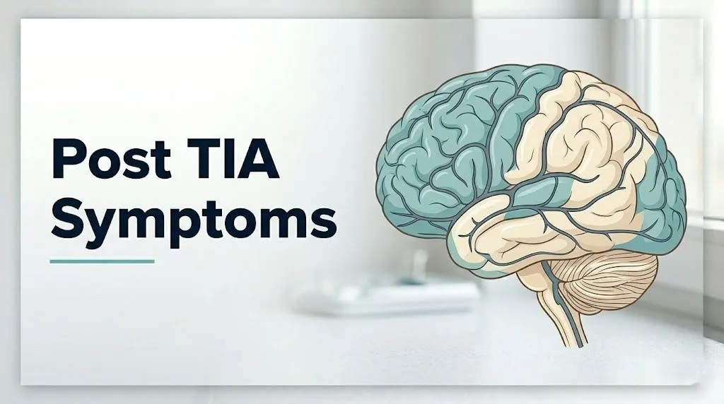
Both of these effects produce symptoms that outlast the acute event. The direct ischemic effect produces the neurological residuals: cognitive fog, word-finding difficulty, subtle motor or sensory changes. The systemic stress response produces the fatigue, sleep disruption, emotional lability, and heightened anxiety that patients often feel embarrassed to report, not understanding why they feel so drained and emotionally raw after an event that has, clinically speaking, “resolved.”
The American Stroke Association recognizes that post-TIA patients frequently report a constellation of symptoms in the days following the acute event that are often underreported to clinicians because patients assume these are not medically relevant once the acute signs have passed. Reporting all post-TIA symptoms to a board-certified neurologist at the earliest scheduled follow-up, which should occur within 24 hours of the TIA per AHA/ASA guidelines, is not optional.
Post-TIA symptoms organized by system:
- Neurological: persistent mild word-finding difficulty, subtle facial asymmetry, mild limb heaviness
- Cognitive: slowed processing speed, short-term memory difficulty, reduced concentration span
- Physical: fatigue, sleep disturbance, headache, balance impairment
- Emotional: anxiety, tearfulness, irritability, low mood, fear of recurrence
Key Takeaway: Post-TIA symptoms extend well beyond the 24-hour resolution window of the acute event and include cognitive fog, fatigue, emotional changes, and subtle neurological residuals that reflect ongoing penumbral recovery and the brain’s stress response to ischemia, all of which should be reported to a neurologist at the earliest possible appointment.
Cognitive Symptoms After a TIA
Cognitive symptoms after a TIA include reduced processing speed, short-term memory difficulty, word-finding problems, and impaired concentration, produced by synaptic dysfunction in the ischemic penumbral zone that persists beyond the clinical resolution of motor or speech deficits.
Think of the ischemic penumbra as a ring of partially stunned neurons surrounding the area where blood flow was most severely reduced. These neurons did not die. But their synaptic machinery took a hit. The ion channels that maintain membrane potential, the mitochondria that generate ATP for synaptic vesicle release, and the receptor systems that receive incoming signals are all operating below normal capacity for hours to days after the ischemic event. The result is a brain that, from the outside, appears to have fully recovered, but from the inside, is running at reduced processing efficiency.
The hippocampus deserves specific mention. This paired structure in the medial temporal lobe, essential for forming new memories and consolidating short-term information into long-term storage, is one of the most metabolically demanding and ischemia-vulnerable structures in the brain. Even brief ischemia in the posterior cerebral artery or anterior choroidal artery territory can produce hippocampal synaptic dysfunction that manifests as the “memory blankness” or “brain fog” patients describe in the days after a TIA.
According to a study published in Stroke reviewing cognitive outcomes after TIA, a measurable proportion of TIA patients demonstrate detectable cognitive deficits on formal neuropsychological testing even when standard bedside neurological examination findings are normal, with processing speed and attention showing the most consistent impairment.
Patients with prior cognitive impairment or early dementia are at higher risk for more pronounced post-TIA cognitive deficits. Because their cognitive reserve (the brain’s functional capacity to compensate for neuronal loss or dysfunction) is already reduced, the same penumbral injury that produces only mild foggy thinking in a cognitively healthy 60-year-old may produce a more visible and longer-lasting functional deficit in a person whose reserve was already compromised.
Fatigue After a TIA
Fatigue after a TIA is one of the most frequently reported and most underestimated post-TIA symptoms, produced by the combined metabolic cost of neuronal recovery in the penumbral zone, sustained activation of the hypothalamic-pituitary-adrenal (HPA) axis, and the significant energetic demand that neurological repair places on the body’s systemic resources.
Brain tissue recovery after ischemia is energetically expensive. Neurons that experienced partial energy failure during the TIA require elevated ATP production to restore their membrane ionic gradients, replenish depleted neurotransmitter stores, and repair mitochondrial function. This elevated neuronal metabolic demand competes with the body’s other energy systems and produces a fatigue that feels disproportionate to physical activity because it is neurological in origin, not muscular.
The HPA axis component compounds this. During the acute TIA, the brain activates an emergency stress response: cortisol and adrenaline surge, the cardiovascular system is placed on high alert, and the immune system enters a state of preparedness. This response does not simply switch off when symptoms resolve. The post-TIA days are often characterized by sustained cortisol elevation, which itself produces fatigue, sleep disruption, and mood disturbance through the same mechanisms seen in any state of prolonged physiological stress.
Clinicians generally observe that post-TIA fatigue is often worst in the first two to seven days after the event and typically improves progressively over two to four weeks, though it may persist longer in older patients or those with larger ischemic burdens on imaging. Pushing through fatigue aggressively during this window, by resuming full work schedules or physical demands, should be discussed with a primary care physician or neurologist before doing so, because the physiological rationale for rest during neuronal recovery is well-established.
Men and women may experience post-TIA fatigue differently in its subjective quality and reporting patterns. Clinical observation suggests women are more likely to describe post-TIA fatigue as a global exhaustion affecting cognitive and emotional function simultaneously, while men more often describe it as physical tiredness. Neither pattern is abnormal.
Emotional and Psychological Symptoms After a TIA
Emotional and psychological symptoms after a TIA, including anxiety, depression, irritability, emotional lability, and post-event fear, are produced by a combination of ischemic involvement of the limbic system, altered neurotransmitter function in peri-ischemic cortical and subcortical regions, and the psychologically distressing experience of having had a potentially life-threatening neurological event.
The limbic system, which includes the amygdala, hippocampus, anterior cingulate cortex, and hypothalamus, regulates emotional processing, stress responses, and mood stability. Ischemic disruption of these structures or of the white matter tracts connecting them to the prefrontal cortex can produce genuine neurobiological changes in emotional regulation that are not simply “anxiety about what happened.” They are the neurological result of what happened.
The amygdala, in particular, has a well-established sensitivity to ischemic injury. Its role in threat detection and the generation of fear responses means that ischemic or peri-ischemic dysfunction in the amygdala can produce a state of heightened vigilance and anxiety that feels disproportionate to the person’s understanding of their medical situation. This is not a character weakness. It is amygdala hyperactivation from ischemic stress.
Research published in Stroke and in The Lancet Neurology has documented that clinically measurable depression affects a substantial minority of TIA patients in the months following the event, with estimates ranging from 20 to 30 percent of patients in some prospective cohort studies. Anxiety affects an even higher proportion.
People who experience post-TIA emotional symptoms should discuss them explicitly with their neurologist or primary care physician rather than dismissing them as understandable worry. These symptoms have both neurological and psychological dimensions, and both can be addressed with appropriate support.
If you are experiencing overwhelming anxiety, hopelessness, or thoughts of self-harm after a TIA, contact the 988 Suicide and Crisis Lifeline by calling or texting 988 at any time. This service is free, confidential, and available 24 hours a day, 7 days a week.
Headache After a TIA
Headache after a TIA is produced by post-ischemic changes in cerebrovascular reactivity, regional hyperemia (increased blood flow) following reperfusion of the ischemic territory, and the neuroinflammatory response that activates pain-sensitive meningeal blood vessel sensory nerves in the dura mater.
Not everyone develops headache after a TIA. The prevalence varies by the vascular territory involved: posterior circulation TIAs (affecting the vertebrobasilar system, cerebellum, and occipital lobes) are more likely to produce headache than anterior circulation TIAs. This makes anatomical sense because the posterior fossa contains a higher density of pain-sensitive dural structures and because the basilar and posterior cerebral arteries supply regions with more direct connections to trigeminovascular pain pathways.
The headache mechanism in post-TIA presentations involves two phases. The first is the acute headache that can accompany the TIA itself, produced by sudden changes in cerebrovascular pressure and the activation of perivascular nociceptors. The second is the post-reperfusion headache, which occurs hours after the event resolves and results from post-ischemic hyperemia (excessive blood flow returning to the previously ischemic territory) and the release of inflammatory mediators including prostaglandins and substance P from activated perivascular immune cells.
Key points for patients about post-TIA headache:
- A mild headache in the days following a TIA that is consistent in location and quality and improving over time is generally a post-ischemic recovery symptom
- A sudden, severe headache described as “the worst headache of my life” at any time after a TIA requires immediate emergency room evaluation to rule out subarachnoid hemorrhage or hemorrhagic transformation
- Any new headache accompanied by neck stiffness, fever, photophobia, or vomiting after a TIA warrants same-day emergency evaluation
- Older adults with post-TIA headache may underreport or attribute it to tension, making it important for caregivers to ask specifically about headache quality and onset during the post-TIA monitoring period
Key Takeaway: Post-TIA headache that is mild and progressively improving over days is a recognized post-ischemic recovery phenomenon, but a sudden severe headache after a TIA is a neurological emergency requiring immediate 911 activation and emergency room evaluation.
Balance and Coordination Problems After a TIA
Balance and coordination problems after a TIA reflect ischemic or post-ischemic dysfunction in the cerebellum, vestibular nuclei in the brainstem, or the corticospinal tracts of the posterior limb of the internal capsule, depending on which vascular territory was involved in the TIA.
Posterior circulation TIAs, which involve the vertebral arteries, basilar artery, or posterior cerebral arteries, directly affect the cerebellum and brainstem. These structures govern balance, gait coordination, eye movement coordination, and the integration of vestibular, visual, and proprioceptive signals into a coherent sense of position and movement. When the posterior circulation is transiently ischemic, the patient experiences vertigo, unsteadiness, and difficulty walking in a straight line.
Even after the acute episode resolves, cerebellar and vestibular pathway neurons in the penumbral zone may remain functionally impaired. The result is a walking pattern that feels subtly “off,” a tendency to reach for walls or furniture for support, and a subjective sensation of the ground being unsteady. These are post-TIA symptoms, not anxiety symptoms, though patients often interpret them as anxiety because they are difficult to describe precisely.
The NINDS notes that vertebrobasilar TIA (posterior circulation) is associated with a different and sometimes more urgent immediate stroke risk profile than anterior circulation TIA, because basilar artery occlusion can produce catastrophic locked-in syndrome or bilateral cerebellar infarction without adequate collateral flow. Any balance or coordination problem returning or worsening after a posterior circulation TIA should be treated as a potential recurrent TIA until a neurologist or emergency physician evaluates the patient.
Patients recovering from balance problems after a TIA should not attempt to drive until cleared by a neurologist, regardless of how mild the balance impairment seems. The integration speed required for safe driving is impaired even when walking-level balance appears functional.
Sensory and Vision Symptoms After a TIA
Sensory and vision symptoms after a TIA, including persistent tingling, numbness, abnormal sensations on one side of the body, blurring of vision, or transient monocular visual loss, reflect ischemic dysfunction in the somatosensory cortex of the parietal lobe, the optic radiations, or the occipital cortex of the posterior cerebral artery territory, and the ophthalmic artery branch of the internal carotid artery.
Transient monocular blindness, also called amaurosis fugax, occurs when an embolus temporarily occludes the ophthalmic artery (the first branch of the internal carotid artery). Patients describe it as a curtain or shade descending over one eye, lasting seconds to minutes before clearing completely. This is a form of TIA localized to the retinal circulation. After the episode, patients may notice mild visual blurring in the affected eye, reduced contrast sensitivity, or mild photosensitivity that persists for hours.
Persistent hemisensory symptoms, where one side of the body feels numb, tingly, or “different” from the other side after a TIA has otherwise resolved, reflect incomplete recovery in the ventral posterolateral nucleus of the thalamus or the parietal somatosensory cortex. These structures integrate sensory signals from the contralateral body half, and their penumbral dysfunction produces sensory awareness distortions that patients often struggle to describe precisely.
According to clinical guidelines from the American Heart Association, any visual symptom after a TIA, including even mild or transient visual disturbance, should be documented precisely and reported to the treating neurologist. The pattern of visual involvement provides diagnostic information about which vascular territory was affected and whether the TIA was anterior or posterior circulation in origin, which directly influences the investigative pathway (carotid imaging versus vertebrobasilar imaging).
Patients with diabetes mellitus may experience more pronounced post-TIA sensory symptoms because pre-existing diabetic peripheral neuropathy alters the baseline sensory threshold, making it harder to distinguish TIA-related sensory changes from the underlying neuropathic pattern. This population requires explicit discussion of sensory changes with both their neurologist and their diabetes care team.
Key Takeaway: Sensory and vision symptoms that persist or fluctuate after a TIA are not anxiety or imagination: they reflect real, measurable penumbral dysfunction in specific cerebrovascular territories, and their pattern tells the neurologist which artery was involved and which investigations are most urgently needed.
How Long Do Post-TIA Symptoms Last
Most post-TIA neurological symptoms, including cognitive fog, mild sensory changes, and balance disturbances, improve substantially within one to two weeks as the ischemic penumbra recovers, but fatigue and emotional symptoms may persist for weeks to months in some patients.
The timeline of post-TIA symptom resolution follows the biology of penumbral recovery. Neuronal synaptic function in the penumbra begins restoring within hours of reperfusion but reaches full functional recovery more slowly. The rate depends on the extent of the penumbral zone, the patient’s age, the adequacy of collateral circulation, and the presence of any chronic cerebrovascular disease.
| Post-TIA Symptom | Typical Duration | Individual Variation |
|---|---|---|
| Acute neurological deficit | Resolves within 24 hours by definition | Persistent deficit beyond 24 hours reclassifies as stroke |
| Cognitive fog | Days to 1 to 2 weeks | Longer in older adults, those with prior cognitive impairment |
| Fatigue | 1 to 4 weeks | Longer with larger ischemic burden, older age, poor sleep |
| Headache | Days to 1 week | Longer in posterior circulation TIA, migraine history |
| Balance disturbance | Days to 2 weeks | Longer in posterior circulation TIA, vestibular involvement |
| Emotional symptoms (anxiety, depression) | Weeks to months | Requires assessment; may need psychological or psychiatric support |
| Sensory changes | Days to 2 weeks | Longer with thalamic or parietal cortex involvement |
Any symptom that is not improving within this timeframe, or any symptom that worsens after initially improving, requires urgent re-evaluation by a board-certified neurologist. Worsening or new symptoms at any point after a TIA should be treated as a potential new TIA or stroke until proven otherwise.
Warning Signs of Stroke After a TIA
Warning signs of stroke after a TIA are any new or returning focal neurological symptoms, including sudden face drooping, arm weakness, speech difficulty, vision loss, sudden severe headache, sudden loss of balance, or sudden confusion, that occur after the initial TIA symptoms have resolved and require immediate 911 activation.
The post-TIA period is not the time to wait and see. The risk of stroke in the 48 hours following a TIA is substantially higher than at any other time in that person’s life. A study published in the New England Journal of Medicine documented that the highest single risk period for TIA-to-stroke progression occurs within the first 24 to 48 hours after the index TIA event.
Think of the post-TIA brain as a fire that appears to have been extinguished but still has smoldering embers. The vessel disease or cardiac source that produced the first event is still present. The platelet aggregation cascade that formed the original clot is still active. The atherosclerotic plaque that released the embolus is still there, and it may be unstable. The first TIA is the fire alarm. A second event is the fire.
The BE-FAST framework helps identify stroke signs returning after a TIA:
- Balance: sudden loss of balance or coordination
- Eyes: sudden vision changes, double vision, or loss of vision in one or both eyes
- Face: drooping or numbness on one side of the face
- Arm: weakness or inability to raise one arm
- Speech: slurred speech, inability to find words, or inability to understand speech
- Time: call 911 immediately and note the time symptoms began
Do not drive yourself to the hospital. Do not wait for symptoms to worsen. Do not take additional medications at home first. Call 911 immediately and tell the dispatcher that you have had a TIA and are experiencing new neurological symptoms.
Atypical TIA Symptoms in Women
Women are more likely than men to present with atypical TIA symptoms, including headache, altered consciousness, pain, nausea, and confusion without the classic limb weakness or speech disturbance, a pattern that leads to higher rates of misdiagnosis and delayed treatment in female patients with TIA.
The physiological basis for this sex difference is incompletely understood but involves several contributing factors. First, women have a higher prevalence of cardioembolic TIA from atrial fibrillation, which tends to produce larger emboli that may affect multiple smaller cerebral territories simultaneously rather than producing the clean focal deficit of a single large vessel occlusion. Second, women have higher rates of migraine with aura, which can produce TIA-like symptoms and which also increases their baseline TIA and stroke risk. Third, hormonal influences on cerebrovascular reactivity and thrombotic risk (estrogen’s complex effects on coagulation and vessel tone) alter the pathophysiology in ways that produce less stereotyped symptom presentations.
A landmark analysis published in Stroke found that women with TIA were more likely to report headache, altered consciousness, and facial pain as their presenting symptoms compared to men, who were more likely to present with limb weakness and speech difficulty. Because emergency providers are trained to recognize the classic FAST pattern, women’s atypical presentations are more likely to be misattributed to migraine, anxiety, or other diagnoses, delaying the urgent neurological workup that determines stroke risk and initiates secondary prevention.
Women who have experienced a TIA should know:
- Atypical symptoms are not less serious than classic motor or speech deficits
- Headache, confusion, or unexplained nausea that came on suddenly and resolved within hours may have been a TIA and warrants neurological evaluation
- Post-TIA management in women should include specific assessment for atrial fibrillation, patent foramen ovale, and estrogen-containing medication use (oral contraceptives, hormone therapy) as contributing factors
TIA Symptoms in Older Adults and People With Cognitive Impairment
Older adults and people with pre-existing cognitive impairment experience more pronounced post-TIA cognitive symptoms and have higher rates of TIA misdiagnosis because their reduced cognitive reserve amplifies the functional impact of even small ischemic events, and because baseline cognitive changes make it harder to identify new deficits against a pre-existing background.
Cognitive reserve refers to the brain’s functional capacity to compensate for neuronal damage or dysfunction by recruiting alternative neural pathways. Younger adults with higher education levels, cognitively active lifestyles, and no prior brain disease have larger cognitive reserves. A TIA in this population may produce only mild, transient fog that resolves completely. The same-sized ischemic event in an older adult with early Alzheimer’s disease, who has already lost substantial functional reserve, may produce a much more pronounced and longer-lasting cognitive decline.
This reserve-dependency has a specific practical implication: a sudden worsening of cognitive function in a person with known mild cognitive impairment or early dementia should prompt TIA evaluation rather than assuming the change is part of dementia progression. A previously stable cognitive baseline that suddenly deteriorates over hours and then partially recovers is a TIA pattern superimposed on dementia, not simply dementia progression.
According to the American Heart Association, atrial fibrillation is more prevalent in older adults and becomes a more dominant TIA etiology in patients over age 75 compared to younger age groups. This means older post-TIA patients are more likely to need anticoagulation therapy (direct oral anticoagulants or warfarin) rather than antiplatelet therapy, and the distinction matters significantly for secondary prevention.
Older adults on anticoagulant or antiplatelet therapy for other conditions (deep vein thrombosis prevention, orthopedic surgery recovery, atrial fibrillation management) should inform their neurologist of their complete medication list at the post-TIA evaluation, because drug interactions and bleeding risk profiles alter which secondary prevention strategy is appropriate.
Key Takeaway: Cognitive reserve determines how severely and how durably a TIA affects cognitive function, meaning older adults and people with pre-existing cognitive impairment deserve earlier, more thorough neurological investigation of any sudden cognitive change because the same small TIA produces a larger functional deficit against a lower-reserve background.
The ABCD2 Score and 90-Day Stroke Risk After TIA
The ABCD2 score is a validated clinical risk stratification tool used by emergency physicians and neurologists to estimate the short-term stroke risk after a TIA, based on five clinical factors: age, blood pressure at presentation, clinical features, duration of TIA symptoms, and presence of diabetes.
Understanding the ABCD2 score matters for patients and families because it is the tool that determines how urgently investigation and treatment are initiated after a TIA. A high ABCD2 score means the treating team is treating the situation as a near-emergency because the probability of stroke within 48 hours is substantially elevated.
The five components and their scoring:
| Factor | Criteria | Score Points |
|---|---|---|
| Age (A) | 60 years or older | 1 |
| Blood pressure (B) | Systolic 140+ or diastolic 90+ mmHg | 1 |
| Clinical features (C) | Unilateral weakness | 2 |
| Clinical features (C) | Speech disturbance without weakness | 1 |
| Duration (D) | 60 minutes or longer | 2 |
| Duration (D) | 10 to 59 minutes | 1 |
| Diabetes (D) | Presence of diabetes mellitus | 1 |
A total score of 0 to 3 is considered lower risk, 4 to 5 intermediate risk, and 6 to 7 higher risk of early stroke. According to research validating the ABCD2 score, published in The Lancet, patients with scores of 6 or 7 face a 2-day stroke risk of approximately 8%, compared to less than 1% for scores of 0 to 3.
The ABCD2 score is not the final word on post-TIA management. It is one input among several. Patients with high-grade carotid stenosis, atrial fibrillation, or multiple prior TIAs face higher stroke risk than the ABCD2 score captures alone. A board-certified neurologist or vascular neurologist integrates the ABCD2 score with imaging findings, cardiac monitoring results, and vascular imaging to make individualized management recommendations.
How a TIA Is Diagnosed and What Tests Are Done After
A TIA is diagnosed through clinical history, neurological examination, and a defined sequence of investigations that include diffusion-weighted MRI (DWI-MRI), CT angiography (CTA) or magnetic resonance angiography (MRA) of the cerebral and neck vessels, cardiac monitoring for atrial fibrillation, and blood tests for coagulation, lipids, and glucose, with the goal of identifying the mechanism of the TIA and initiating secondary prevention before a stroke occurs.
The clinical history is the most important initial diagnostic tool. A neurologist or emergency physician will ask for a precise description of what happened: exactly which symptoms occurred, in what order, at what time, and for how long. The symptom pattern itself localizes the ischemic territory. Sudden left arm weakness and speech difficulty point to the right middle cerebral artery territory. Sudden vision loss in the right eye points to the right ophthalmic artery.
The complete post-TIA diagnostic workup typically includes:
- DWI-MRI of the brain: the most sensitive imaging test for acute ischemia. A positive DWI signal in a clinically relevant territory confirms tissue injury and reclassifies the event as minor stroke rather than pure TIA, which has management implications
- CT angiography (CTA) of the neck and brain: identifies carotid artery stenosis, intracranial large vessel disease, and cerebral aneurysms that could alter management
- Echocardiogram: identifies cardiac sources of embolism including left atrial thrombus, valvular disease, patent foramen ovale, and wall motion abnormalities
- Cardiac rhythm monitoring: a 12-lead ECG at minimum; a 24-hour Holter monitor or longer-term implantable cardiac monitor to detect paroxysmal atrial fibrillation that may not be captured on a short recording
- Blood tests: complete blood count (for thrombocytopenia or polycythemia), coagulation panel, fasting lipid panel, HbA1c (for diabetes), inflammatory markers, and in younger patients, hypercoagulability panel
- Blood pressure measurement: establishes baseline and guides immediate antihypertensive management
Patients who experience a TIA outside of hospital hours and whose symptoms have resolved should still go to the nearest emergency room the same day, not wait for a primary care appointment. The 48-hour post-TIA period is the window where the investigative findings are most actionable in reducing stroke risk.
When to Call 911 or Go to the ER After a TIA
Any new or returning neurological symptom after a TIA, including sudden face drooping, arm weakness, speech difficulty, vision loss, sudden severe headache, sudden loss of balance, or sudden confusion, requires immediate 911 activation and should not be managed at home, monitored for improvement, or driven to the hospital independently.
The fundamental rule after a TIA is that any new focal neurological symptom is a stroke until proven otherwise by emergency evaluation. There is no symptom that should cause a post-TIA patient to think “let me wait and see if this passes.”
Call 911 immediately if you or someone who has had a TIA experiences any of the following:
- Sudden face drooping, arm weakness, or leg weakness, indicating motor cortex or corticospinal tract ischemia in the middle cerebral artery territory
- Sudden inability to speak, slurred speech, or inability to understand spoken words, indicating left hemisphere temporal or frontal lobe ischemia affecting Broca’s or Wernicke’s area
- Sudden loss of vision in one eye or visual field loss in both eyes, indicating ophthalmic artery or posterior cerebral artery territory ischemia
- Sudden severe headache unlike any prior headache, indicating possible subarachnoid hemorrhage or hemorrhagic transformation
- Sudden loss of balance, inability to walk, or sudden vertigo with vomiting, indicating posterior circulation ischemia
- Sudden confusion, altered consciousness, or inability to recognize familiar faces or surroundings
- Any neurological symptom that lasts longer than a few minutes and is new or different from prior TIA symptoms
Do not drive to the hospital. Tell the 911 dispatcher that the patient has had a recent TIA and is experiencing new neurological symptoms. This information allows the receiving emergency room to activate a stroke protocol immediately on arrival, which is the fastest pathway to time-sensitive imaging and treatment.
These presentations can indicate an acute ischemic stroke requiring emergency thrombolysis or mechanical thrombectomy within a narrow time window, and they require emergency medical assessment, not an urgent care appointment or a call to a primary care office.
Frequently Asked Questions About Post TIA Symptoms
How long do symptoms last after a mini stroke?
Most acute post-TIA neurological symptoms resolve within 24 hours by clinical definition, but residual symptoms including cognitive fog, fatigue, and emotional changes can persist for days to several weeks.
The timeline depends on which vascular territory was affected, how much penumbral tissue was involved, the patient’s age, and whether any DWI-MRI signal change (indicating actual tissue injury) was present.
Any symptom that is not improving within two weeks, or that worsens at any point, requires re-evaluation by a board-certified neurologist.
Is it normal to feel tired and foggy after a TIA?
Fatigue and cognitive fog are among the most frequently reported post-TIA symptoms and are physiologically explained by neuronal metabolic recovery demands and sustained HPA axis cortisol elevation after the acute ischemic event.
The American Stroke Association recognizes these as expected post-TIA symptoms that most patients experience in the first one to two weeks after the event.
Persistent or worsening fatigue and cognitive fog beyond two weeks should be reported to a neurologist because they may indicate ongoing cerebrovascular instability or post-TIA depression requiring assessment.
Can a TIA cause lasting brain damage?
A TIA can cause measurable brain tissue injury even when symptoms resolve completely, with approximately 30 to 40 percent of clinically defined TIA patients showing acute ischemic lesions on DWI-MRI according to research published in Stroke.
These lesions, while often small, contribute to cumulative white matter burden and may affect processing speed and attention in subtle but measurable ways over time.
This is one of the reasons that urgent post-TIA investigation and secondary stroke prevention are treated as medical priorities even when the patient feels completely back to normal.
What are the signs that a TIA is turning into a full stroke?
A TIA is turning into a full stroke when focal neurological symptoms that were previously resolving return or worsen, last beyond 24 hours, or appear suddenly at any new point after the initial TIA.
The FAST signs (Face drooping, Arm weakness, Speech difficulty, Time to call 911) apply equally to identifying a new stroke after a TIA as they do to identifying any stroke.
Call 911 immediately if any of these signs return or any new focal neurological symptom appears after a TIA, because the post-TIA period carries substantially elevated stroke risk concentrated in the first 48 hours.
How soon after a TIA can another one happen?
A second TIA or a full stroke can occur within hours of the first event, with the highest risk period documented in the first 24 to 48 hours after the index TIA.
Research published in the New England Journal of Medicine as part of the POINT trial showed that the early post-TIA period carries a stroke risk of 3 to 10 percent within 90 days, with a disproportionate share of that risk concentrated in the earliest days.
This concentration of early risk is why emergency evaluation and rapid initiation of secondary prevention are treated as time-sensitive priorities even when the patient feels well.
Do I need to go to the hospital if my TIA symptoms have already gone away?
A person who has experienced TIA symptoms that have fully resolved still needs to go to the nearest emergency room the same day, not the following morning and not to a scheduled outpatient appointment.
The AHA/ASA guidelines recommend that TIA evaluation, including neuroimaging, vascular imaging, and cardiac monitoring, begin within 24 hours of symptom onset, because the investigations and secondary prevention treatment initiated during this window substantially reduce early stroke risk.
Waiting until symptoms worsen or until a convenient appointment is available gives up the protective value of early intervention during the highest-risk period.
The Window That Matters Most
The days immediately following a TIA are the most clinically consequential period in that person’s vascular history. What happens in those 48 to 96 hours, whether imaging identifies a carotid stenosis treatable with urgent endarterectomy, whether cardiac monitoring catches atrial fibrillation requiring anticoagulation, whether blood pressure is brought under control before another embolic event occurs, determines the probability of a major stroke.
Post-TIA symptoms including cognitive fog, fatigue, headache, emotional changes, and balance disturbance are real, physiologically grounded, and clinically meaningful. Reporting them to a neurologist matters, because the full symptom picture informs the treatment decisions being made in that critical window.
Every person who has had a TIA deserves evaluation by a board-certified neurologist within 24 hours, a completed vascular and cardiac workup within 48 hours, and a clear plan for secondary stroke prevention before they leave the hospital or clinic. If you have not received all three of those things, ask for them specifically and by name