What Causes Heel Spurs? All Causes Explained for 2026
Heel spurs are caused by repetitive tensile stress at the attachment point of the plantar fascia and intrinsic foot muscles on the calcaneus, which triggers a gradual process of fibrocartilage breakdown and new bone deposition that builds into a bony projection over months to years. The most frequent cause is plantar fasciitis, but biomechanical abnormalities, inflammatory arthritis, metabolic conditions, and occupational factors each contribute through distinct physiological pathways.
According to the National Institute of Arthritis and Musculoskeletal and Skin Diseases (NIAMS), heel pain is one of the most common musculoskeletal complaints in adults, with plantar fasciitis and calcaneal spurs accounting for a large proportion of cases seen in primary care and podiatric settings. Research published in Foot and Ankle International has found that calcaneal spurs are present on imaging in approximately 50 to 75% of people who have plantar fasciitis, though importantly, spurs are also identified in people with no heel pain at all.
This article covers every recognized cause of heel spurs with the specific anatomical and physiological mechanism behind each, distinguishes the two main types of calcaneal spurs, identifies which populations face the highest risk, and gives you the clinical thresholds for when physician evaluation is necessary.
What Causes Heel Spurs? A Clinical Overview
What causes heel spurs is, at its core, a question about how bone forms where it normally should not: at the junction between the plantar fascia and the heel bone, in response to sustained mechanical stress, inflammatory signaling, or both.
Heel spurs are bony projections that grow from the calcaneus in one of two distinct locations: the anterior-inferior surface of the calcaneal tuberosity (where the plantar fascia and flexor digitorum brevis originate, producing the inferior or plantar calcaneal spur) and the posterior calcaneal surface (where the Achilles tendon inserts, producing the posterior calcaneal spur). These two types have different causes and different clinical implications, and conflating them is one of the most common errors in heel spur patient education.
| Spur Type | Location | Primary Cause | Associated Condition |
|---|---|---|---|
| Inferior (plantar) calcaneal spur | Anterior-inferior calcaneal tuberosity | Plantar fascial traction | Plantar fasciitis |
| Posterior calcaneal spur | Posterior calcaneal surface | Achilles tendon traction | Insertional Achilles tendinopathy |
| Inflammatory entheseal spur | Either location | Cytokine-driven enthesopathy | Ankylosing spondylitis, psoriatic arthritis |
The most important clinical fact: most calcaneal spurs are asymptomatic. Research published in the Journal of Bone and Joint Surgery has documented that calcaneal spurs can be identified radiologically in a substantial portion of middle-aged and older adults with no history of heel pain. The spur itself is not always the source of pain. Frequently, the pain comes from the inflamed soft tissue at the enthesis rather than the bony projection itself.
Postmenopausal women have a higher prevalence of calcaneal spurs compared to age-matched men, likely due to estrogen-related changes in connective tissue elasticity and bone metabolism that occur after menopause, increasing susceptibility to entheseal microtrauma at lower mechanical loads.
What Are Heel Spurs and How Do They Form?
A heel spur is an abnormal bony outgrowth from the calcaneus, formed through a process called heterotopic ossification, in which bone develops at a site where it would not normally exist, driven by the body’s response to chronic mechanical stress or inflammatory signaling at the enthesis.
The enthesis is the specialized junction between a tendon or ligament and bone. At the calcaneus, the plantar fascial enthesis is not simply a direct fibrous attachment. It contains a zone of fibrocartilage that acts as a transition layer between the flexible fascial tissue and the rigid cortical bone of the calcaneus. This fibrocartilage zone is the anatomical site where microtrauma accumulates and where heterotopic ossification eventually begins.
Think of the plantar fascial enthesis like the hinge on a repeatedly slammed door. Each slam produces tiny stress at the hinge point. Over time, the metal at the hinge starts to deform and eventually crack. The body’s response to the equivalent cracking at the enthesis is to lay down more material at that point. The difference is that the body lays down bone rather than metal, and the result is a spur.
The process is not instantaneous. Research in the Journal of Foot and Ankle Research indicates that measurable calcaneal spurs typically require six months to several years of sustained mechanical loading to develop. This timeline explains why heel spurs are rare in adolescents and progressively more common with each decade of adult life.
Adults with pre-existing osteoarthritis of the foot and ankle have altered subchondral bone remodeling that can accelerate spur formation at adjacent entheseal sites, making this population more susceptible to calcaneal spur development even at lower activity levels than younger adults.
The Biology of Calcaneal Bone Spur Formation
The cellular biology of calcaneal spur formation centers on the activation of osteoblasts (bone-forming cells) at the periosteal surface of the calcaneus by molecular signals released during chronic entheseal microtrauma and tissue repair.
When repetitive tensile stress exceeds the repair capacity of the fibrocartilage at the plantar fascial enthesis, the tissue undergoes fibrocartilage metaplasia: the fibrous collagen architecture is replaced by fibrocartilage, which has a higher propensity for mineralization. Local macrophages and platelets recruited to the injury site release bone morphogenetic proteins (BMP-2 and BMP-4), which are among the most potent known inducers of osteoblast differentiation. Concurrently, transforming growth factor-beta (TGF-beta), released from the bone matrix and inflammatory cells, stimulates periosteal osteoblast precursors to proliferate and begin depositing calcium hydroxyapatite crystals in the extracellular matrix.
According to research published in Arthritis and Rheumatism, the same BMP signaling pathway activated by mechanical microtrauma at the enthesis is also activated by inflammatory cytokines, specifically tumor necrosis factor-alpha (TNF-alpha) and interleukin-1 (IL-1), in patients with inflammatory arthritis. This explains why patients with ankylosing spondylitis or psoriatic arthritis develop calcaneal spurs through an inflammatory rather than purely mechanical mechanism.
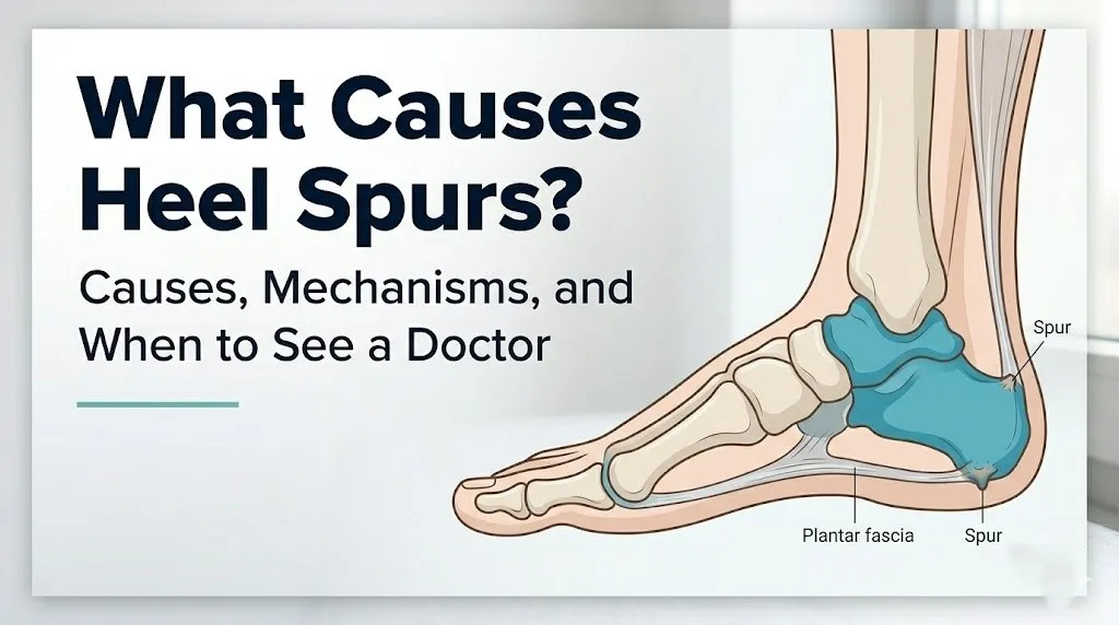
Over months to years, the mineralized deposits coalesce into a mature bony projection extending from the calcaneal surface. The spur’s direction of growth reflects the direction of the tensile force: plantar spurs grow anteriorly along the plantar fascia axis, while posterior spurs grow superiorly along the Achilles tendon axis.
People with genetic variants affecting collagen type I synthesis (the primary structural collagen of tendons and fascial tissue) may have a predisposition to entheseal microtrauma at lower mechanical loads, making them susceptible to spur formation even without the high-risk occupational or body weight factors seen in other populations, according to preliminary research published in the Journal of Orthopaedic Research.
Key Takeaway: Heel spurs form through osteoblast activation driven by BMP and TGF-beta signaling at the plantar fascial enthesis, triggered by either repetitive mechanical microtrauma or inflammatory cytokines from arthritis. Understanding which pathway applies to your situation determines whether the cause is mechanical and lifestyle-addressable or inflammatory and requiring specialist evaluation.
Plantar Fasciitis as the Primary Cause of Heel Spurs
Plantar fasciitis is the single most common cause of inferior calcaneal spur formation, occurring when chronic inflammation and microtrauma at the origin of the plantar fascia on the anterior-inferior calcaneal tuberosity stimulates the heterotopic ossification process that produces the spur.
The plantar fascia is a thick, multilayered fibrous aponeurosis running from the calcaneal tuberosity to the bases of the proximal phalanges of all five toes, forming the structural foundation of the medial longitudinal arch. During each step, ground reaction force compresses the heel while toe dorsiflexion (extension) during push-off simultaneously stretches the plantar fascia through a mechanism called the windlass mechanism. The calcaneal enthesis bears the full tensile load of this stretch with every step.
According to the American Academy of Orthopaedic Surgeons (AAOS), plantar fasciitis is the most common cause of heel pain in adults, affecting approximately 2 million Americans each year. The chronically inflamed plantar fascial enthesis in plantar fasciitis is the exact anatomical site where BMP-driven osteoblast activation produces calcaneal spur formation over time.
The clinical relationship between plantar fasciitis and heel spurs is important to understand correctly. Plantar fasciitis causes the spur; the spur does not cause plantar fasciitis. Pain in plantar fasciitis arises from the inflamed fascial tissue and enthesis, not from the bony spur itself. This is why surgical removal of the spur without addressing the underlying fascial pathology does not reliably resolve pain.
People with a history of bilateral plantar fasciitis (occurring in both feet) face a higher probability of bilateral calcaneal spur development. Identifying and correcting the biomechanical factors driving plantar fasciitis (foot posture, footwear, activity load) is the most effective way to reduce the progression of spur formation, according to clinical guidelines from the American Academy of Family Physicians (AAFP).
Repetitive Mechanical Stress and Heel Spur Development
Repetitive mechanical stress at the plantar fascial enthesis is the biomechanical driver behind the majority of heel spur cases, producing heel spurs through cumulative microtrauma that exceeds the enthesis’s repair capacity over a sustained period.
Every walking step generates a ground reaction force roughly equal to body weight at the calcaneus. Running generates ground reaction forces of two to three times body weight. Over a typical day of walking 8,000 steps, the calcaneal enthesis experiences millions of cycles of tensile loading annually. When the loading frequency or magnitude consistently exceeds the tissue’s repair rate, net tissue damage accumulates, driving the BMP-mediated osteogenic response described in the previous section.
The relationship between repetitive stress and spur formation follows a dose-response pattern. Clinical observation reported in Foot and Ankle International consistently shows that individuals who spend more hours per day on their feet, who run higher weekly mileage, or who work on hard surfaces have higher rates of calcaneal spur formation compared to more sedentary individuals.
Key factors that amplify the mechanical stress at the calcaneal enthesis:
- Running or walking on hard, non-cushioned surfaces (concrete, asphalt, warehouse floors)
- Sudden increases in activity level without progressive tissue adaptation time
- Walking barefoot on hard floors, which removes the heel cushion provided by footwear
- Carrying heavy loads regularly, which increases ground reaction force at the calcaneus
- Activities involving prolonged standing without adequate arch support
Military recruits are a well-studied high-risk population. Research published in the Journal of Bone and Joint Surgery has documented elevated rates of plantar fascial stress injuries and calcaneal spur development in military trainees, attributed to sudden high-volume marching and running activity on hard terrain without adequate footwear adaptation time.
Flat Feet and High Arches as Heel Spur Causes
Both pes planus (flat feet) and pes cavus (high arches) cause heel spurs through distinct but equally damaging biomechanical pathways that concentrate tensile stress at the calcaneal enthesis in different anatomical patterns.
In pes planus, the medial longitudinal arch collapses partially or fully during weight-bearing, causing the foot to overpronate (roll inward excessively). This overpronation places the plantar fascia under greater medial tensile stretch than normal arch mechanics would produce. The medial calcaneal enthesis, which bears the brunt of this excess stretch, accumulates microtrauma at an accelerated rate. According to the Mayo Clinic, flat feet are among the most consistently identified structural risk factors for plantar fasciitis and associated calcaneal spur formation.
High arches produce the opposite biomechanical problem. The rigid, elevated arch in pes cavus reduces the foot’s ability to absorb ground reaction force through normal arch flattening. Instead, the force is transmitted more directly to the calcaneal enthesis and Achilles tendon insertion, producing high tensile loading at both the plantar and posterior calcaneal sites.
The clinical implication is that both foot postures require different corrective approaches. Flat feet benefit from medial arch support to reduce overpronation and plantar fascial stretch. High arches benefit from cushioning and lateral posting to improve shock absorption and reduce direct calcaneal impact loading. A podiatrist or board-certified orthopedic surgeon specializing in foot and ankle can assess foot posture through clinical gait analysis and recommend specific orthotic modifications based on the individual’s specific arch structure and spur location.
Children and adolescents with symptomatic flat feet who are not assessed and managed appropriately face a higher lifetime risk of developing calcaneal spurs as adults, because the abnormal mechanical loading pattern accumulates over decades if the foot posture is not corrected during the growth years.
Obesity and Excess Body Weight as a Heel Spur Cause
Excess body weight causes heel spurs by proportionally increasing the ground reaction force transmitted to the calcaneal enthesis with every step, accelerating the cumulative microtrauma that drives heterotopic ossification at the plantar fascial attachment.
The relationship between body weight and plantar fascial loading is direct and measurable. Each kilogram of body weight adds approximately ten newtons of force to the ground reaction force at the foot. An adult with a body mass index (BMI) of 35 generates substantially higher calcaneal loading per step than an adult with a BMI of 22, even when walking the same distance at the same speed. This excess loading multiplied across thousands of daily steps produces a measurably higher rate of entheseal microtrauma.
According to a review published in the Journal of Bone and Joint Surgery, obesity (BMI above 30) is one of the most consistently identified risk factors for plantar fasciitis and calcaneal spur formation in epidemiological studies of foot pain in adults. The association holds after controlling for physical activity level, meaning that excess weight contributes to heel spur risk independently of exercise volume.
Adipose tissue also has an inflammatory role that compounds the mechanical effect. Adipocytes (fat cells), particularly visceral adipocytes, produce pro-inflammatory cytokines including TNF-alpha and interleukin-6 (IL-6) that can sustain low-grade systemic inflammation. This chronic inflammatory state may lower the threshold for entheseal inflammation in response to mechanical loading, making the tissue more reactive to the same amount of mechanical stress that a non-obese individual would tolerate without developing enthesopathy.
Postmenopausal women with obesity carry a compounded risk. Estrogen loss after menopause reduces collagen elasticity in fascial tissue, while excess body weight simultaneously increases mechanical loading. The combination produces a particularly high-risk state for plantar fascial enthesopathy and calcaneal spur formation.
Key Takeaway: Obesity increases heel spur risk through two simultaneous mechanisms: direct proportional increase in calcaneal loading per step and low-grade systemic inflammation from adipose-derived cytokines that sensitizes the enthesis to mechanical damage. Both mechanisms compound each other in postmenopausal women.
Improper Footwear and Its Role in Causing Heel Spurs
Improper footwear causes heel spurs by either removing the mechanical support structures that protect the calcaneal enthesis from excessive loading (in the case of worn-out or unsupportive shoes) or by altering the normal load distribution across the foot (in the case of high heels or completely flat shoes).
The footwear-heel spur connection operates through two distinct biomechanical pathways. First, shoes with inadequate heel cushioning fail to attenuate the ground reaction force at the calcaneus, transmitting the full impact directly to the calcaneal enthesis at each heel strike. Second, shoes with inadequate arch support allow overpronation, which increases medial plantar fascial tension as described in the pes planus section.
High-heeled shoes present a different but equally problematic pathway. Chronic high-heel use keeps the Achilles tendon in a shortened position. When the wearer switches to flat shoes or goes barefoot, the now-shortened Achilles tendon pulls on its calcaneal insertion with greater-than-normal tension, producing high tensile stress at the posterior calcaneal enthesis. This is a recognized cause of posterior calcaneal spur formation and insertional Achilles tendinopathy in people who wear high heels regularly.
Quick Tip:
- Replace athletic footwear every 400 to 500 miles of use (or every six to eight months for daily walkers), because midsole cushioning degrades substantially before the upper shows visible wear
- Choose shoes with at least 10 mm of heel-to-toe drop if you have a history of Achilles tightness or posterior heel pain, to reduce Achilles tendon loading at the calcaneal insertion
- Women who wear high heels regularly should include daily Achilles tendon stretching (gastrocnemius and soleus stretch) to prevent chronic Achilles shortening that increases posterior calcaneal enthesis loading
People with jobs requiring safety footwear (steel-toed boots, work boots) should be aware that the rigid structure of many safety shoes provides poor arch support and significant heel cushioning degradation over months of heavy use. Workers in these occupations should discuss orthotic insole options with a podiatrist.
Age and Occupation as Heel Spur Risk Factors
Age increases heel spur risk through cumulative lifetime mechanical loading at the calcaneal enthesis combined with age-related reductions in the regenerative capacity of fibrocartilaginous tissues, meaning older adults accumulate entheseal microtrauma faster and repair it more slowly than younger adults.
Research in Foot and Ankle International has documented that the radiological prevalence of calcaneal spurs increases with each decade of adult life. Studies using weight-bearing lateral foot X-rays have found calcaneal spur prevalence of approximately 10 to 15% in adults in their third and fourth decades, rising to over 50% in adults in their seventh decade and beyond. The increase reflects both lifetime accumulation of mechanical loading and the declining collagen repair capacity of aging connective tissue.
Occupational risk is concentrated in roles requiring prolonged standing or walking on hard surfaces. Specific occupations with elevated heel spur rates based on clinical epidemiological data include:
- Retail workers and cashiers who stand on concrete or tile floors for 6 to 10 hours per shift
- Healthcare workers (nurses, surgical technicians) who walk long distances daily on hospital floors
- Manufacturing and warehouse workers who stand on concrete or asphalt surfaces
- Construction and agricultural workers who stand on uneven, hard terrain
- Teachers and school staff who stand and walk for extended periods daily
According to the American Academy of Family Physicians (AAFP), occupational heel pain is a frequently underreported work-related musculoskeletal complaint. Workers in these roles who develop heel pain should report symptoms early to their employer and request ergonomic assessment, as early intervention with appropriate footwear and anti-fatigue matting can reduce the mechanical loading that drives spur progression.
Older adults in physically demanding occupations face a compounded risk profile. Declining tissue repair capacity combined with high daily mechanical loading produces accelerated entheseal microtrauma accumulation. These individuals should discuss heel pain and any confirmed calcaneal spurs with a primary care physician who can coordinate referral to a podiatrist or foot and ankle specialist for individualized management.
Inflammatory Conditions That Cause Calcaneal Spurs
Inflammatory systemic conditions cause calcaneal spurs through a fundamentally different mechanism than mechanical overuse: cytokine-driven entheseal inflammation activates the same BMP-mediated osteoblast pathway as mechanical microtrauma, producing spur formation without high mechanical loading, which is why younger, physically active adults with inflammatory arthritis can develop heel spurs that have no relationship to their activity level or foot structure.
The primary inflammatory conditions associated with calcaneal spur and enthesopathy formation include:
- Ankylosing spondylitis (AS): A seronegative spondyloarthropathy driven by HLA-B27-associated immune dysregulation. TNF-alpha and IL-17 drive entheseal inflammation throughout the axial skeleton and peripheral entheses, including the calcaneus. Calcaneal spurs in AS are often bilateral and accompanied by posterior heel pain at the Achilles insertion.
- Reactive arthritis: Formerly called Reiter’s syndrome. Triggered by prior urogenital or gastrointestinal infection. Produces entheseal inflammation at the calcaneus alongside urogenital, eye, and joint symptoms.
- Psoriatic arthritis: Approximately 30% of people with psoriasis develop psoriatic arthritis, which includes enthesopathy at the calcaneus as a recognized feature. The IL-17 pathway drives bone remodeling at entheseal sites.
- Diffuse idiopathic skeletal hyperostosis (DISH): A condition of abnormal bone formation at entheseal sites throughout the skeleton, producing large, flowing calcaneal spurs and ossification of spinal ligaments. Associated with older age, metabolic syndrome, and type 2 diabetes.
- Gout: Monosodium urate crystal deposition at the calcaneal enthesis activates the NLRP3 inflammasome, triggering acute and chronic inflammatory episodes that can stimulate spur formation.
According to the National Institute of Arthritis and Musculoskeletal and Skin Diseases (NIAMS), enthesopathy is a hallmark feature of the seronegative spondyloarthropathies and should be considered in any patient under 45 with heel spurs who does not have obvious mechanical risk factors. A rheumatologist should evaluate patients with bilateral calcaneal spurs, inflammatory back pain, skin or eye symptoms, or a family history of AS or psoriasis.
Key Takeaway: Inflammatory arthritis, including ankylosing spondylitis, psoriatic arthritis, and reactive arthritis, causes calcaneal spurs through cytokine-driven entheseal inflammation in younger adults who often have no biomechanical risk factors. Any adult under 45 with bilateral heel spurs should be evaluated by a rheumatologist to rule out an inflammatory cause.
Posterior Calcaneal Spur Causes at the Achilles Tendon Insertion
The posterior calcaneal spur forms at the insertion of the Achilles tendon on the posterior calcaneal surface through a traction mechanism and inflammatory response at the calcaneal apophysis, producing a bony projection at the back of the heel rather than the bottom, and is therefore caused by different factors than the more commonly discussed plantar (inferior) calcaneal spur.
The Achilles tendon, composed of the merged tendons of the gastrocnemius and soleus muscles, inserts on the posterior calcaneal tuberosity. Every time the calf muscles contract (during walking, running, jumping, or climbing stairs), the Achilles tendon transmits that force as a tensile pull on the posterior calcaneus. When the cumulative tensile loading at this insertion site exceeds repair capacity, fibrocartilage metaplasia and heterotopic ossification produce a bony spur projecting upward from the posterior calcaneal surface.
Insertional Achilles tendinopathy is the most common condition associated with posterior calcaneal spur formation. Specific causes of posterior calcaneal spur formation include:
- Chronic overloading of the Achilles tendon from high-volume running, jumping sports, or stair climbing
- Tight gastrocnemius or soleus muscles that increase resting tension on the Achilles insertion
- Haglund’s deformity, an anatomical variant where the posterior-superior calcaneal angle is more prominent than normal, causing friction between the calcaneus and the Achilles tendon insertion
- High-heeled shoe use causing adaptive Achilles shortening (described in the footwear section)
- Seronegative spondyloarthropathy-driven posterior entheseal inflammation
A clinically important distinction: posterior calcaneal spurs produce pain at the very back of the heel, worsened by shoe counter pressure and Achilles tendon loading. Inferior calcaneal spurs (plantar spurs) produce pain on the bottom of the heel, characteristically worst with the first steps after rest. A clinician can differentiate the two by palpation, but imaging confirmation with a weight-bearing lateral foot X-ray is standard.
Older men have a higher rate of posterior calcaneal spur formation compared to older women, in contrast to the overall calcaneal spur prevalence pattern, likely because of higher rates of insertional Achilles tendinopathy associated with sports participation patterns in older male populations, based on clinical observational data.
Diabetes and Metabolic Conditions Linked to Heel Spurs
Type 2 diabetes mellitus is associated with an elevated prevalence of calcaneal spur formation through three distinct pathophysiological pathways: abnormal collagen cross-linking that impairs tendon and fascial repair, peripheral neuropathy that eliminates protective pain signaling, and the overlapping association with diffuse idiopathic skeletal hyperostosis (DISH).
In type 2 diabetes, elevated blood glucose promotes advanced glycation end product (AGE) accumulation in collagen fibers of tendons and fascial tissue. AGEs stiffen the collagen architecture, reducing the tissue’s ability to absorb and distribute mechanical stress. The resulting increase in stress concentration at the calcaneal enthesis accelerates the fibrocartilage metaplasia and spur formation process even at normal activity levels.
Peripheral neuropathy, affecting approximately 50% of people with long-standing diabetes according to the American Diabetes Association, eliminates the normal pain signals that alert a person to excess heel loading. Without protective pain, individuals with diabetic neuropathy continue walking on already-damaged entheseal tissue, accelerating the cumulative microtrauma that drives spur formation.
According to clinical observation documented in Foot and Ankle International, people with type 2 diabetes have higher rates of plantar fascial thickening (a sonographic marker of plantar fasciopathy) compared to age-matched non-diabetic adults, and this thickening is associated with a higher prevalence of calcaneal spur on imaging.
DISH, which is strongly associated with type 2 diabetes and metabolic syndrome, produces particularly large and exuberant calcaneal spurs through pathological bone formation at entheseal sites, and is identified by its characteristic appearance on lateral foot X-ray alongside spinal and peripheral joint ossification.
People with type 2 diabetes who have heel pain should receive a comprehensive foot evaluation by a podiatrist, who can assess for both calcaneal spur and the neuropathic foot complications that can mask the protective pain signals that would otherwise prompt earlier self-assessment and treatment.
Heel Spur Causes in Athletes and Runners
Heel spurs in athletes and distance runners develop primarily from the combination of high-volume repetitive calcaneal loading and training errors that prevent adequate entheseal tissue recovery between loading sessions, with running-specific biomechanical factors compounding the baseline risk.
Distance runners subject the plantar fascial enthesis to ground reaction forces of approximately 2.5 times body weight with each running step, at step frequencies of 150 to 180 steps per minute. A runner logging 50 miles per week subjects the calcaneal enthesis to several million loading cycles annually at forces exceeding body weight. The enthesis’s repair capacity, which operates optimally during rest and sleep, is routinely outpaced by this loading volume when training load increases faster than tissue adaptation allows.
Specific training and biomechanical factors in runners that elevate calcaneal spur risk:
- Rapid weekly mileage increases exceeding the “10% rule” (increasing total weekly distance by more than 10% per week) that prevents adequate entheseal adaptation
- Transitioning rapidly from cushioned to minimalist footwear, which removes heel cushioning protection before the foot has adapted to the higher mechanical demand
- Running on cambered road surfaces, which produces asymmetric foot loading and concentrates stress on the downhill-side calcaneal enthesis
- Insufficient rest days, which eliminate the recovery window during which BMP-mediated entheseal repair normally occurs
- Running with excessive heel strike pattern at high speeds, which amplifies calcaneal impact loading compared to midfoot striking
According to the American Academy of Orthopaedic Surgeons (AAOS), plantar fasciitis and calcaneal spur formation are among the most common overuse injuries in runners, particularly those training for half-marathon and marathon distances. Runners who experience persistent heel pain should have a gait assessment performed by a sports medicine physician or podiatrist, with weight-bearing lateral foot X-ray to evaluate for calcaneal spur, before returning to full training.
Female runners should be aware that the female athlete triad (low energy availability, menstrual dysfunction, and low bone density) impairs entheseal tissue repair, increasing susceptibility to plantar fasciopathy and calcaneal spur formation at lower training volumes than are required to produce the same injury in female athletes with adequate energy availability.
Key Takeaway: In runners, calcaneal spurs develop when training load increases faster than entheseal tissue can adapt, and are accelerated by hard surfaces, rapid footwear transitions, and insufficient rest. Female runners with menstrual irregularities face elevated risk at lower training volumes due to the female athlete triad’s effect on tissue repair capacity.
How Heel Spurs Are Diagnosed
Calcaneal spurs are diagnosed primarily through weight-bearing lateral foot X-ray, which allows visualization of the bony projection on the calcaneal surface and assessment of its size, location, and direction of growth relative to the plantar fascia axis and Achilles tendon insertion.
The weight-bearing requirement is clinically important. X-rays taken with the foot non-weight-bearing can miss subtle spur formation and do not allow accurate assessment of the relationship between the spur and the functional loading axis of the foot. A standard lateral view of the foot taken while the patient stands is the imaging gold standard for initial calcaneal spur evaluation.
Additional diagnostic tools used in specific clinical circumstances include:
- Ultrasound imaging of the plantar fascia: Allows assessment of plantar fascial thickness (greater than 4 mm is associated with plantar fasciitis in most published reference ranges), fascial fiber integrity, and the soft tissue changes at the enthesis that coexist with the bony spur. Preferred for dynamic assessment in runners and athletes.
- MRI of the foot: Used when the diagnosis is uncertain, when soft tissue pathology (partial plantar fascia tear, fat pad atrophy, nerve entrapment) needs to be distinguished from spur-related pain, or when stress fracture is a clinical concern.
- Bone scan: Rarely needed but used when infection, tumor, or occult stress fracture is being considered alongside a calcaneal abnormality.
To describe your heel pain accurately to a primary care physician or podiatrist:
- Note the exact location of pain: bottom of the heel (inferior spur likely), back of the heel (posterior spur likely), or diffuse
- Describe the timing: worst with first steps in the morning (classic plantar fasciitis and inferior spur), worsening through the day (fat pad atrophy or nerve entrapment more likely), or worsening with specific activities
- Report how long symptoms have been present and whether they are improving, stable, or worsening
- List all footwear types you wear regularly and how long you’ve had them
- Bring a list of all medical conditions and medications, particularly corticosteroids and fluoroquinolone antibiotics, which are associated with increased tendon and fascial fragility
Older adults with calcaneal spurs on imaging should have the finding interpreted in clinical context by a primary care physician, as incidental spurs without pain do not require treatment and should not be the basis of a clinical diagnosis of heel spur syndrome without a thorough clinical examination.
When to See a Doctor for Heel Spurs
See a primary care physician for heel pain if discomfort has been present for more than four weeks without improvement, if walking is limited, or if you have any systemic symptoms alongside heel pain, because these presentations can indicate conditions requiring diagnosis beyond what physical examination alone can confirm.
The clinical threshold for specialist referral is lower than most people assume. A podiatrist or board-certified orthopedic surgeon specializing in foot and ankle should evaluate:
- Heel pain that persists beyond six to eight weeks of conservative home management (rest, ice, stretching, footwear modification)
- Severe pain with weight-bearing that limits daily activity
- Bilateral heel pain in an adult under 45 without clear mechanical explanation (possible inflammatory arthritis)
- Heel pain in a person with known psoriasis, inflammatory bowel disease, or family history of ankylosing spondylitis
- Heel pain in a person with type 2 diabetes, where neuropathy may mask the true severity of underlying structural damage
A rheumatologist should evaluate adults under 45 with calcaneal spurs who also have morning stiffness lasting more than 30 minutes, inflammatory back pain that improves with activity, eye inflammation (uveitis), skin plaques, or a history of sexually transmitted or gastrointestinal infection preceding joint symptoms.
Things to bring to your appointment:
- Your regular footwear for the physician or podiatrist to examine
- A symptom log noting pain location, intensity (rated 1 to 10), time of day, and activity relationship
- Any prior imaging (X-rays, ultrasound, MRI) of the affected foot
- A complete list of medications including corticosteroids and fluoroquinolone antibiotics, which affect tendon and fascial tissue integrity
People with diabetes who develop new heel pain should schedule an appointment with their podiatrist within one to two weeks rather than waiting, because peripheral neuropathy may be masking more severe structural damage than the pain level suggests.
How to Reduce the Risk of Developing Heel Spurs
Reducing the risk of calcaneal spur development requires addressing the specific biomechanical and physiological mechanisms that drive entheseal microtrauma accumulation, rather than simply resting or avoiding activity.
The most evidence-supported risk reduction strategies, based on clinical guidelines from the American Academy of Orthopaedic Surgeons and research published in Foot and Ankle International, include:
- Daily plantar fascia and Achilles tendon stretching: The plantar fascia stretch (pulling the toes into dorsiflexion and holding for 30 seconds, repeated three times before the first step of the morning) has the strongest evidence for reducing plantar fascia tension at the calcaneal enthesis. The gastrocnemius stretch (straight-leg calf stretch against a wall) and soleus stretch (bent-knee calf stretch) reduce Achilles tendon resting tension.
- Maintaining body weight within a healthy BMI range: Reduces calcaneal loading per step, slowing the cumulative microtrauma rate at the enthesis.
- Progressive activity increases: Increasing running or walking volume by no more than 10% per week allows entheseal tissue to adapt between loading sessions rather than accumulating net damage.
- Footwear with adequate cushioning and arch support: Replacing athletic shoes before cushioning is fully compressed (typically every 400 to 500 miles) and choosing occupational footwear with appropriate arch support for daily standing work.
- Anti-fatigue matting in standing workplaces: Evidence from occupational health research supports that compliant floor surfaces reduce peak calcaneal loading in workers who stand for extended periods.
Athletes who develop plantar fascia tightness or early heel pain should reduce training volume by 25 to 50% and consult a sports medicine physician or podiatrist before attempting to run through the discomfort, as continuing high-volume loading on an irritated enthesis accelerates the progression toward spur formation.
Postmenopausal women with a history of plantar fasciitis should discuss bone health and connective tissue support with their primary care physician or gynecologist, as optimizing calcium and vitamin D intake supports both bone metabolism and the entheseal repair processes that protect against spur formation.
Frequently Asked Questions About Heel Spurs
What is the main cause of heel spurs?
The main cause of heel spurs is chronic repetitive tensile stress at the origin of the plantar fascia on the calcaneal tuberosity, which is most commonly produced by plantar fasciitis.
This stress triggers fibrocartilage metaplasia and bone morphogenetic protein-driven osteoblast activation at the enthesis, producing calcium hydroxyapatite deposits that consolidate into a bony projection over months to years.
Inflammatory arthritis conditions, including ankylosing spondylitis and psoriatic arthritis, can also produce calcaneal spurs through a cytokine-driven mechanism without the mechanical trigger.
Can heel spurs go away on their own?
Established calcaneal spurs, which are mature bony projections, do not resorb and disappear on their own because they are composed of cortical bone.
The associated pain, however, often does resolve with conservative management including stretching, footwear modification, and activity adjustment, because the pain in most cases comes from inflamed soft tissue at the enthesis rather than the spur itself.
According to the American Academy of Orthopaedic Surgeons, approximately 90% of patients with plantar fasciitis and associated heel spurs experience pain resolution with non-surgical treatment within 10 months.
What is the difference between a heel spur and plantar fasciitis?
Plantar fasciitis is inflammation of the plantar fascia at its calcaneal attachment, while a heel spur is the bony projection that forms on the calcaneus as a result of chronic plantar fascia inflammation.
Plantar fasciitis causes the spur; the spur does not cause plantar fasciitis, which is why surgically removing the spur without treating the fascial inflammation does not reliably resolve pain.
Both conditions produce inferior heel pain that is characteristically worst with the first steps after rest, but they are diagnosed differently: plantar fasciitis is a clinical diagnosis based on examination, while a calcaneal spur requires weight-bearing lateral foot X-ray for confirmation.
Do flat feet cause heel spurs?
Flat feet (pes planus) are a recognized biomechanical risk factor for calcaneal spur formation because overpronation increases medial tensile stress on the plantar fascial enthesis at the calcaneal tuberosity.
This excess mechanical stress accelerates the fibrocartilage microtrauma and osteoblast activation sequence that produces heel spurs over time.
Not every person with flat feet develops heel spurs, and not every person with heel spurs has flat feet, but a gait and foot posture assessment by a podiatrist is recommended for anyone with flat feet and heel pain to determine whether orthotic arch support would reduce their entheseal loading.
Can inflammatory arthritis cause heel spurs in young people?
Yes, inflammatory arthritis conditions including ankylosing spondylitis, reactive arthritis, and psoriatic arthritis can cause calcaneal spurs in adults as young as their 20s and 30s through cytokine-driven entheseal inflammation rather than mechanical overuse.
These inflammatory conditions produce calcaneal enthesopathy via TNF-alpha and IL-17 signaling that activates the same osteoblast pathway as mechanical microtrauma, without requiring high activity levels or biomechanical risk factors.
Any adult under 45 with bilateral calcaneal spurs and no clear mechanical explanation should be evaluated by a rheumatologist to rule out an HLA-B27-associated seronegative spondyloarthropathy.
How long does it take for a heel spur to form?
Calcaneal spurs typically require at least six months to several years of sustained mechanical loading or chronic entheseal inflammation to develop into a radiologically detectable bony projection.
Research published in the Journal of Foot and Ankle Research indicates that the duration depends on the intensity of the causative mechanical or inflammatory stimulus, the individual’s baseline tissue repair capacity, and the presence of amplifying factors such as obesity, inflammatory arthritis, or diabetes.
This timeline explains why heel spurs are rarely seen in adolescents and progressively more common with each decade of adult life, with radiological prevalence exceeding 50% in adults over 70.
Closing
The cause of your heel spur determines what can actually be done about it. Mechanical causes tied to footwear, body weight, foot posture, and activity level can be addressed with specific, evidence-supported interventions that reduce the entheseal loading driving further spur progression. Inflammatory causes require a different approach entirely, one that involves a rheumatologist and potentially systemic treatment.
The most actionable step after reading this article: if you’ve had heel pain for more than four weeks, schedule an appointment with a primary care physician and ask specifically for a weight-bearing lateral foot X-ray. Bring your regular footwear to the appointment for examination, and note whether your pain is worst on the bottom of the heel (inferior spur) or the back of the heel (posterior spur), as that distinction guides the diagnostic and management pathway.
If you’re under 45, have bilateral heel pain, or have any inflammatory symptoms in other joints, skin, or eyes alongside your heel pain, request a referral to a rheumatologist. The treatment for an inflammatory calcaneal spur is fundamentally different from the treatment for a mechanical one, and getting the cause right is the essential first step.