Causes of Sagging Breasts at Young Age: What’s Really Happening
The causes of sagging breasts at young age include structural fatigue of the breast’s internal support ligaments, hormonal remodeling from pregnancy or contraceptive use, significant weight loss, smoking, UV skin damage, genetics, and inadequate breast support during high-impact physical activity. These are not random or cosmetic trivialities; each cause works through a specific, named physiological or anatomical mechanism that permanently alters the connective tissue architecture of the breast.
Breast ptosis in younger women is more common than most people expect. A study published in the Aesthetic Surgery Journal found that women under 35 represent a growing proportion of mastopexy consultations, with pregnancy, weight fluctuation, and sport-related mechanical loading identified as the leading documented causes in younger age groups. Understanding the specific mechanism behind each cause is what tells you whether a contributing factor in your life is modifiable, and when a breast shape change deserves a clinical evaluation rather than watchful waiting.
This article explains each cause with precise anatomical and physiological detail, ranks them by how commonly they affect young women, identifies the one red flag scenario that requires prompt medical attention, and gives you a practical framework for what you can and cannot change.
What Causes Sagging Breasts at Young Age
The primary causes of sagging breasts at young age are mechanical fatigue and biochemical degradation of the suspensory ligaments of the breast (Cooper’s ligaments), combined with loss of skin elasticity and volume changes within the breast parenchyma itself.
Unlike a joint or a muscle, breast tissue has no active structural support mechanism. It relies entirely on passive support: the fibrous bands of Cooper’s ligaments, the skin envelope, and the breast’s own internal fibrous stroma. When any of these passive supports are weakened, stretched, or chemically degraded, ptosis results. The process can begin in the late teens or early 20s if the right combination of contributing factors is present.
The causes fall into two broad categories: non-modifiable (genetics, breast size, number of pregnancies, age) and modifiable (smoking, UV exposure, unsupported exercise, weight cycling). Most young women experiencing early ptosis have a combination of both.
| Cause Category | Examples | Modifiable? | Onset Speed |
|---|---|---|---|
| Structural/genetic | Cooper’s ligament laxity, breast size, connective tissue disorders | No | Gradual, lifelong |
| Hormonal/reproductive | Pregnancy, breastfeeding, contraceptive hormones | Partially | Months to years |
| Lifestyle | Smoking, UV exposure, unsupported exercise | Yes | Months to years |
| Weight-related | Weight loss, weight cycling | Partially | Weeks to months |
Individual variation note: Young women with underlying connective tissue disorders, including Marfan syndrome and Ehlers-Danlos syndrome, may experience premature and more severe breast ptosis because their collagen and fibrillin architecture is inherently compromised. A primary care physician who identifies signs of these conditions should note breast support as one of several structural concerns requiring monitoring.
The Anatomy of Breast Support: Cooper’s Ligaments
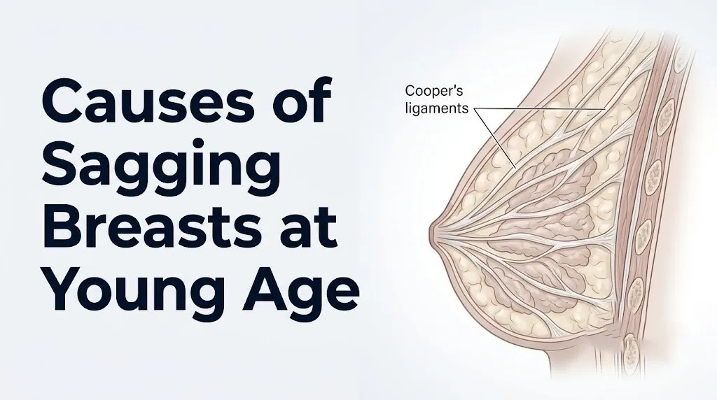
Cooper’s ligaments (formally, the suspensory ligaments of the breast) are fibrous bands of dense connective tissue that run from the deep dermis of the breast skin, through the breast parenchyma, and anchor to the pectoralis major fascia below, functioning as the breast’s primary internal suspension system.
Imagine Cooper’s ligaments as a hammock suspended between two fixed points: the skin above and the chest wall fascia below. The breast’s glandular and adipose tissue rests in this hammock. When the hammock’s fibers stretch, fray, or lose tensile strength, the tissue it supports descends under gravitational load. That descent is what ptosis looks and feels like from the outside.
These ligaments are composed primarily of collagen type I and type III, the same structural proteins that provide tensile strength to tendons and skin. They are not elastic in the way that muscle tissue is. Once stretched beyond their physiological limit, they do not recoil. This is why established ptosis does not reverse with exercise, weight gain, or any non-surgical intervention.
The specific anatomical arrangement matters for understanding why breasts sag in the direction they do. Cooper’s ligaments are most dense in the upper pole of the breast. As the upper pole ligaments fatigue, the nipple-areolar complex descends relative to the inframammary fold, which is the defining clinical marker of ptosis grade. The inframammary fold itself is a distinct anatomical structure formed by the adherence of breast skin to the underlying chest wall fascia.
The American Society of Plastic Surgeons identifies Cooper’s ligament integrity as the central determinant of breast projection and position across all ages and body types.
Individual variation note: Women with naturally larger breast volume place greater sustained gravitational load on Cooper’s ligaments from early adolescence. This accelerated mechanical loading means that a 22-year-old with a large natural breast size may show Cooper’s ligament fatigue patterns that would typically appear a decade later in women with smaller breast volume.
Genetics and Breast Ptosis in Young Women
Genetics is a non-modifiable primary cause of breast ptosis in young women, operating through inherited differences in collagen fiber density, Cooper’s ligament tensile strength, skin elasticity, and baseline adipose-to-glandular tissue ratio within the breast.
If a young woman’s mother and maternal grandmother experienced early breast ptosis without obvious lifestyle or reproductive causes, genetic predisposition is the most probable explanation. Heritability of connective tissue characteristics, including collagen architecture and elastic fiber density in skin and ligaments, is well-documented in dermatological and plastic surgery literature.
Several specific genetic factors contribute:
- Inherited variations in collagen synthesis genes (COL1A1, COL1A2 for type I collagen; COL3A1 for type III collagen) affect the tensile strength of Cooper’s ligaments from the moment of breast development
- Skin elastin gene variants (ELN gene) determine the baseline elastic recoil capacity of breast skin, affecting how well skin accommodates volume changes without permanent stretch
- Familial patterns of adipose distribution within the breast (higher adipose-to-glandular ratio) result in a heavier, more gravitationally challenged breast that loads Cooper’s ligaments more than a primarily glandular breast of the same external size
- Connective tissue disorders with autosomal dominant inheritance patterns, including Marfan syndrome (FBN1 gene) and hypermobile Ehlers-Danlos syndrome, produce systemic ligament laxity that includes Cooper’s ligaments
Research published in the Journal of Plastic and Reconstructive Surgery identifies family history as an independent predictor of early breast ptosis in multivariate analyses that controlled for BMI, parity, and lifestyle factors.
Individual variation note: Women of African ancestry have, on average, denser fibroglandular breast tissue and different elastic fiber composition in breast skin compared to women of European ancestry, according to observational data from breast imaging studies. These differences affect ptosis risk and pattern, though individual variation within any ethnic group is wide.
Key Takeaway: Cooper’s ligaments are the breast’s only structural suspension system, are made of collagen that does not regenerate once permanently stretched, and their inherent strength is substantially determined by genetics before any lifestyle factor ever acts on them.
Hormonal Causes of Sagging Breasts at Young Age
Hormonal fluctuations cause breast ptosis in young women by repeatedly expanding and then contracting the breast parenchyma through cyclic glandular tissue remodeling, driven by estrogen, progesterone, and their downstream effects on breast connective tissue.
The breast is an estrogen-sensitive organ throughout reproductive life. Estrogen, produced by the ovaries under the direction of the hypothalamic-pituitary-ovarian axis, promotes collagen synthesis in dermal fibroblasts via estrogen receptor-alpha (ERa) signaling. This means estrogen is, paradoxically, both a driver of breast glandular growth (which loads Cooper’s ligaments) and a promoter of the collagen that supports breast structure.
When estrogen levels fluctuate significantly, as they do across the menstrual cycle, with hormonal contraceptive use, and with conditions like polycystic ovary syndrome (PCOS), the breast undergoes repeated cycles of mild glandular expansion and contraction. Over years, these cycles impose cumulative mechanical stress on Cooper’s ligaments.
Hormonal factors specifically affecting young women:
- Combined oral contraceptives with high progestin content can temporarily increase breast volume. Stopping them after years of use may reveal underlying ptosis previously masked by that volume.
- Women with PCOS experience irregular estrogen patterns and elevated androgens. Androgen excess partially counteracts estrogen’s collagen-protective effect in skin fibroblasts, potentially accelerating connective tissue laxity.
- Young women with eating disorders who experience hypothalamic amenorrhea (loss of menstrual periods from low body weight or excessive exercise) undergo prolonged estrogen deficiency. Estrogen deficiency accelerates collagen loss in skin and ligaments at an age when estrogen-mediated collagen support should be at its peak.
According to the American College of Obstetricians and Gynecologists, estrogen’s role in maintaining skin and connective tissue integrity is well-established, and conditions that chronically suppress estrogen production in reproductive-age women carry connective tissue implications beyond reproductive health alone.
Individual variation note: Adolescent girls who begin hormonal contraception before breast development is fully complete (typically not fully complete until age 18 to 21) expose developing breast tissue and Cooper’s ligaments to synthetic hormone fluctuations during the most architecturally sensitive period of breast formation.
Pregnancy and Breast Ptosis
Pregnancy causes breast structural changes through a sequence of hormonally driven tissue expansion followed by post-delivery involution, and the net effect on Cooper’s ligaments and breast skin depends more on the magnitude of these changes than on pregnancy itself.
During pregnancy, progesterone from the corpus luteum and then the placenta drives glandular lobule proliferation throughout the breast. Prolactin from the anterior pituitary gland prepares the ductal system for lactation. Relaxin, produced by the corpus luteum and placenta, promotes systemic connective tissue laxity to allow pelvic expansion for delivery, but this effect is not limited to the pelvis. Cooper’s ligaments, as fibrous connective tissue, are subject to relaxin-mediated laxity during pregnancy.
Breast volume can increase by one to two cup sizes during pregnancy in many women. This increased volume imposes significantly greater gravitational load on Cooper’s ligaments throughout the final trimester. After delivery, if the woman does not breastfeed (or after breastfeeding ends), glandular tissue involutes and is replaced by adipose tissue, but the stretched skin envelope and mechanically fatigued ligaments remain.
A study published in Plastic and Reconstructive Surgery by Rinker and colleagues, examining factors associated with breast ptosis, found that the number of pregnancies was a statistically independent predictor of ptosis grade, with each pregnancy increasing ptosis risk independently of breastfeeding status. This is the research that disproved the longstanding clinical assumption that breastfeeding is the primary culprit: pregnancy itself, regardless of whether breastfeeding follows, is a substantial driver.
Individual variation note: Women who experience pregnancy before age 20 (adolescent pregnancy) have breast tissue that is still architecturally maturing. Relaxin-mediated ligament laxity acting on incompletely developed Cooper’s ligaments during adolescent pregnancy may produce greater long-term structural change than the same process occurring in a fully developed adult breast.
Breastfeeding and Breast Sagging
Breastfeeding does not directly cause breast sagging; the primary driver of post-pregnancy ptosis is the pregnancy-associated volume expansion and hormonal ligament laxity, not the act of lactation itself.
This is one of the most clinically important distinctions in the entire breast ptosis literature, and it is consistently misrepresented in general health content. The Rinker study published in Plastic and Reconstructive Surgery, which examined 132 women seeking mastopexy or augmentation mastopexy, found no statistically independent association between breastfeeding and breast ptosis after controlling for number of pregnancies, age, BMI, and smoking status.
What breastfeeding does contribute is prolonged elevation of prolactin from the anterior pituitary, which suppresses estrogen production by inhibiting the hypothalamic-pituitary-ovarian axis. This estrogen suppression during lactation accelerates the post-pregnancy glandular involution process, where lactating glandular tissue is replaced by adipose tissue. Volume loss from this involution, occurring inside an already-stretched skin envelope, contributes to the breast’s apparent deflated appearance post-weaning.
Key clinical distinctions for women who are pregnant or breastfeeding:
- Wearing a supportive, well-fitted bra throughout pregnancy and during breastfeeding reduces the gravitational load on Cooper’s ligaments during the period of greatest breast weight
- Avoiding rapid post-pregnancy weight loss reduces the speed of volume reduction inside the breast skin envelope
- The degree of post-breastfeeding ptosis correlates more strongly with the magnitude of pregnancy-related volume increase than with duration of breastfeeding, according to clinical observations published in the Aesthetic Surgery Journal
Individual variation note: Women who breastfeed multiple children in sequence, particularly without returning to their pre-pregnancy weight between pregnancies, experience cumulative cycles of Cooper’s ligament loading and involution that produce greater ptosis than any single pregnancy-breastfeeding episode would produce alone.
Key Takeaway: Pregnancy itself, not breastfeeding, is the primary reproductive driver of breast ptosis, operating through relaxin-mediated Cooper’s ligament laxity and the mechanical load of increased breast volume during the third trimester.
Weight Loss and Sagging Breasts in Young Women
Weight loss causes breast sagging in young women by reducing the adipose tissue volume within the breast faster than the surrounding skin envelope can contract, leaving excess skin and partially unsupported Cooper’s ligaments visible as ptosis.
Breasts are largely composed of adipose (fatty) tissue, with the proportion varying by individual and hormonal status. In women with higher adipose-to-glandular ratios, a substantial portion of breast volume is adipose tissue that responds to body-wide fat loss. When a young woman loses weight, breast volume decreases as adipose stores are mobilized. If the skin envelope does not contract at the same rate as the volume it contains, the excess skin collapses downward, carrying the nipple-areolar complex with it.
The speed and magnitude of weight loss are the key determinants of how much ptosis results:
- Rapid weight loss (more than 1 to 1.5 pounds per week) outpaces the skin’s adaptive capacity for collagen remodeling and contraction
- Large total weight loss (more than 50 pounds) consistently produces greater skin excess than the skin can reabsorb non-surgically in most individuals
- Age modifies the response: young women in their 20s have better skin contractile capacity than women in their 30s and 40s, but this advantage does not fully compensate for very rapid or very large losses
- Pre-existing Cooper’s ligament laxity (from genetics, prior pregnancy, or prior weight cycling) worsens the ptosis outcome of any given weight loss episode
The Mayo Clinic notes that the skin’s ability to contract following weight loss is determined by the individual’s baseline collagen architecture, age, sun damage history, and smoking history, none of which are controllable at the time of weight loss.
Individual variation note: Young women who achieve weight loss through bariatric surgery, which produces more rapid and more total weight reduction than behavioral weight loss programs in most cases, experience the most severe breast volume loss and associated ptosis in the shortest timeframe. A board-certified plastic surgeon should be part of the care team for women planning bariatric surgery who have concerns about breast shape.
Rapid Weight Cycling and Breast Skin Damage
Repeated cycles of weight gain followed by weight loss, a pattern clinically termed weight cycling or “yo-yo dieting,” damage breast skin elasticity more severely than any single weight loss episode of equivalent total magnitude.
The skin’s dermis contains a lattice of collagen type I and type III fibers interwoven with elastin fibers. This lattice allows skin to stretch as breast volume increases and contract as volume decreases, within limits. Each stretch-and-contract cycle in weight cycling exceeds the skin’s elastic limit slightly more than the last. The elastin fibers, which provide recoil, are progressively fatigued and fragmented. Collagen fiber alignment becomes disorganized. The cumulative result is skin that retains less contractile capacity with each cycle.
This is mechanically similar to repeatedly stretching a rubber band beyond its elastic limit. Each stretch leaves it slightly longer than before. Ten stretches leave it substantially longer than its original length, even though no single stretch produced catastrophic failure.
Research published in the Journal of Women’s Health has found an association between weight cycling history and skin laxity independent of current BMI, suggesting that the cycling process itself, not simply the current weight, causes the structural skin changes associated with breast ptosis.
Individual variation note: Young women with a history of disordered eating patterns, including cycles of restrictive eating followed by weight restoration, may experience breast skin and Cooper’s ligament changes disproportionate to their current body weight and age. A primary care physician evaluating breast ptosis in this population should ask about weight history specifically, not only current BMI.
Quick Tip:
- Aiming for gradual, sustained weight loss at a rate of no more than 0.5 to 1 pound per week allows more time for skin collagen remodeling to follow volume reduction
- Wearing a well-fitted, supportive bra during weight loss periods reduces the gravitational acceleration of ptosis as breast volume decreases
- Women with Ehlers-Danlos syndrome who also have a history of weight cycling face compounded skin laxity risk and should discuss breast support strategies specifically with their primary care physician
Smoking and Breast Sagging
Smoking accelerates breast ptosis by suppressing collagen synthesis in dermal and ligament fibroblasts and simultaneously activating the enzymatic collagen-degradation machinery, producing a net loss of structural collagen in Cooper’s ligaments and breast skin.
The mechanism is specific and well-characterized. Nicotine is metabolized in the body to cotinine, which reaches dermal fibroblasts through the bloodstream. Cotinine suppresses fibroblast proliferation and reduces the expression of procollagen genes, decreasing the rate of new collagen synthesis. At the same time, smoking-generated oxidative stress activates matrix metalloproteinases, specifically MMP-1 and MMP-3, which enzymatically cleave and degrade the existing collagen framework of skin and connective tissue.
The net effect is a double attack on the structural substrate of Cooper’s ligaments: less new collagen being made, and more existing collagen being destroyed.
The Cleveland Clinic reports that smokers show measurably accelerated skin aging compared to non-smokers of the same chronological age, with dermatological research consistently documenting reduced skin collagen density and elastin fiber fragmentation in smokers’ skin biopsies. These changes are not limited to facial skin. The dermis of the breast undergoes identical structural degradation from the same systemic exposure to cotinine and oxidative stress.
For young women who smoke: the ptosis-promoting effect of smoking begins accumulating from the first years of smoking, not after decades. A 22-year-old who has smoked since 16 has already accumulated six years of collagen suppression and MMP activation in her breast skin and Cooper’s ligaments.
Individual variation note: Young women who smoke and also use combined oral contraceptives receive a warning from the American College of Obstetricians and Gynecologists about cardiovascular risk. They also carry an underappreciated compounded structural risk: contraceptive-related estrogen supports collagen, but nicotine/cotinine actively suppresses it. The net effect on breast connective tissue depends on which mechanism predominates in a given individual.
Key Takeaway: Smoking causes breast ptosis through two simultaneous named mechanisms: cotinine-mediated suppression of fibroblast collagen synthesis and MMP-mediated enzymatic degradation of existing collagen in breast skin and Cooper’s ligaments.
UV Radiation and Breast Skin Elasticity
Ultraviolet A (UVA) radiation accelerates breast ptosis by activating matrix metalloproteinases in dermal fibroblasts of breast skin, producing photodamage to the collagen and elastin matrix that the skin requires to maintain its structural support of breast tissue.
The upper and lateral breast skin, particularly in women who wear low-cut or strapless clothing, is exposed to cumulative UVA radiation throughout the year. UVA penetrates glass and is present even on overcast days. It reaches the dermis, where it generates reactive oxygen species (free radicals). These reactive molecules activate MMP-1 (collagenase) and MMP-3 (stromelysin) in dermal fibroblasts, which then enzymatically degrade the collagen network of the dermis.
Over years, this produces photoaging: thinning of the dermal collagen layer, fragmentation of elastin fibers, and loss of the skin’s capacity to retract following stretch. In the breast, where the skin is expected to support significant tissue weight against gravity, photoaged skin fails this mechanical role earlier and more severely than non-photoaged skin of the same chronological age.
Research published in the Journal of Investigative Dermatology has documented that UVA-induced MMP activation in dermis is quantitatively similar to the MMP activation produced by inflammatory cytokines, confirming that sun-exposed skin undergoes connective tissue degradation through the same enzymatic pathway as inflamed skin.
Individual variation note: Women with Fitzpatrick skin types I and II (lighter skin tones with less melanin) have less inherent UV protection from melanin and accumulate UV-induced dermal damage faster per unit of sun exposure than women with Fitzpatrick types IV through VI. For lighter-skinned young women, consistent broad-spectrum sunscreen application to chest and breast skin is a mechanistically grounded, not purely cosmetic, intervention.
High-Impact Exercise and Breast Sagging
Unsupported or poorly supported high-impact exercise causes breast ptosis by imposing repetitive, multidirectional mechanical loading on Cooper’s ligaments that exceeds their fatigue tolerance over time, producing irreversible elongation of these fibrous suspension bands.
During running, jumping, and other high-impact activities, the breast moves in three planes simultaneously: vertically (up and down), horizontally (side to side), and in an anterior-posterior direction (forward and back). Research published by Professor Joanna Scurr at the University of Portsmouth, whose laboratory has produced extensive peer-reviewed research on breast biomechanics and published findings in the Journal of Sports Sciences, has documented that the breast of an unrestrained D-cup woman moves up to 15 centimeters vertically during running.
Each repetitive loading cycle stretches Cooper’s ligaments. Ligamentous connective tissue subjected to repetitive loading beyond its physiological range undergoes creep deformation, where permanent elongation accumulates with each loading cycle even if no single cycle produces visible failure.
The critical protective factor is a properly fitted sports bra that encapsulates and immobilizes breast tissue during movement. Encapsulation sports bras (which encase each breast in a shaped cup) are biomechanically superior to compression sports bras (which press both breasts flat against the chest) for women with cup sizes C and above, according to research from the University of Portsmouth breast biomechanics group.
To assess sports bra adequacy during exercise:
- Perform 30 seconds of jumping in place while wearing the bra
- Observe whether the breasts move independently of the torso or move with it as a unit
- Any independent upward and downward motion of more than approximately 2 centimeters suggests inadequate support for that activity level
- Replace sports bras every 6 to 12 months or after 30 to 50 washes, as elastic fibers in sports bra fabric fatigue with washing and wearing
- Get professionally fitted for sports bras when breast size changes (after pregnancy, after weight change)
Individual variation note: Adolescent girls who participate in high-impact sports during breast development, when Cooper’s ligaments are still architecturally immature, may be particularly susceptible to mechanical loading fatigue. Sports medicine physicians and school athletic trainers should include sports bra fitting guidance as standard equipment advice for female athletes.
Collagen and Elastin Loss in Young Women
Collagen and elastin loss in young women begins earlier than most people expect, with measurable reductions in skin collagen density detectable from the mid-20s, driven by declining synthesis rates and increasing enzymatic degradation accelerated by the lifestyle factors described in prior sections.
Collagen is the primary structural protein of Cooper’s ligaments and breast skin dermis. The human body’s peak collagen synthesis rate occurs during the late teenage years and begins a gradual decline from approximately age 25 onward. From age 25, the dermis loses an estimated 1 percent of its collagen content per year under baseline conditions, according to research published in the British Journal of Dermatology. This rate accelerates with smoking, UV exposure, and oxidative stress from any source.
Elastin provides the recoil capacity that allows skin to return to its original dimensions after stretch. Unlike collagen, elastin is produced almost exclusively during fetal development and early childhood. By adulthood, the body has essentially its full elastin complement. Elastin fibers damaged by UV radiation, oxidative stress, or mechanical fatigue are not replaced by newly synthesized elastin in meaningful quantities. They are replaced by poorly organized scar-like protein deposits that lack elastic recoil.
The interplay between these two proteins determines breast skin’s ability to accommodate volume changes without permanent shape alteration:
- Healthy collagen provides tensile strength: the skin does not tear under load
- Healthy elastin provides recoil: the skin returns to its pre-stretch dimensions when load is removed
- When collagen is reduced and elastin is damaged, skin stretches under load but fails to retract, permanently changing breast shape
The National Institutes of Health has funded research confirming that skin collagen synthesis and degradation are dynamically regulated throughout adult life and are responsive to both protective behaviors (sunscreen, smoking cessation) and damaging exposures.
Individual variation note: Young women with inflammatory skin conditions including moderate to severe acne (particularly if treated with long-term antibiotics or retinoids) experience altered skin barrier function and inflammatory cytokine activity that may affect dermal collagen remodeling in ways not yet fully characterized in the clinical literature.
Key Takeaway: Elastin fibers damaged by UV exposure or mechanical fatigue in young women are not meaningfully replaced by the body, making the prevention of elastin damage the more clinically effective strategy compared to any attempt to reverse it after the fact.
Regnault Classification of Breast Ptosis Severity
The Regnault classification is the standard clinical grading system for breast ptosis severity, dividing ptosis into three grades based on the position of the nipple-areolar complex relative to the inframammary fold, with an additional category for pseudoptosis.
This classification, developed by French plastic surgeon Paulette Regnault and published in Plastic and Reconstructive Surgery in 1976, remains the primary framework used by plastic surgeons and obstetrician-gynecologists to communicate about ptosis severity and surgical planning. Understanding it gives a young woman a clinically accurate way to describe and think about her own anatomy.
| Grade | Clinical Description | Nipple Position | Typical Management Considered |
|---|---|---|---|
| Pseudoptosis | Breast volume falls below fold, but nipple remains at or above fold | At or above inframammary fold | Supportive bra; augmentation may address volume |
| Grade I (Minor ptosis) | Nipple at the level of the inframammary fold | At fold level | Supportive measures; may consider mastopexy if desired |
| Grade II (Moderate ptosis) | Nipple below the inframammary fold but still pointing forward | Below fold, pointing forward | Mastopexy typically discussed |
| Grade III (Severe ptosis) | Nipple well below the inframammary fold, pointing downward | Well below fold, pointing down | Mastopexy, often with augmentation |
Plastic surgeons use the Regnault grade not only to describe severity but to plan the type of surgical correction that will restore the nipple-areolar complex to an anatomically appropriate position above the inframammary fold.
The American Society of Plastic Surgeons reports that mastopexy is among the top five surgical procedures requested by women under 35, with the majority of young candidates having Grade I or Grade II ptosis attributable to post-pregnancy changes, significant weight loss, or a combination of genetic predisposition and lifestyle factors.
Individual variation note: Grade I ptosis in a young woman with very large natural breast volume may produce significantly more functional discomfort (inframammary skin irritation, bra strap shoulder grooving, chronic upper back strain from pectoralis major compensatory effort) than Grade II ptosis in a woman with a smaller breast volume. The Regnault grade alone does not capture functional impact.
Breast Size, BMI, and Ptosis Risk
Larger natural breast volume and higher body mass index are independent risk factors for early breast ptosis in young women, operating through the greater sustained gravitational mechanical load they impose on Cooper’s ligaments throughout the day.
The gravitational force acting on breast tissue is proportional to breast mass. A woman with naturally larger breasts begins loading her Cooper’s ligaments more heavily from the onset of breast development in adolescence. This chronically elevated mechanical load produces Cooper’s ligament creep deformation earlier in life than the same process occurring in a woman with smaller natural breast volume.
Higher BMI amplifies this in two ways: it increases breast adipose volume (adding to gravitational load) and it adds to the adipose tissue envelope surrounding the chest wall, changing the biomechanical angles at which Cooper’s ligaments are loaded.
Key points supported by research in the Plastic and Reconstructive Surgery literature:
- Pre-pregnancy bra cup size is a significant predictor of post-pregnancy ptosis grade, independent of pregnancy number or breastfeeding status
- BMI at any given age correlates with ptosis severity in women who have never been pregnant, confirming that reproductive history is not required for weight-related ptosis risk to manifest
- Women with BMI above 30 kg/m² who also have large cup sizes face the highest cumulative gravitational loading on Cooper’s ligaments before any reproductive or lifestyle factor is added
For young women with larger natural breast volume, wearing a properly fitted supportive bra during waking hours, not only during exercise, reduces the cumulative daily gravitational load on Cooper’s ligaments. This is one of the few genuinely evidence-grounded non-surgical strategies for reducing the rate of mechanical ptosis progression.
Individual variation note: Adolescents with rapid breast development during puberty, particularly those developing C cup or larger volumes between ages 12 and 15, may benefit from early professional bra fitting guidance. Pediatricians and family physicians who care for adolescent girls are in a position to address breast support as a practical health topic at annual wellness visits.
When Breast Shape Change Needs Medical Evaluation
New, unexplained, unilateral breast shape change or progressive ptosis in a young woman without a clear cause, such as recent pregnancy or major weight loss, requires prompt clinical evaluation by a primary care physician or obstetrician-gynecologist because it can be an early sign of an underlying breast mass.
This is the red flag that no competitor article addresses. Breast ptosis is almost always bilateral and develops gradually over months to years. When ptosis or shape change is:
- Unilateral (affecting one breast while the other is unchanged)
- Rapid in onset (developing over weeks rather than months)
- Associated with other changes: skin dimpling, skin thickening resembling an orange peel (peau d’orange texture), nipple retraction, nipple discharge, or a palpable lump
…the clinical picture shifts from a benign structural process to a presentation that requires tissue diagnosis. Inflammatory breast cancer, though rare in young women, can present with skin thickening, breast enlargement, and shape change without a palpable lump. A growing benign or malignant mass within the breast can tether Cooper’s ligaments and the overlying skin, producing skin dimpling and apparent localized shape change.
What to tell your primary care physician or OB-GYN at your evaluation:
- Which breast is affected and whether both breasts are involved equally or unequally
- How long you have noticed the change and whether it appeared suddenly or gradually
- Whether you have had any recent pregnancy, breastfeeding, or major weight change
- Whether you noticed any associated changes: skin texture change, nipple retraction, discharge, or a palpable area of firmness
- Your family history of breast conditions, including any first-degree relative with breast or ovarian cancer
- Your current medications, including hormonal contraceptives
According to the American College of Obstetricians and Gynecologists, clinical breast examination remains a standard component of reproductive health care and should be performed at preventive visits. Any new breast asymmetry or shape change noted by the clinician during this examination warrants further assessment, which may include diagnostic breast ultrasound or mammography depending on age and clinical findings.
Individual variation note: Young women under 30 are below standard mammographic screening age. For this population, diagnostic breast ultrasound is typically the first-line imaging modality when a clinical concern warrants imaging, because ultrasound does not involve ionizing radiation and has good sensitivity for solid masses in the dense breast tissue common in younger women.
Key Takeaway: New unilateral breast shape change, skin dimpling, nipple retraction, or breast asymmetry that develops rapidly without a clear cause (pregnancy, weight change) in a young woman needs a clinical breast examination from a primary care physician or OB-GYN within weeks, not months.
Reducing Breast Ptosis Risk in Young Women
Reducing breast ptosis risk in young women involves consistently addressing the modifiable causes through specific, evidence-grounded behaviors targeting the named physiological mechanisms of collagen preservation, Cooper’s ligament mechanical protection, and skin elasticity maintenance.
No intervention can guarantee that a young woman will not develop breast ptosis. Genetics, breast size, and reproductive choices are not fully modifiable. But the lifestyle-related causes described in this article are genuinely addressable, and each has a named mechanism through which the protective behavior works.
Modifiable risk reduction strategies with named mechanisms:
- Wear a properly fitted, encapsulation sports bra during all high-impact exercise: reduces repetitive multidirectional Cooper’s ligament loading. Encapsulation bras outperform compression styles for C cup and larger in independent biomechanical testing from the University of Portsmouth breast research group.
- Wear a supportive bra during waking hours if you have large natural breast volume: reduces cumulative daily gravitational Cooper’s ligament load during the years of maximum ligament vulnerability
- Cease smoking: stops cotinine-mediated fibroblast collagen suppression and reduces MMP activation, preserving the collagen substrate of Cooper’s ligaments and breast skin
- Apply broad-spectrum SPF 30 or higher sunscreen to chest and upper breast skin on days with sun exposure: blocks UVA-mediated MMP activation in dermal fibroblasts, slowing photodamage to breast skin collagen and elastin
- Avoid rapid weight cycling: reduces repetitive stretch-and-contraction loading of breast skin that fragments elastin fibers and disorganizes collagen alignment
- Maintain a stable BMI: reduces the gravitational load variation on Cooper’s ligaments from weight fluctuation
For women who have established ptosis that is causing functional symptoms including shoulder pain from bra strap compensation, chronic inframammary rash, or neck strain, a referral to a board-certified plastic surgeon for a mastopexy consultation is appropriate. Mastopexy, which surgically repositions the nipple-areolar complex and removes excess skin, is not a cosmetic triviality. It is the only intervention with a defined anatomical mechanism for correcting established Cooper’s ligament and skin envelope changes.
Individual variation note: Women currently breastfeeding should wear a properly fitted nursing bra with adequate support throughout lactation and during nighttime rest if breast volume is large. Reducing gravitational loading on already relaxin-laxened Cooper’s ligaments during breastfeeding may reduce the degree of ptosis observed after weaning, though controlled research on this specific intervention is limited.
Frequently Asked Questions About Causes of Sagging Breasts at Young Age
Why are my breasts sagging at 20?
Breast sagging at 20 is most commonly caused by genetic predisposition, large natural breast volume imposing gravitational load on Cooper’s ligaments since puberty, a prior pregnancy, rapid weight loss, smoking, or inadequate breast support during high-impact exercise.
Each of these causes operates through a specific physiological mechanism: Cooper’s ligament mechanical fatigue, cotinine-mediated collagen suppression, or skin elastin fragmentation from stretch-and-contraction cycles.
A primary care physician or obstetrician-gynecologist can review your specific history and confirm whether the pattern is bilateral and gradual, as expected in benign ptosis, or requires imaging assessment.
Does breastfeeding cause breasts to sag permanently?
Breastfeeding is not the primary cause of permanent breast ptosis; pregnancy itself, through relaxin-mediated Cooper’s ligament laxity and the mechanical load of increased breast volume during the third trimester, is the stronger independent driver.
Research published in Plastic and Reconstructive Surgery by Rinker and colleagues found that number of pregnancies predicted ptosis grade independently of breastfeeding status after controlling for BMI, age, and smoking.
The post-weaning volume loss from glandular involution inside a pregnancy-stretched skin envelope is what produces the deflated appearance often attributed to breastfeeding specifically.
Can losing weight cause breast sagging in young women?
Weight loss causes breast sagging when breast adipose volume decreases faster than the surrounding skin envelope can contract to follow it, leaving excess skin and exposed Cooper’s ligament laxity.
The speed and total magnitude of weight loss determine how much ptosis results, with rapid loss exceeding 1 to 1.5 pounds per week outpacing the skin’s collagen remodeling capacity.
Young women planning substantial weight loss should discuss breast support strategies and realistic skin remodeling expectations with their primary care physician before beginning.
Is breast sagging at a young age normal?
Breast sagging at a young age is physiologically explainable and not rare, though the word “normal” depends on the specific cause and pattern.
Bilateral, gradual ptosis with a clear contributing cause (pregnancy, large breast size, weight loss, smoking history) follows a known and benign structural mechanism.
New, rapid, or unilateral breast shape change without a clear cause in a young woman is not a pattern to normalize without clinical evaluation.
What is the Regnault classification for breast ptosis?
The Regnault classification grades breast ptosis by the position of the nipple-areolar complex relative to the inframammary fold: Grade I places the nipple at fold level, Grade II places it below the fold but pointing forward, and Grade III places it well below the fold pointing downward.
Pseudoptosis is an additional category where breast volume falls below the fold but the nipple remains at or above it.
Plastic surgeons use this grading system to plan the type and extent of mastopexy correction required for each individual’s anatomy.
When should I see a doctor about breast shape changes?
Any new, unilateral breast shape change, skin dimpling, nipple retraction, or breast asymmetry that develops rapidly without a clear cause warrants evaluation by a primary care physician or obstetrician-gynecologist within weeks.
Inflammatory breast cancer, though rare in young women, can present as breast enlargement and shape change without a palpable lump, making clinical examination necessary to rule out non-structural causes.
For breast changes that are bilateral, gradual, and clearly linked to a known cause like pregnancy or weight loss, discuss the findings at your next scheduled preventive health visit with your primary care physician or OB-GYN.
Closing
The causes of sagging breasts at young age are specific, mechanistically grounded, and largely distinct from the vague “aging and gravity” explanation that most content on this topic offers. Cooper’s ligaments do the structural work of suspending breast tissue, and their integrity is shaped by genetics, hormonal exposure, pregnancy, weight history, smoking, UV exposure, and mechanical loading from unsupported exercise. None of these factors operate vaguely; each has a named pathway through which it damages or preserves the collagen and elastin architecture of breast support.
The practical priorities are these: stop smoking, protect breast skin from UV exposure, wear an adequate sports bra during exercise, and avoid rapid weight cycling. These four behaviors address the four most clearly modifiable mechanisms in this article.
If you’ve noticed a breast shape change that is unilateral, rapid, or associated with skin or nipple changes, schedule a clinical breast examination with your primary care physician or OB-GYN this month. That appointment is the one step no amount of reading can replace.