What Causes Pleural Effusion: All Key Causes in 2026
Pleural effusion is caused by an abnormal disruption in the balance of fluid entering and leaving the pleural space, the thin gap between the two membranes that wrap around the lungs, and the underlying cause determines both the type of effusion and the urgency of treatment. More than 50 different medical conditions can produce this fluid buildup, but heart failure, cancer, and pneumonia account for the majority of cases diagnosed in adults.
According to the National Heart, Lung, and Blood Institute (NHLBI), approximately 1.5 million cases of pleural effusion are diagnosed in the United States each year, making it one of the most common respiratory complications encountered across cardiology, oncology, and pulmonary medicine. The condition is not a disease in itself but a symptom of another underlying process, which means identifying what causes pleural effusion in any given patient is the first and most medically consequential step.
This article covers every major cause of pleural effusion with the specific physiological mechanism behind each, explains the clinical classification system that doctors use to narrow down the cause, identifies which populations face the greatest risk, and tells you exactly when symptoms require same-day medical evaluation or emergency care.
What Causes Pleural Effusion
Pleural effusion is caused when one or more of four core physiological mechanisms produces more fluid in the pleural space than the body’s lymphatic drainage system can remove.
The four mechanisms are: (1) elevated hydrostatic pressure in the pulmonary or systemic capillaries, which forces fluid across the pleural membrane; (2) reduced plasma oncotic pressure, which decreases the force that normally pulls fluid back into the bloodstream; (3) increased capillary permeability from inflammation or malignancy, which allows protein-rich fluid to leak into the pleural cavity; and (4) impaired lymphatic drainage, which prevents normal fluid clearance regardless of how much fluid enters.
Understanding which mechanism is active matters because it determines the type of fluid that accumulates and, therefore, the type of diagnostic tests needed to find the cause.
Key causes grouped by mechanism:
- Elevated hydrostatic pressure: congestive heart failure, constrictive pericarditis, superior vena cava obstruction
- Reduced oncotic pressure: cirrhosis with hypoalbuminemia, nephrotic syndrome, severe protein malnutrition
- Increased permeability: bacterial pneumonia, cancer metastases to pleura, lupus pleuritis, drug-induced pleuritis
- Impaired lymphatic drainage: malignant lymph node infiltration, thoracic duct injury, post-surgical lymphatic disruption
Older adults over 65 are disproportionately affected because they carry higher rates of the three most common causes: heart failure, cancer, and bacterial pneumonia. Research published in the CHEST journal has estimated that these three conditions together account for approximately 75% of all pleural effusion cases evaluated in hospital settings.
What Is Pleural Effusion and How Does It Develop
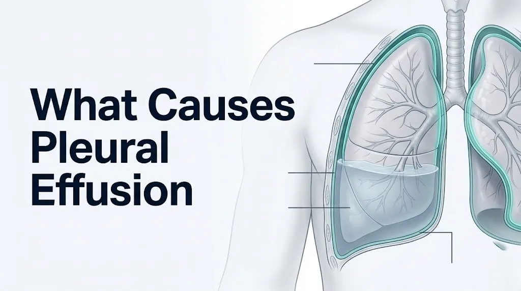
Pleural effusion is defined as an abnormal accumulation of fluid in the pleural space, the normally near-dry potential space between the visceral pleura (the membrane adhered to the lung surface) and the parietal pleura (the membrane lining the inner thoracic wall, diaphragm, and mediastinum).
In a healthy person, this space contains between 5 and 15 milliliters of fluid. That small volume acts as a lubricant, allowing the lungs to slide freely against the chest wall with each breath. Fluid enters through the parietal pleural capillaries, where hydrostatic pressure exceeds oncotic pressure slightly, and is continuously absorbed through pleural lymphatics draining into the thoracic duct and subpleural lymphatic networks.
Think of this system like a slow drip into a sink with a constantly running drain. Under normal conditions, the drain keeps pace with the drip perfectly. Pleural effusion is what happens when the drip speeds up, the drain slows down, or both happen at once. The fluid level rises steadily, and the lung has less room to expand.
When the fluid volume exceeds roughly 300 milliliters, it becomes detectable on a chest X-ray as blunting of the costophrenic angle. Volumes exceeding 1,500 to 2,000 milliliters begin compressing the lower lobe of the lung on the affected side, reducing gas exchange and producing the shortness of breath that most patients notice first.
The Mayo Clinic states that small pleural effusions may produce no symptoms at all, while large ones cause progressive dyspnea (shortness of breath), a dry cough, and a dull aching heaviness on the affected side of the chest.
Pediatric patients develop pleural effusion through similar mechanisms, but the most common cause in children is parapneumonic effusion from bacterial pneumonia, not heart failure or malignancy, reflecting the age-specific disease burden in this population.
Transudative vs Exudative Pleural Effusion Causes
The single most clinically important distinction in pleural effusion is whether the fluid is transudative or exudative, because this classification directly identifies which category of cause is responsible.
A transudative effusion forms when systemic forces push fluid into the pleural space without any local pleural disease. The fluid is low in protein and low in the enzyme lactate dehydrogenase (LDH) because the pleural membrane itself is intact and not inflamed. The leak is passive, driven by pressure imbalance.
An exudative effusion forms when the pleural membrane or nearby tissue is directly diseased, inflamed, or malignantly invaded. Inflammatory mediators increase capillary permeability, allowing protein, cells, and enzymes to leak into the pleural cavity along with fluid. The resulting fluid is protein-rich and LDH-rich.
| Feature | Transudative Effusion | Exudative Effusion |
|---|---|---|
| Pleural fluid protein | Less than 3.0 g/dL | Greater than 3.0 g/dL |
| Pleural/serum protein ratio | Less than 0.5 | Greater than 0.5 |
| Pleural fluid LDH | Less than 200 IU/L | Greater than 200 IU/L |
| Pleural/serum LDH ratio | Less than 0.6 | Greater than 0.6 |
| Common causes | Heart failure, cirrhosis, nephrotic syndrome | Pneumonia, cancer, autoimmune disease, TB |
| Pleural membrane involvement | None: membrane is intact | Direct disease or inflammation present |
This classification is performed using Light’s criteria, a biochemical standard established by Dr. Richard Light in 1972 and published in the Annals of Internal Medicine, which remains the gold standard diagnostic tool for pleural fluid analysis worldwide.
People taking diuretics for heart failure present a diagnostic challenge because diuresis concentrates the pleural fluid protein, sometimes causing a true transudate to falsely classify as an exudate by Light’s criteria. In this population, clinicians check the serum to pleural fluid albumin gradient; a gradient greater than 1.2 g/dL confirms a transudative cause despite the elevated protein reading.
Heart Failure as a Cause of Pleural Effusion
Congestive heart failure is the single most common cause of pleural effusion in adults, producing fluid accumulation through elevated pulmonary capillary hydrostatic pressure that overwhelms the oncotic pressure holding fluid within the vascular space.
The mechanism follows the Starling equation directly. In left ventricular failure, the left ventricle cannot pump blood forward efficiently, causing blood to back up into the pulmonary veins and pulmonary capillary beds. Pulmonary capillary wedge pressure rises. When this hydrostatic pressure exceeds plasma oncotic pressure (normally maintained by albumin at approximately 25 mmHg), fluid is pushed across the thin capillary walls of the visceral pleura into the pleural space.
The American Heart Association states that pleural effusion occurs in approximately 80% of patients hospitalized with decompensated heart failure, making it far more common in this disease context than any other single condition. In heart failure, effusions are typically bilateral (occurring on both sides of the chest) because the elevated pulmonary pressure affects both lung fields, though right-sided effusion is often larger.
To prepare for a cardiology or pulmonology appointment when heart failure is suspected as the cause:
- Document when shortness of breath started and whether it worsens lying flat (orthopnea)
- Note any ankle swelling, weight gain over days, or reduced exercise tolerance
- Bring a list of all current medications including diuretics and their doses
- Report any recent changes in sodium or fluid intake
- Ask specifically whether an echocardiogram has been ordered to assess left ventricular ejection fraction
Women with heart failure may present with atypical symptoms including fatigue and nausea more than classic chest heaviness, according to the American Heart Association, making the initial diagnosis of cardiac-related effusion sometimes delayed in female patients compared to male patients with the same degree of left ventricular dysfunction.
Key Takeaway: Congestive heart failure is the leading cause of pleural effusion in adults, producing a transudative fluid buildup through elevated pulmonary capillary pressure according to the Starling equation, and bilateral effusion in a patient with known cardiac disease should prompt same-day cardiology evaluation.
Cancer and Malignant Pleural Effusion Causes
Malignant pleural effusion occurs when cancer cells invade the pleural membrane or its lymphatic drainage, triggering a combination of increased vascular permeability and impaired fluid clearance that produces large, protein-rich exudative effusions.
Cancer is the second most common cause of pleural effusion in adults overall, and the most common cause of large unilateral exudative effusions. The four cancers most frequently responsible are lung cancer, breast cancer, lymphoma, and ovarian cancer, together accounting for approximately 75% of all malignant pleural effusions, according to research published in the Journal of Thoracic Disease.
The mechanism has two components. First, malignant cells invading the pleural surface secrete vascular endothelial growth factor (VEGF), a protein that directly increases the permeability of nearby capillaries. This allows large protein molecules and inflammatory cells to escape into the pleural cavity, creating an exudate. Second, tumor cells infiltrate the mediastinal lymph nodes that normally drain pleural fluid, obstructing lymphatic return and preventing clearance of even normal fluid volumes.
Mesothelioma deserves separate mention. Malignant pleural mesothelioma (ICD-10: C45.0) is a cancer arising from the mesothelial cells of the pleural lining itself, most commonly caused by asbestos exposure with a latency period of 20-50 years from first exposure to diagnosis. It produces a particularly aggressive and rapidly reaccumulating effusion that is nearly always unilateral and exudative.
Women with ovarian tumors or pelvic fibromas can develop a specific syndrome called Meigs syndrome, in which a benign or malignant pelvic tumor produces both ascites (abdominal fluid) and a right-sided pleural effusion through mechanisms involving diaphragmatic lymphatic transport. Meigs syndrome is frequently misattributed to heart failure or liver disease in women who have not yet had their pelvic pathology identified, making it a clinically important cause for physicians to consider in women with unexplained right-sided effusion and pelvic symptoms.
Pneumonia and Parapneumonic Pleural Effusion
Pneumonia causes pleural effusion when the inflammatory process in the adjacent lung parenchyma spreads to the visceral pleura, increasing pleural capillary permeability through the release of pro-inflammatory cytokines including interleukin-6 (IL-6) and tumor necrosis factor-alpha (TNF-alpha).
This type of effusion is called a parapneumonic effusion (para meaning “alongside” pneumonia) and it is the most common cause of exudative pleural effusion worldwide. According to the American Thoracic Society, parapneumonic effusion complicates approximately 20-40% of bacterial pneumonia cases requiring hospitalization. Not all of these require drainage; small parapneumonic effusions typically resolve as the underlying pneumonia is treated with antibiotics.
The critical concern with parapneumonic effusion is progression to empyema thoracis, a condition in which the pleural fluid becomes frankly purulent (filled with pus). This progression occurs when bacteria cross the visceral pleural membrane directly into the pleural space, converting the effusion from a sterile inflammatory reaction to an active infection within the pleural cavity. Empyema produces a pleural fluid pH below 7.20, glucose below 40 mg/dL, and LDH exceeding 1,000 IU/L, markers that indicate the effusion requires immediate drainage rather than antibiotic therapy alone.
Common bacteria causing parapneumonic effusion include Streptococcus pneumoniae, Staphylococcus aureus (including methicillin-resistant strains), Klebsiella pneumoniae, and anaerobic organisms in aspiration pneumonia. The bacterial species involved helps predict the risk of empyema: Staphylococcus aureus pneumonia carries a higher empyema risk than pneumococcal pneumonia.
Immunocompromised individuals, including people receiving chemotherapy, those with HIV infection, and organ transplant recipients taking immunosuppressive medications, face an expanded spectrum of infectious causes for exudative effusion. These include Pneumocystis jirovecii pneumonia, fungal infections such as Aspergillus, Histoplasma, and Cryptococcus, and mycobacterial infections including Mycobacterium tuberculosis and nontuberculous mycobacteria, all of which produce pleural inflammation through mechanisms distinct from typical community-acquired bacterial pneumonia.
Key Takeaway: Parapneumonic effusion complicates 20-40% of hospitalized bacterial pneumonia cases, and progression to empyema requires immediate pleural drainage: a pleural fluid pH below 7.20 is the threshold at which drainage becomes necessary rather than antibiotics alone.
Liver Disease and Pleural Effusion
Liver disease causes pleural effusion through two simultaneous mechanisms: reduced albumin synthesis by the failing liver decreases plasma oncotic pressure, and portal hypertension from cirrhosis drives ascitic fluid through microscopic defects in the diaphragm directly into the pleural space.
This specific condition is called hepatic hydrothorax, defined as a transudative pleural effusion occurring in a patient with cirrhosis in the absence of primary cardiopulmonary disease. It occurs in approximately 5-10% of patients with cirrhosis and is almost exclusively right-sided (occurring on the right), because the right hemidiaphragm has more naturally occurring porous defects than the left.
The oncotic pressure mechanism works through albumin. The liver synthesizes the majority of circulating albumin, and in cirrhosis, progressive hepatocyte loss reduces albumin production dramatically. Serum albumin levels below 2.5 g/dL (normal is 3.5-5.0 g/dL) reduce plasma oncotic pressure enough that even normal capillary hydrostatic pressure is sufficient to drive fluid into the pleural space.
The portal hypertension mechanism is more direct. In cirrhosis, scarring of liver tissue obstructs portal venous flow, raising portal venous pressure. This pressure drives fluid from the portal circulation into the peritoneal cavity (producing ascites). That ascitic fluid then moves upward through small fenestrations (pore-like defects) in the diaphragm, particularly on the right side, directly entering the pleural space. This explains why hepatic hydrothorax can be massive and refractory even in patients with relatively small amounts of ascites.
Men with alcohol-related cirrhosis represent the largest single demographic group affected by hepatic hydrothorax in the United States, though non-alcoholic fatty liver disease (NAFLD)-related cirrhosis is a growing cause across all demographics, including younger adults and women. A hepatologist (liver disease specialist) should be involved in the care of any patient with cirrhosis and new-onset pleural effusion.
Kidney Disease and Pleural Effusion
Kidney disease causes pleural effusion primarily through nephrotic syndrome, a condition in which damaged glomeruli in the kidneys allow massive protein loss into the urine, collapsing plasma oncotic pressure and driving fluid into body cavities including the pleural space.
The glomerulus is the filtration unit of the kidney, normally impermeable to large proteins like albumin. In nephrotic syndrome, glomerular basement membrane disease (from causes including diabetic nephropathy, minimal change disease, focal segmental glomerulosclerosis, and membranous nephropathy) allows albumin to pour into the urine in volumes exceeding 3.5 grams per day. This severe protein loss depletes serum albumin rapidly.
With serum albumin reduced to levels below 2.5 g/dL, the oncotic pressure that normally holds fluid within blood vessels falls dramatically. Fluid moves passively out of capillaries into interstitial spaces and body cavities. The pleural effusion in nephrotic syndrome is therefore transudative: the pleural membrane itself is healthy, but the systemic oncotic pressure failure makes fluid accumulation inevitable.
According to the National Institute of Diabetes and Digestive and Kidney Diseases (NIDDK), diabetic nephropathy is the most common cause of nephrotic syndrome in adults in the United States, meaning that patients with long-standing poorly controlled diabetes are at elevated risk for this cause of pleural effusion in addition to the cardiovascular causes.
Chronic kidney disease patients on hemodialysis can also develop uremic pleuritis, an inflammatory effusion produced by uremic toxin accumulation irritating the pleural membrane directly. This is an exudative effusion with a distinct mechanism from the oncotic pressure failure of nephrotic syndrome, and it typically responds to improved dialysis adequacy rather than diuresis.
Key Takeaway: Kidney disease causes pleural effusion through two separate mechanisms: oncotic pressure failure from protein loss in nephrotic syndrome and direct pleural inflammation from uremic toxins in advanced renal failure, and these require entirely different treatments.
Pulmonary Embolism and Pleural Effusion
Pulmonary embolism causes pleural effusion in approximately 30-50% of cases through ischemic injury to the lung parenchyma distal to the clot, which triggers an inflammatory response that increases pleural capillary permeability and produces a hemorrhagic exudative effusion.
A pulmonary embolism (PE) occurs when a blood clot (most commonly originating in the deep veins of the legs or pelvis as deep vein thrombosis) travels through the venous circulation and lodges in a pulmonary artery branch, cutting off blood supply to a segment of lung tissue. The affected lung tissue undergoes ischemic inflammation. Inflammatory mediators released from the ischemic zone reach adjacent pleural surfaces, increasing permeability of pleural capillaries and allowing protein-rich fluid to accumulate.
The effusion from pulmonary embolism is typically small, unilateral, and on the same side as the affected lung segment. It is often bloody (hemorrhagic) because the inflammatory disruption of pleural capillaries allows red blood cells to enter the effusion alongside the exudative fluid. Research published in the Annals of the American Thoracic Society has found that the presence of a new unilateral effusion in a patient with unexplained pleuritic chest pain and leg swelling should be considered a PE presentation until proven otherwise.
The clinical challenge is that PE-related effusion can closely mimic parapneumonic effusion in its presentation, particularly when pulmonary infarction (actual death of lung tissue from PE) produces fever, leukocytosis, and a pleural effusion simultaneously. CT pulmonary angiography (CTPA) is the definitive diagnostic test to confirm PE in this setting.
Patients with hypercoagulable states face particular risk of PE-related effusion. This includes individuals with factor V Leiden mutation, antiphospholipid syndrome, malignancy (especially pancreatic and lung cancer), prolonged immobilization, pregnancy, and postpartum state. Women in the third trimester of pregnancy and the first six weeks postpartum are at the highest physiological risk for PE of any identifiable group.
Autoimmune Disease Causes of Pleural Effusion
Autoimmune diseases cause pleural effusion through immune complex deposition and cytokine-mediated inflammation of the pleural mesothelium, increasing capillary permeability and producing an exudative effusion that reflects the underlying systemic inflammatory process.
Systemic lupus erythematosus (SLE) is the autoimmune disease most strongly associated with pleural involvement. Pleuritis (inflammation of the pleural lining) occurs in 40-60% of SLE patients at some point during their disease course, according to the American College of Rheumatology. In lupus, immune complexes containing anti-double-stranded DNA antibodies deposit in the pleural membrane microvasculature. This triggers complement activation (via the classical complement pathway, releasing C3a and C5a), which recruits neutrophils and macrophages to the pleural surface. The resulting inflammatory cascade increases vascular permeability sharply, producing an exudative effusion with a characteristically low glucose level and elevated LDH.
Rheumatoid arthritis (RA) produces a pleural effusion with a distinctive biochemical signature: very low pleural fluid glucose (often below 30 mg/dL), very high LDH, high protein, and a low pH (often below 7.20). This profile reflects intense local metabolic consumption by the dense inflammatory infiltrate within the pleural cavity. RA-related effusion occurs more commonly in men despite RA itself being more prevalent in women, because the severe seropositive RA phenotype (with high rheumatoid factor and anti-CCP antibody titers) that most commonly causes pleural involvement is more clinically aggressive in male patients.
Other autoimmune causes of pleural effusion include mixed connective tissue disease, Sjögren syndrome, dermatomyositis, and granulomatosis with polyangiitis (formerly Wegener’s granulomatosis). In each case, the mechanism involves inflammatory disruption of pleural capillary integrity mediated by condition-specific immune pathways.
Quick Tip:
- Tell your rheumatologist specifically if new breathlessness develops in the context of a known autoimmune disease flare
- Low-grade fever accompanying pleuritic chest pain in an SLE patient warrants same-day evaluation to rule out lupus pleuritis from infectious pleuritis
- Men with seropositive RA should ask their rheumatologist whether chest imaging has been performed to screen for subclinical pleural disease
Drug-Induced Pleural Effusion Causes
Drug-induced pleural effusion occurs when specific medications produce direct pleural toxicity, an immune-mediated pleuritis, or a drug-induced lupus-like syndrome that inflames the pleural membrane and generates an exudative effusion.
Drug-induced pleural disease is underdiagnosed because clinicians and patients alike attribute new breathlessness or effusion to the underlying condition being treated rather than to the medication itself. Amiodarone, a widely used antiarrhythmic drug, is one of the most common causes of drug-induced pleural effusion. Amiodarone and its active metabolite desethylamiodarone accumulate in lung and pleural tissue due to their highly lipophilic nature, producing a direct toxic inflammatory response in the pleural lining that generates an exudative effusion. The effusion may not appear for months to years after starting the drug.
Methotrexate, used for rheumatoid arthritis and cancer treatment, produces pleural effusion through both direct pulmonary toxicity and the inhibition of folate-dependent pathways in pleural mesothelial cells, which disrupts pleural membrane integrity. Nitrofurantoin, used for urinary tract infection treatment, causes a hypersensitivity pleuritis through a T-cell-mediated immune response to nitrofurantoin antigens expressed on pleural macrophage surfaces.
Other medications with documented pleural effusion risk:
- Phenytoin (anti-epileptic): drug-induced lupus with pleural involvement
- Hydralazine (antihypertensive): drug-induced lupus syndrome with pleuritis
- Procainamide (antiarrhythmic): drug-induced lupus with high anti-histone antibody titers
- Dasatinib (tyrosine kinase inhibitor for leukemia): direct pleural permeability increase, in approximately 35% of treated patients
- Clozapine (atypical antipsychotic): rare but documented pleural effusion via unknown mechanism
Older adults taking multiple medications (polypharmacy) are at elevated risk for drug-induced pleural effusion because cumulative drug exposure increases over time. Any patient who develops a new effusion while stable on a long-term medication should have that medication assessed as a potential cause by their prescribing physician or a clinical pharmacologist.
Key Takeaway: Drug-induced pleural effusion is systematically underdiagnosed because symptoms are attributed to the underlying disease rather than the medication: amiodarone, methotrexate, and dasatinib are among the most common offending drugs, and reviewing the medication list is a required step in any new effusion workup.
Pleural Effusion Risk Factors and Who Is Most Affected
The risk factors for pleural effusion reflect the prevalence of the conditions that cause it, with cardiovascular disease, malignancy, and chronic systemic illness driving the majority of cases across different demographic groups.
According to epidemiological data published in the CHEST journal, the annual incidence of pleural effusion in the United States is approximately 1.5 million cases, with congestive heart failure accounting for roughly 500,000 of those cases, malignancy accounting for approximately 200,000, and parapneumonic effusion accounting for approximately 300,000.
Specific risk factors by category:
- Cardiovascular: prior myocardial infarction, reduced left ventricular ejection fraction below 40%, atrial fibrillation (associated with diastolic heart failure), hypertensive heart disease
- Malignancy: current diagnosis of lung, breast, ovarian, or lymphoid cancer; history of asbestos exposure (mesothelioma risk); prior mediastinal radiation therapy (lymphatic disruption)
- Hepatic: cirrhosis of any etiology, portal hypertension, serum albumin below 2.5 g/dL
- Renal: nephrotic syndrome, hemodialysis-dependent kidney failure, diabetic nephropathy
- Infectious: recurrent bacterial pneumonia, HIV infection, immunosuppressive therapy, tuberculosis exposure in endemic regions
- Autoimmune: established diagnosis of SLE, RA, or other systemic inflammatory disease
Biological sex influences risk profile meaningfully. Premenopausal women face a specific risk from lupus pleuritis (SLE affects women at approximately nine times the rate of men) and from Meigs syndrome associated with ovarian pathology. Postmenopausal women carry elevated risk for malignant effusion from breast and ovarian cancer. Men over 60 face the highest rate of mesothelioma from occupational asbestos exposure, with symptoms typically appearing 20-50 years after exposure.
Bilateral vs Unilateral Pleural Effusion Causes
Whether a pleural effusion occurs on one side (unilateral) or both sides (bilateral) of the chest is a clinically meaningful diagnostic indicator that narrows the range of probable causes before any fluid is even sampled.
Bilateral pleural effusion strongly suggests a systemic cause: a process affecting the entire body’s fluid dynamics rather than one localized to a specific area of the thorax. Congestive heart failure is the prototypical bilateral effusion cause because elevated pulmonary capillary pressure affects both lung fields simultaneously. Other causes of bilateral effusion include nephrotic syndrome (systemic oncotic pressure failure), cirrhosis with severe hypoalbuminemia, hypothyroidism (rare, producing a chylothorax-like effusion through unknown mechanism), and constrictive pericarditis.
Unilateral pleural effusion suggests a local cause: a process affecting one side of the thorax, one lung, or the adjacent structures on that side. Unilateral effusion narrows the differential considerably:
| Unilateral Effusion Location | Most Common Causes |
|---|---|
| Right-sided only | Hepatic hydrothorax, Meigs syndrome, right-sided pneumonia |
| Left-sided only | Pancreatitis (amylase-rich effusion), left-sided aortic dissection, post-cardiac surgery |
| Either side, but unilateral | Malignant effusion, parapneumonic effusion, PE, autoimmune, TB |
Pancreatitis deserves specific mention as a cause of left-sided unilateral effusion. The pancreas sits in the retroperitoneum, and acute pancreatitis releases amylase-rich fluid that can track through the aortic or esophageal hiatus of the diaphragm into the left pleural space. The resulting effusion has a characteristically high amylase concentration (greater than the upper limit of normal serum amylase), which distinguishes it from other exudative causes.
People with hemothorax (blood in the pleural space) produce a unilateral effusion in which the pleural fluid hematocrit exceeds 50% of peripheral blood hematocrit. This occurs after thoracic trauma, rupture of an aortic aneurysm, or hemorrhage from a pleural tumor, and requires emergency evaluation regardless of which side is affected.
Light’s Criteria and Pleural Effusion Diagnosis
Light’s criteria are a set of three biochemical thresholds applied to pleural fluid analysis that reliably classify a pleural effusion as transudative or exudative, which is the foundational step in identifying what is causing it.
Established by Dr. Richard W. Light and published in the Annals of Internal Medicine in 1972, Light’s criteria state that a pleural effusion is classified as an exudate if it meets at least one of the following three conditions on analysis of fluid obtained by thoracentesis (the procedure in which a needle is inserted through the chest wall under ultrasound guidance to sample or drain the pleural fluid):
- Pleural fluid protein divided by serum protein is greater than 0.5
- Pleural fluid LDH divided by serum LDH is greater than 0.6
- Pleural fluid LDH is greater than two-thirds of the upper limit of normal serum LDH
An effusion meeting none of these criteria is classified as a transudate. A 2014 meta-analysis published in the CHEST journal confirmed that Light’s criteria have a sensitivity of approximately 98% for identifying exudates and a specificity of approximately 83%, making them the most clinically dependable initial classification tool available.
To understand what happens at a pleural effusion diagnostic workup:
- A chest X-ray confirms the presence and approximate size of the effusion
- Thoracic ultrasound is performed to guide needle placement safely for thoracentesis
- Pleural fluid is sent for protein, LDH, glucose, pH, cell count and differential, culture, and cytology
- Light’s criteria are applied to protein and LDH measurements with simultaneous serum values
- Additional tests (ADA for TB, amylase for pancreatitis, triglycerides for chylothorax, cytology for malignancy) are ordered based on the clinical picture
- Results direct the next step: treat the underlying cause (transudates) or pursue further pleural investigation (exudates with unknown origin)
CT scanning of the chest with contrast is typically performed alongside or following thoracentesis to visualize the pleural surfaces, identify thickening or nodularity suggesting malignancy or mesothelioma, and assess the lung parenchyma and mediastinum for primary disease.
Key Takeaway: Light’s criteria are applied to fluid obtained by thoracentesis and classify any effusion as transudative or exudative with 98% sensitivity: understanding this process helps you ask the right questions when your doctor discusses your test results.
Pleural Effusion Complications If Left Untreated
Untreated pleural effusion produces complications that range from chronic lung compression and fibrosis to life-threatening cardiovascular collapse, depending on the volume of fluid, the rate of accumulation, and the underlying cause.
The most immediate mechanical complication is progressive atelectasis: compression collapse of lung tissue on the affected side as the effusion volume grows. With the lower lobe compressed, ventilation-perfusion mismatch develops, meaning blood flowing through the compressed lung is not being oxygenated properly. This produces progressive hypoxemia (low blood oxygen) that worsens with exertion and eventually at rest.
Fibrothorax is a chronic complication of untreated or recurrent exudative effusion, particularly in empyema, tuberculosis pleuritis, and hemothorax. When protein-rich exudative fluid sits in the pleural space for weeks to months without drainage, the fibrinogen in the fluid polymerizes into fibrin strands that form adhesions between the visceral and parietal pleura. Over time, these fibrin deposits calcify into a rigid fibrous peel that permanently traps and compresses the underlying lung, producing a fixed restrictive ventilation defect that does not improve with fluid drainage and may require surgical decortication.
Tension hydrothorax is the most immediately dangerous complication and represents a medical emergency. It occurs when a rapidly accumulating or very large effusion shifts the mediastinum (the central compartment containing the heart, great vessels, trachea, and esophagus) away from the affected side. This mediastinal shift compresses the heart and kinks the superior and inferior vena cava, catastrophically reducing cardiac venous return and cardiac output. Without immediate drainage, tension hydrothorax produces cardiovascular collapse.
People with known malignant effusion who delay treatment face accelerated reaccumulation, as malignant cells continue producing VEGF-driven vascular permeability, and the window for effective pleurodesis (a procedure to fuse the pleural layers and prevent reaccumulation) narrows as the lung becomes increasingly trapped.
When to See a Doctor for Pleural Effusion Symptoms
You should see a primary care physician or pulmonologist within 24-48 hours if you have new unexplained shortness of breath, a persistent dry cough that does not improve over one to two weeks, or a dull aching heaviness on one side of the chest that worsens when lying flat.
These symptoms reflect the two most common ways pleural effusion makes itself known: reduced lung expansion from fluid compression producing breathlessness, and irritation of the parietal pleura (which contains pain-sensitive nerve fibers from the intercostal nerves) producing chest discomfort. The shortness of breath may initially be noticeable only with exertion, but it progresses to rest-level dyspnea as the effusion grows.
Things to tell your doctor or bring to your appointment:
- How long the breathlessness has been present and whether it is getting worse
- Whether you have a history of heart failure, cancer, liver disease, or kidney disease
- A complete list of all medications, including those prescribed for other conditions
- Whether you have had recent surgery, trauma, or prolonged bed rest
- Whether you have had tuberculosis exposure, recent international travel, or contact with anyone diagnosed with TB
- Whether the breathlessness is worse lying flat (orthopnea, suggesting cardiac cause) or with effort on either side (suggesting effusion without a cardiac pattern)
Pregnant women who develop new breathlessness should seek evaluation with their obstetrician-gynecologist immediately, as pulmonary embolism (a common cause of PE-related effusion) occurs more frequently during pregnancy and postpartum, and the presentation can overlap with normal pregnancy breathlessness in ways that make self-assessment unreliable.
People with a known cancer diagnosis who develop new or worsening breathlessness should contact their oncologist the same day, as malignant pleural effusion can develop rapidly and may require urgent drainage to restore comfortable breathing and prevent further lung compression.
Emergency Symptoms: When to Call 911 or Go to the ER
Certain symptoms associated with pleural effusion require immediate emergency evaluation. Do not wait to see if these resolve on their own.
Call 911 or go to the nearest emergency room immediately if you experience:
- Severe sudden shortness of breath at rest with an oxygen saturation below 90%: this indicates the effusion is large enough to compromise gas exchange at the level of respiratory failure
- Bluish discoloration of the lips or fingernails (cyanosis): cyanosis signals that blood oxygen levels have fallen to a critically dangerous threshold requiring immediate respiratory support
- Rapid heart rate above 120 beats per minute combined with shortness of breath and chest pressure: this combination can indicate tension hydrothorax with cardiovascular compromise or a pulmonary embolism producing both effusion and acute right heart strain
- Sudden severe chest pain with breathlessness in a patient known to have a pleural effusion: rapid reaccumulation or tension hydrothorax can present this way and requires emergency drainage
- Altered consciousness, extreme agitation, or confusion combined with respiratory distress: these neurological signs indicate hypoxemia has progressed to cerebral oxygen deprivation
- Fever above 103°F (39.4°C) combined with worsening chest symptoms and a known or suspected pleural effusion: this pattern raises concern for empyema thoracis, which requires emergency drainage and intravenous antibiotics
These presentations can indicate tension hydrothorax, respiratory failure, septic empyema, or massive pulmonary embolism and require emergency medical assessment, not an urgent care appointment or a phone call.
Frequently Asked Questions About Pleural Effusion Causes
What is the most common cause of pleural effusion?
Congestive heart failure is the most common cause of pleural effusion in adults, accounting for approximately 500,000 of the estimated 1.5 million annual cases in the United States.
Heart failure produces a transudative effusion by elevating pulmonary capillary hydrostatic pressure beyond the oncotic pressure threshold described by the Starling equation, forcing fluid into the pleural space.
A pulmonologist or cardiologist should evaluate any new pleural effusion in a patient with known or suspected heart failure to confirm the cause and assess for additional contributing conditions.
Can pleural effusion go away on its own?
Small transudative pleural effusions from heart failure can reduce or resolve when the underlying heart failure is treated effectively with diuretics and other cardiac medications.
Exudative effusions from infection, malignancy, or autoimmune disease do not typically resolve without treatment of the underlying cause, and some require direct drainage by thoracentesis to relieve symptoms.
Any effusion that is causing breathlessness or has not resolved within two to four weeks of treating the suspected cause warrants evaluation by a pulmonologist to determine whether drainage is needed.
What does pleural effusion feel like?
Pleural effusion typically feels like a dull, aching heaviness on one side of the chest combined with progressive breathlessness that worsens with exertion and sometimes with lying flat.
Some people notice a dry, persistent cough that does not produce mucus, caused by the fluid pressing the compressed lower lobe of the lung inward against the bronchi.
Sharp pleuritic chest pain that worsens with deep breathing indicates that the parietal pleura (which contains pain-sensitive intercostal nerve fibers) is inflamed, a pattern more common in infectious and autoimmune causes than in cardiac or oncologic causes.
Is pleural effusion a sign of cancer?
Pleural effusion is not always a sign of cancer, but malignancy is the second most common cause in adults and the most common cause of large unilateral exudative effusions.
According to research in the Journal of Thoracic Disease, lung cancer, breast cancer, lymphoma, and ovarian cancer together cause approximately 75% of all malignant pleural effusions.
A new unilateral exudative effusion without an obvious infectious cause in an adult over 50 warrants urgent cancer screening by a pulmonologist and oncologist, as malignant effusion may be the first clinical sign of an undiagnosed malignancy.
What is the difference between transudative and exudative pleural effusion?
A transudative effusion forms when systemic pressure or oncotic imbalance forces fluid through an otherwise healthy pleural membrane, producing low-protein, low-LDH fluid; an exudative effusion forms when local disease inflames or invades the pleural membrane, producing protein-rich, LDH-rich fluid.
The distinction is made clinically using Light’s criteria applied to pleural fluid obtained by thoracentesis, a classification system with approximately 98% sensitivity for identifying exudates.
Transudative effusions are treated by addressing the systemic cause (heart failure, cirrhosis, nephrotic syndrome), while exudative effusions require further investigation to identify whether infection, malignancy, or autoimmune disease is responsible.
How serious is a pleural effusion?
The seriousness of a pleural effusion depends entirely on its underlying cause, its size, and how rapidly it is accumulating.
A small transudative effusion from well-controlled heart failure may be clinically minor, while a large malignant effusion or an infected empyema can be life-threatening without prompt drainage.
The American Thoracic Society guidelines recommend that any symptomatic effusion causing breathlessness, oxygen desaturation, or fever be evaluated by a pulmonologist within 24 hours and that any effusion producing cardiovascular instability be treated as a medical emergency requiring immediate hospitalization.
Closing
Pleural effusion always has a cause, and identifying that cause as specifically as possible is the only path to effective treatment. Heart failure, cancer, and pneumonia together account for the majority of cases, but liver disease, kidney failure, autoimmune conditions, and medication toxicity each operate through distinct mechanisms that require different diagnostic and therapeutic approaches.
If you or someone you care for has been told a pleural effusion is present, the most productive next step is to request thoracentesis with fluid analysis, including Light’s criteria measurements, to classify the effusion as transudative or exudative. That single result narrows the cause dramatically and guides the rest of the workup. A pulmonologist is the appropriate specialist to coordinate that process.
New unexplained breathlessness with chest heaviness always deserves a chest X-ray and clinical evaluation, not watchful waiting. The earlier the cause is identified, the wider the treatment options available