Symptoms of a Blocked Bile Duct: 2026 Clinical Guide
The symptoms of a blocked bile duct include yellowing of the skin and eyes, intense skin itching, dark amber urine, clay-colored stools, and upper right abdominal pain, each produced by a specific and well-understood biochemical failure in the body’s bile drainage system. When something obstructs the pathway that bile uses to travel from the liver to the small intestine, bile and its components back up into the bloodstream and surrounding tissues, producing this characteristic cluster of signs.
This matters beyond simple discomfort. According to the National Institute of Diabetes and Digestive and Kidney Diseases (NIDDK), bile duct obstruction is a condition with a spectrum that ranges from a temporarily blocked gallstone to bile duct cancer, and the specific symptom pattern helps determine both the underlying cause and the urgency of the response. Unrecognized or untreated obstruction can progress to acute bacterial cholangitis, a potentially life-threatening septic emergency that requires hospitalization within hours.
This article covers every clinically recognized symptom of bile duct blockage, explains the biochemical mechanism behind each one, distinguishes early from advanced presentations, addresses how symptoms differ by cause, sex, age, and prior surgical history, and gives you the specific pattern to recognize an emergency.
Symptoms of Blocked Bile Duct: What You Need to Know
The symptoms of a blocked bile duct are jaundice, pruritus (intense skin itching), dark urine, pale or clay-colored stools, upper right abdominal pain, nausea, vomiting, fever, and in chronic cases, fat malabsorption and weight loss.
These symptoms do not all appear simultaneously. The pattern and timing depend on whether the obstruction is complete or partial, acute or gradual, caused by a gallstone or by a growing tumor, and located in the common bile duct, the hepatic ducts, or at the ampulla of Vater. A sudden gallstone obstruction can produce jaundice within 24 hours. A slowly growing cholangiocarcinoma may cause only mild, intermittent symptoms for months before the clinical picture becomes obvious.
Here is a broad overview before each symptom is examined in detail:
| Symptom | Underlying Mechanism | Typical Onset Timing | Urgency Level |
|---|---|---|---|
| Jaundice (skin/eye yellowing) | Conjugated bilirubin accumulation in tissues | Hours to days (acute); weeks (chronic) | Urgent: same-day evaluation |
| Dark urine | Conjugated bilirubin filtered by kidneys | Same timeframe as jaundice | Urgent |
| Pale/clay-colored stools | Absent stercobilin from bile absence in gut | Days after obstruction | Urgent |
| Skin pruritus (itching) | Bile salt deposition in cutaneous nerve endings | Days to weeks; often worse at night | Urgent |
| Right upper quadrant pain | Bile duct distension and pressure | Variable; often colicky in gallstone cause | Urgent to emergency |
| Nausea and vomiting | Gastrointestinal reflex to biliary distension | Concurrent with pain | Seek care |
| Fever and rigors | Bacterial infection in obstructed duct | Can appear rapidly in cholangitis | Emergency |
| Fat malabsorption, weight loss | Absent bile acids in duodenum | Weeks to months of chronic obstruction | Urgent specialist referral |
Individuals over age 60 and those with a prior history of gallstone disease, liver disease, or abdominal surgery involving the biliary system are at higher risk for missing early symptoms because they may attribute them to other known conditions. Any new yellowing of the skin or eyes in these groups warrants same-day evaluation by a primary care physician.
What Is a Blocked Bile Duct and How Does It Develop
A blocked bile duct, clinically termed biliary obstruction, occurs when any of the tubular structures that carry bile from the liver to the small intestine become partially or completely occluded, preventing normal bile drainage and causing bile and its components to accumulate in the liver and bloodstream.
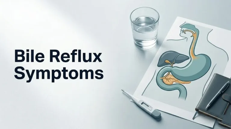
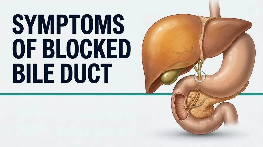
Understanding the anatomy clarifies why this condition produces the symptoms it does. The liver produces approximately 500 to 1,000 milliliters of bile per day, according to MedlinePlus of the U.S. National Library of Medicine. Hepatocytes (liver cells) secrete bile into microscopic intrahepatic bile ductules, which drain into the right and left hepatic ducts. These merge into the common hepatic duct, which joins the cystic duct (coming from the gallbladder) to form the common bile duct (also called the choledochus). The common bile duct passes through the head of the pancreas and enters the duodenum (the first segment of the small intestine) through the ampulla of Vater, regulated by the sphincter of Oddi.
Think of this system like a river with tributaries feeding into a main channel. A blockage anywhere along that channel stops the flow and forces the water back into the tributaries. The liver keeps producing bile, the ducts keep accumulating it, and the pressure builds until bile components force their way into the bloodstream. That backed-up bile is exactly what produces every symptom of a blocked bile duct.
The most common causes of blockage include choledocholithiasis (gallstones that have migrated into the common bile duct), biliary strictures from previous surgery or inflammation, malignancy (particularly cholangiocarcinoma or pancreatic cancer at the ampulla), and primary sclerosing cholangitis (PSC), a chronic inflammatory condition of the bile ducts. The cause determines how rapidly symptoms develop and how the obstruction progresses.
Bile Duct Symptoms: The Full Clinical Picture
The full clinical picture of bile duct symptoms spans a range from subtle early signs that are easily missed to a classic triad of findings that most clinicians can identify immediately. Not every patient develops every symptom, and the pattern your body produces depends on how much bile is being blocked and for how long.
The American College of Gastroenterology recognizes the core symptom complex of biliary obstruction as jaundice, pruritus, pale stools, and dark urine, collectively termed cholestatic symptoms, because they reflect the failure of normal cholestasis: the regulated flow of bile from liver to gut. These four symptoms share a common mechanism: bile components that would normally travel through the bile duct into the intestine are instead accumulating where they should not be.
Full symptom inventory with clinical significance:
- Jaundice: yellowing of skin and sclera (whites of the eyes), most reliably seen first in the sclera under natural light
- Pruritus: generalized itching without visible rash, often worse at night and on the palms and soles
- Dark urine: tea-colored or amber-brown, produced by conjugated bilirubin filtered through the kidneys
- Pale or acholic stools: clay-gray or putty-colored, from absence of stercobilin pigment
- Upper right quadrant abdominal pain: dull, cramping, or colicky depending on cause
- Nausea and vomiting: reflex response to biliary pressure and gut disruption
- Fever and chills: indicates bacterial contamination of the obstructed bile duct
- Fatigue: from impaired liver metabolic function and nutrient absorption
- Weight loss: from fat malabsorption in prolonged obstruction
- Easy bruising or bleeding: from vitamin K deficiency affecting coagulation factor synthesis
Patients with liver disease or cirrhosis already present with baseline jaundice, making new-onset worsening or a sudden change in stool color particularly important to report to their managing hepatologist or primary care physician promptly.
Key Takeaway: Pale stools and dark urine appearing together are a more specific paired indicator of bile duct obstruction than jaundice alone, because they reflect the same bile flow failure from two different directions simultaneously.
Jaundice as a Symptom of Bile Duct Blockage
Jaundice in the context of a blocked bile duct occurs when conjugated bilirubin, the water-soluble form of bilirubin processed by the liver, backs up into the bloodstream and deposits in the skin, subcutaneous tissue, and the sclera of the eyes, producing a yellow discoloration that ranges from faint lemon-yellow in mild cases to deep amber-orange in severe or prolonged obstruction.
The biochemical pathway is precise. Red blood cells are broken down continuously throughout life, releasing hemoglobin. The heme component of hemoglobin is converted by macrophages into unconjugated (indirect) bilirubin, which is fat-soluble and toxic to the nervous system at high levels. The liver’s hepatocytes take up unconjugated bilirubin and convert it to conjugated (direct) bilirubin using the enzyme UDP-glucuronosyltransferase, making it water-soluble and ready for excretion into the bile. When the bile duct is blocked, this conjugated bilirubin cannot exit into the gut. It accumulates in the liver and spills back into the bloodstream, then deposits in tissues with high collagen content, particularly the skin and the sclera of the eyes.
The sclera turns yellow before the skin does in most cases. This is because scleral icterus (yellowing of the eye whites) becomes visible at serum bilirubin levels of approximately 2.5 to 3 mg/dL, while skin jaundice typically requires levels above 4 to 5 mg/dL. Checking the sclera in natural daylight is the most sensitive bedside assessment for early jaundice.
According to the NIDDK, jaundice from biliary obstruction is classified as post-hepatic jaundice (also called obstructive jaundice) to distinguish it from hepatic jaundice (caused by liver cell dysfunction) and pre-hepatic jaundice (caused by excessive red blood cell breakdown). The distinction matters because the treatment pathway differs completely. Post-hepatic jaundice requires addressing the physical obstruction, not treating the liver cells directly.
Individuals with darker skin tones may not show visible skin jaundice until bilirubin levels are substantially elevated. The sclera, palate, and fingernail beds are the most reliable examination sites in darker-skinned individuals and should be the first areas assessed by both patients and clinicians.
Bile Blockage Symptoms: Pruritus, Dark Urine, and Pale Stools
The three symptoms that most reliably confirm bile is failing to reach the intestine are pruritus (cholestatic itching), dark amber urine, and pale or clay-colored stools, and each has a distinct biochemical explanation that tells you something different about the anatomy of the failure.
Cholestatic pruritus is one of the most distressing symptoms of bile duct blockage, affecting a substantial proportion of patients with obstructive jaundice. It is not caused simply by bile salts irritating the skin, though that older explanation still appears in some resources. More recent research, including studies published in the journal Gastroenterology, points to bile acids activating TGR5 receptors (bile acid-sensitive G protein-coupled receptors) on cutaneous sensory nerve endings, triggering itch signals that travel through unmyelinated C-fibers to the spinal cord. The itch is classically worst at night, worse on the palms and soles, and not accompanied by a visible rash or skin lesion. Scratching provides no lasting relief, and the itching can become so severe it disrupts sleep and impairs daily function.
Dark urine in bile duct blockage is caused by conjugated bilirubin, being water-soluble, passing through the glomeruli of the kidneys and appearing in urine. Normal urine contains no bilirubin. The presence of bilirubin turns urine a deep amber to brown color, sometimes described as resembling tea or cola. This symptom often appears at the same time as scleral icterus and may precede visible skin jaundice.
Pale or clay-colored stools result from the absence of bile in the intestine. Normally, conjugated bilirubin secreted into the gut is converted by intestinal bacteria into stercobilin, the pigment that gives stool its characteristic brown color. Without bile reaching the gut, stercobilin is not produced. Stools become pale gray, putty-colored, or clay-white.
Quick Tip:
- Photograph pale stools or dark urine to show your doctor: it helps establish when this change started and how pronounced it is
- The combination of dark urine and pale stools together is more clinically specific for obstructive disease than either finding alone
- Patients taking iron supplements may have darker stools unrelated to bile, so disclose all supplements to your primary care physician when describing stool color changes
Abdominal Pain and Nausea from Bile Duct Obstruction
Abdominal pain from bile duct obstruction is most commonly felt in the right upper quadrant of the abdomen, the area beneath the right ribcage where the liver and gallbladder sit, and its character ranges from a dull, constant ache in chronic obstruction to severe, cramping, wave-like colicky pain in acute gallstone obstruction.
The pain mechanism differs by cause. When a gallstone lodges in the common bile duct, the duct wall muscle contracts repeatedly in an attempt to push the stone through, producing biliary colic: a cramping, severe pain that builds rapidly to a peak and may radiate to the right shoulder or mid-back via referred pain pathways through the phrenic nerve and T7-T10 thoracic dermatomes. The pain of biliary colic typically lasts 30 minutes to several hours. When obstruction is from a stricture or malignancy, the pain is typically duller, more constant, and develops more gradually as the duct dilates slowly under accumulating pressure.
The Mayo Clinic notes that pain from common bile duct obstruction often worsens after fatty meals because food in the duodenum triggers cholecystokinin (CCK) release from the duodenal mucosa, which stimulates gallbladder contraction and bile duct peristalsis. When the duct is already obstructed, this increased pressure translates directly into increased pain.
Nausea and vomiting accompany the pain in many cases, particularly in acute gallstone obstruction. They result from a combination of the visceral pain reflex (the same autonomic reflex that causes nausea with severe pain from any organ) and the disruption of normal gut motility that bile flow interruption produces in the duodenum.
Pregnant women in the third trimester may experience bile duct-related right upper quadrant pain that is attributed to normal pregnancy discomfort or to the growing uterus displacing abdominal organs, delaying recognition of biliary pathology. Any right upper quadrant pain in pregnancy accompanied by jaundice or itching warrants evaluation by an obstetrician and gastroenterologist on the same day.
Dilated Bile Duct Symptoms
A dilated bile duct discovered on imaging does not always produce obvious symptoms in the early stages, and this is one of the most clinically underappreciated aspects of biliary obstruction: the duct can be significantly dilated on ultrasound while the patient reports only vague fatigue or mild right upper quadrant discomfort.
Bile duct dilation is the body’s physical response to sustained obstructive pressure. The normal common bile duct measures 6 to 8 millimeters in internal diameter in adults who have not had their gallbladder removed, and up to 10 millimeters in those who have. When obstruction occurs, bile backed up against the blockage distends the duct wall progressively. At mild dilation (8 to 10 mm with gallbladder intact), many patients have no symptoms or only subtle fatigue. At moderate dilation (10 to 15 mm), right upper quadrant fullness or discomfort often appears. At severe dilation above 15 mm, jaundice, pruritus, and dark urine are typically present.
According to a clinical review published in Gastroenterology, the presence of a dilated bile duct on abdominal ultrasound in a patient without prior biliary surgery is an indication for further investigation regardless of whether jaundice is present, because malignant causes of obstruction can produce significant ductal dilation before producing visible jaundice.
| Duct Diameter (adults with gallbladder) | Classification | Typical Symptoms | Recommended Action |
|---|---|---|---|
| Up to 6 mm | Normal | None | No action required |
| 6 to 8 mm | Borderline | Usually none; monitor if risk factors present | Follow-up imaging if symptomatic |
| 8 to 10 mm | Mild dilation | Possible fatigue, mild right upper quadrant fullness | Gastroenterologist evaluation |
| 10 to 15 mm | Moderate dilation | RUQ pain, possible early jaundice | Prompt gastroenterologist evaluation; MRCP |
| Above 15 mm | Severe dilation | Jaundice, pruritus, dark urine likely present | Urgent evaluation; rule out malignancy |
Patients who have had their gallbladder removed (cholecystectomy) normally have a somewhat wider common bile duct as the duct takes over a compensatory storage role, so the upper limit of normal in post-cholecystectomy patients is approximately 10 mm. A radiologist interpreting the imaging should adjust interpretation accordingly.
Key Takeaway: A dilated bile duct found incidentally on an ultrasound ordered for unrelated reasons is never a finding to ignore, even without jaundice, because malignant obstruction can produce significant dilation before causing visible yellowing.
Partial Versus Complete Bile Duct Blockage Symptoms
Partial bile duct blockage and complete bile duct blockage produce distinctly different symptom patterns, and distinguishing between them helps predict how rapidly the clinical situation will progress and how urgently treatment is needed.
In partial obstruction, some bile continues to flow past the blockage into the duodenum. Jaundice may be mild or intermittent. Stools may be lighter than normal but not fully pale or clay-colored. Dark urine may be present but not deeply amber. Pruritus may be mild and intermittent. The patient often describes symptoms that come and go, improving somewhat after certain meals or body positions. This fluctuating pattern is particularly characteristic of a gallstone that partially blocks the common bile duct but shifts position intermittently, allowing some bile flow at certain times.
Complete obstruction eliminates all bile flow to the intestine. Jaundice develops rapidly and deepens progressively. Stools become fully acholic (clay-colored) within a few days. Dark urine is pronounced. Pruritus can become severe within a week. Biochemical liver tests show progressively rising alkaline phosphatase (ALP), gamma-glutamyl transferase (GGT), and direct bilirubin. Coagulation can begin to be affected within days to weeks as vitamin K absorption fails.
The American College of Gastroenterology guidelines emphasize that fluctuating symptoms in biliary disease do not indicate a benign or self-resolving condition. Intermittent partial obstruction from a common bile duct gallstone can progress to complete obstruction without warning, and the same fluctuating pattern can be seen in cholangiocarcinoma as tumor growth alternately compresses and partially releases the duct.
Patients with partial obstruction from known common bile duct gallstones who experience a sudden worsening of symptoms, particularly new-onset fever or shaking chills, should be evaluated in an emergency department, not a scheduled outpatient appointment, because this transition pattern is consistent with the onset of acute bacterial cholangitis.
Bile Duct Injury Symptoms
Bile duct injury symptoms represent a specific and clinically distinct presentation of biliary pathology that most online health resources fail to address adequately: injury to the bile duct itself, most commonly as a complication of laparoscopic cholecystectomy (gallbladder removal surgery), produces a different symptom onset pattern from obstruction caused by a gallstone or tumor.
According to the American College of Gastroenterology, bile duct injury occurs in approximately 0.3 to 0.5 percent of laparoscopic cholecystectomy procedures, making it the most common serious complication of one of the most frequently performed abdominal surgeries in the United States. When the common bile duct or a hepatic duct is clipped, cut, or cauterized during surgery, bile can leak into the abdominal cavity or a stricture can form that progressively narrows the duct over weeks to months.
Symptoms of acute bile duct injury presenting within days of surgery include:
- Right upper quadrant or generalized abdominal pain that is increasing rather than improving after surgery
- Abdominal distension and tenderness from bile accumulating in the peritoneal cavity (bile peritonitis)
- Fever, often starting 3 to 5 days post-operatively
- Nausea and reduced appetite persisting beyond the expected recovery window
- Jaundice developing days after a surgery where it was not present before
Symptoms of delayed bile duct stricture, developing weeks to months after surgery, include:
- Gradually increasing jaundice with pruritus
- Recurrent bouts of right upper quadrant pain
- Episodic fever and chills from cholangitis developing above the stricture
Any patient who develops jaundice, fever, or worsening abdominal pain in the days to weeks following cholecystectomy or any upper abdominal surgery should contact their surgeon immediately and be evaluated in an emergency department if fever and jaundice are both present. The managing surgeon or a board-certified hepatobiliary surgeon should coordinate further evaluation.
Symptoms of Blocked Bile Duct in Females and During Pregnancy
The symptoms of a blocked bile duct follow the same general pattern in females as in males, but females face a distinctly higher prevalence of the most common cause (gallstone-related obstruction) and a unique pregnancy-related form of bile duct dysfunction that presents differently from any other biliary condition.
Choledocholithiasis (gallstones in the common bile duct) is substantially more prevalent in females. The NIDDK reports that gallstones are approximately twice as common in women as in men, driven by estrogen’s effect on increasing cholesterol secretion into bile (raising bile cholesterol saturation and promoting stone formation) and progesterone’s effect on slowing gallbladder emptying (allowing bile to stagnate and nucleate crystals). Females between ages 40 and 60 with prior pregnancies and obesity carry the highest prevalence risk. Their bile duct obstruction symptoms follow the classic pattern of jaundice, dark urine, pale stools, and right upper quadrant pain, often with a history of prior gallbladder attacks.
Intrahepatic cholestasis of pregnancy (ICP) is a separate and unique condition in which bile flow within the liver becomes impaired during pregnancy, typically in the third trimester, without a physical bile duct obstruction. The NIDDK estimates ICP affects approximately 1 to 2 percent of pregnancies in the United States, though rates are higher in women of South American descent (up to 5 to 15 percent in certain Chilean populations) and women carrying multiple fetuses. ICP produces severe pruritus, particularly on the palms and soles, that is disproportionately intense relative to the degree of jaundice (which may be mild or absent early). Elevated serum bile acids, not just bilirubin, are the defining biochemical marker.
ICP carries fetal risk: elevated maternal bile acids are associated with an increased risk of stillbirth, preterm birth, and fetal distress, according to research published in Gastroenterology. Pregnant women experiencing intense generalized itching, particularly on the palms and soles at night, should be evaluated by their obstetrician and a gastroenterologist without delay.
Key Takeaway: Intense nighttime palm and sole itching in a pregnant woman in the third trimester is intrahepatic cholestasis of pregnancy until proven otherwise, and it carries fetal risks that make same-day obstetric evaluation non-negotiable.
Bile Duct Obstruction Symptoms in Elderly Patients
Elderly patients, generally defined as those over age 65, present with bile duct obstruction symptoms that are frequently blunted, atypical, or attributed to other pre-existing conditions, which delays diagnosis and increases complication risk in this age group.
The classical symptom triad of jaundice, right upper quadrant pain, and fever may not be fully present in older patients. Pain perception is often reduced in elderly individuals due to age-related changes in visceral nociception (pain signaling from internal organs). An elderly patient with a common bile duct gallstone may report only mild fatigue, loss of appetite, or confusion rather than the severe colicky pain that would drive a younger adult to seek evaluation immediately. Jaundice may be less visually prominent if age-related skin changes have altered baseline skin tone.
The most clinically important concern in elderly patients is malignant bile duct obstruction. Cholangiocarcinoma (bile duct cancer) and pancreatic cancer invading or compressing the bile duct at the ampulla of Vater occur with substantially higher frequency in adults over 65. Both conditions tend to produce painless, progressive jaundice as their presenting feature, without the colicky pain pattern associated with gallstone obstruction. The classic teaching for painless jaundice in an elderly patient is to assume malignant obstruction until imaging proves otherwise.
According to the American Association for the Study of Liver Diseases (AASLD), older adults are also at higher risk for acute cholangitis complications including septic shock, partly because they may not develop the full fever and rigors that would otherwise signal the emergency. An elderly patient with new jaundice, mild confusion, and low blood pressure may be in septic cholangitis without presenting the textbook fever and shaking chills that younger patients display.
Elderly patients on anticoagulant medications such as warfarin or direct oral anticoagulants (DOACs) face compounded bleeding risk if chronic bile duct obstruction develops, because vitamin K deficiency from impaired fat absorption combines with anticoagulant therapy to suppress coagulation factor synthesis further. Their managing physician should monitor coagulation parameters carefully alongside biliary function tests.
Acute Cholangitis: The Emergency Presentation of Bile Duct Blockage
Acute bacterial cholangitis is the most dangerous complication of bile duct obstruction and represents a medical emergency that requires hospital admission, intravenous antibiotics, and bile duct drainage, typically within hours of recognition.
Cholangitis occurs when bacteria colonize bile that has become stagnant behind an obstruction. Normally, flowing bile carries bacteria out of the biliary system into the gut. When flow is blocked, bacteria, most commonly Escherichia coli, Klebsiella pneumoniae, and Enterococcus species from the gut, ascend into the biliary tree and proliferate rapidly in the stagnant bile. The infected bile is under pressure from the upstream obstruction, which drives bacteria directly into the hepatic sinusoids and from there into the systemic circulation, producing bacteremia and potentially septic shock within hours.
Charcot’s triad, named after the 19th-century neurologist Jean-Martin Charcot who first described it in the surgical context, refers to the three classic findings of acute cholangitis:
- Right upper quadrant pain (from obstructed, infected bile duct distension)
- Fever with rigors (violent shaking chills, from bacteremia and systemic inflammatory response)
- Jaundice (from the underlying biliary obstruction)
According to clinical guidelines published by the American College of Gastroenterology, all three features of Charcot’s triad are present in only approximately 50 to 70 percent of acute cholangitis cases, which means the absence of one feature does not rule out the diagnosis. Any two of the three findings in a patient with known or suspected biliary disease warrant emergency evaluation.
Reynolds’ pentad adds two additional findings that indicate the condition has progressed to septic shock: hypotension (low blood pressure) and altered mental status (confusion, disorientation, or reduced responsiveness). The presence of Reynolds’ pentad indicates a life-threatening emergency with a mortality rate that rises rapidly without immediate intervention.
Immunocompromised patients, including those receiving chemotherapy, long-term corticosteroids, or living with HIV, may not mount the expected fever response even in the presence of severe cholangitis. In these patients, confusion, low blood pressure, and deteriorating liver function tests may be the only early signs of a condition that is progressing to septic shock.
Bile Duct Symptoms After Gallbladder Removal
Bile duct symptoms after gallbladder removal, a condition encountered frequently enough that it has its own clinical designation, post-cholecystectomy syndrome, are experienced by a meaningful proportion of the approximately 750,000 Americans who undergo laparoscopic cholecystectomy each year, according to the NIDDK.
The gallbladder’s removal does not eliminate the bile duct system. The liver continues to produce bile, which flows continuously through the hepatic ducts, the common hepatic duct, and the common bile duct into the duodenum. After cholecystectomy, there is no gallbladder reservoir to store and concentrate bile between meals, which changes the dynamics of bile flow but does not create obstruction under normal circumstances.
Symptoms that develop after gallbladder removal and resemble biliary obstruction can result from several distinct causes:
- Retained common bile duct stone: A gallstone that was already in the common bile duct at the time of surgery but was missed. This is the most common cause of jaundice appearing shortly after cholecystectomy. It produces the classic symptom pattern of jaundice, dark urine, pale stools, and right upper quadrant pain.
- Bile duct stricture: Progressive narrowing of the common bile duct at the surgical clip site, anastomosis, or injury point, producing gradually developing cholestatic symptoms weeks to months after surgery.
- Bile leak: Bile leaking from a divided cystic duct stump or an injured duct, causing abdominal pain, fever, and sometimes jaundice from bile peritonitis.
- Sphincter of Oddi dysfunction: Abnormal contraction or spasm of the sphincter controlling bile entry into the duodenum, producing recurrent episodes of right upper quadrant pain that mimic the original gallbladder attacks.
Any patient who has undergone cholecystectomy and develops new jaundice, fever, or worsening right upper quadrant pain should contact their surgeon or gastroenterologist immediately, not assume the symptoms are expected post-surgical discomfort. New-onset jaundice after gallbladder removal is not normal and requires same-day evaluation.
Key Takeaway: New jaundice, fever, or worsening abdominal pain after gallbladder removal is not expected post-surgical discomfort. It is a clinical sign that requires same-day contact with a surgeon or gastroenterologist, not watchful waiting.
Chronic Bile Duct Blockage Symptoms and Fat Malabsorption
Chronic bile duct blockage produces a second tier of symptoms beyond the acute jaundice and pruritus picture, driven by months of impaired bile flow into the intestine disrupting fat digestion and the absorption of fat-soluble vitamins A, D, E, and K.
Bile acids, secreted into the duodenum during normal digestion, form micelles: tiny spherical structures that surround dietary fat molecules and fat-soluble vitamins, allowing them to be absorbed through the intestinal wall. Without adequate bile acids reaching the gut in chronic obstruction, dietary fat passes largely undigested through the intestine. The clinical result is steatorrhea: loose, foul-smelling, oily or greasy stools that float in the toilet because of their high fat content. This is distinct from the pale, clay-colored acholic stools of acute obstruction, though both can occur in severe chronic cases.
The consequences of fat-soluble vitamin deficiency develop progressively:
| Vitamin | Function Impaired by Deficiency | Clinical Symptom Produced |
|---|---|---|
| Vitamin A | Night vision, immune function, epithelial integrity | Night blindness, dry eyes, increased infection susceptibility |
| Vitamin D | Calcium absorption, bone mineralization | Bone pain, osteomalacia, muscle weakness |
| Vitamin E | Antioxidant protection of nerve membranes | Peripheral neuropathy, balance problems, poor reflexes |
| Vitamin K | Coagulation factor II, VII, IX, X synthesis in the liver | Easy bruising, prolonged bleeding, elevated INR |
Vitamin K deficiency is the most immediately dangerous of these in the context of chronic biliary obstruction. Coagulation factors II (prothrombin), VII, IX, and X all require vitamin K as a cofactor for their activation in the liver. Deficiency prolongs the prothrombin time (PT) and international normalized ratio (INR), creating a coagulopathy that can make even minor injuries bleed excessively and makes any necessary surgical or endoscopic procedure significantly higher risk.
Patients with chronic bile duct obstruction from primary sclerosing cholangitis or malignancy who are on anticoagulant medications face the greatest coagulopathy risk. Their managing gastroenterologist or hepatologist should monitor prothrombin time alongside liver function tests at every follow-up visit.
How Is a Blocked Bile Duct Diagnosed
A blocked bile duct is diagnosed through a combination of blood tests that identify specific liver enzyme and bilirubin elevation patterns, followed by imaging studies that visualize the bile ducts directly to locate the obstruction and identify its cause.
The diagnostic process typically unfolds as follows:
- Initial blood tests (liver function panel): Elevated alkaline phosphatase (ALP) and gamma-glutamyl transferase (GGT) are the most sensitive early blood markers of biliary obstruction, often rising before bilirubin becomes elevated enough to produce visible jaundice. Total and direct bilirubin rise as obstruction progresses. Alanine aminotransferase (ALT) and aspartate aminotransferase (AST) may be elevated but are typically less disproportionately raised than in liver cell-specific disease. A complete blood count (CBC) showing elevated white blood cell count suggests cholangitis.
- Abdominal ultrasound (first-line imaging): The initial imaging study for suspected bile duct obstruction. Ultrasound identifies ductal dilation reliably and can identify gallstones in the gallbladder and common bile duct in many cases. It is non-invasive, widely available, and performed without radiation. Its limitation is that the distal common bile duct is often obscured by bowel gas.
- Magnetic resonance cholangiopancreatography (MRCP): The most detailed non-invasive imaging study for the biliary tree. MRCP produces high-resolution three-dimensional images of the entire bile duct system, identifying strictures, stones, and tumors with high accuracy without radiation and without dye injection. It is the preferred follow-up study when ultrasound confirms dilation but does not define the cause.
- Endoscopic retrograde cholangiopancreatography (ERCP): Both a diagnostic and therapeutic procedure performed by a board-certified gastroenterologist or interventional endoscopist. A flexible endoscope is passed through the mouth to the ampulla of Vater, where contrast dye is injected into the bile ducts under X-ray. ERCP can directly visualize the obstruction, obtain tissue biopsies, and in the same procedure remove gallstones, dilate strictures, or place stents to restore bile flow.
- Endoscopic ultrasound (EUS) and CT scan: Used when MRCP findings are inconclusive or when malignancy staging is needed. EUS allows high-resolution imaging adjacent to the bile ducts from inside the gut. CT with contrast defines the extent of any malignant mass and assesses vascular involvement.
The board-certified gastroenterologist coordinating this diagnostic sequence will determine which combination of tests is appropriate based on the severity of symptoms, the presumed cause, and the patient’s overall health status. Pregnant patients require radiation-free imaging such as MRCP whenever possible; ERCP in pregnancy should be performed only when therapeutically necessary and with appropriate radiation shielding, in consultation with the managing obstetrician.
Emergency Symptoms: When to Call 911 or Go to the ER
Certain symptoms associated with a blocked bile duct require immediate emergency evaluation. Do not wait to see if these resolve on their own.
Call 911 or go to the nearest emergency room immediately if you experience:
- Fever above 38.5 degrees Celsius (101.3 degrees Fahrenheit) combined with jaundice and right upper quadrant pain: This is Charcot’s triad, the defining clinical presentation of acute bacterial cholangitis. Bacteria are actively entering the bloodstream from the infected bile duct. This is not an outpatient appointment situation.
- Rigors (violent, uncontrollable shaking chills) with or without high fever in the context of jaundice: Rigors indicate bacteremia and systemic sepsis response. The bile duct infection has spread beyond the biliary system.
- Confusion, disorientation, or unusual drowsiness combined with jaundice or known bile duct disease: These neurological signs combined with biliary obstruction indicate Reynolds’ pentad, meaning septic shock is developing. This is a surgical and critical care emergency.
- Low blood pressure (feeling faint, lightheaded, or collapsing) in the setting of jaundice or abdominal pain: Hypotension in the context of biliary disease signals septic shock from ascending cholangitis. Mortality rises rapidly without emergency drainage.
- Severe right upper quadrant pain that is constant, worsening, and not relieved by position changes, lasting more than 6 hours: This pattern, particularly if accompanied by tenderness on physical examination over the gallbladder area (Murphy’s sign) or in the epigastrium, may indicate acute cholecystitis, bile duct obstruction with cholangitis, or pancreatitis from an impacted common bile duct stone at the ampulla of Vater.
- Jaundice appearing in the days or weeks after abdominal surgery, particularly gallbladder removal: This is bile duct injury or retained stone until proven otherwise and requires emergency surgical evaluation.
- Worsening jaundice with new-onset confusion in an elderly patient: In older adults, altered mental status may be the only prominent feature of septic cholangitis. Do not wait for fever to appear.
These presentations can indicate acute bacterial cholangitis or biliary sepsis, conditions that require emergency intravenous antibiotics and biliary drainage within hours of recognition, not an urgent care appointment or a scheduled outpatient referral.
Frequently Asked Questions About Blocked Bile Duct Symptoms
What are the first signs of a blocked bile duct?
The first signs of a blocked bile duct are often dark urine and scleral icterus (yellowing of the whites of the eyes), which appear before skin jaundice becomes visible because the sclera changes color at lower serum bilirubin levels than the skin.
Mild right upper quadrant discomfort, fatigue, and lighter-than-normal stool color may accompany these early findings, particularly in gradual-onset obstruction from a stricture or tumor.
Any new yellowing of the eyes or skin warrants same-day evaluation by a primary care physician regardless of whether other symptoms are present.
Can a blocked bile duct clear on its own?
A blocked bile duct caused by a gallstone can occasionally shift position and temporarily allow bile flow to resume, producing an apparent improvement in symptoms, but this does not represent resolution of the underlying problem.
The stone remains in the common bile duct and can re-obstruct at any time, including progressing to acute cholangitis, which is why even a temporary improvement does not mean medical evaluation can be deferred.
Obstruction from a stricture, malignancy, or bile duct injury does not clear without medical or surgical intervention, and delaying evaluation in these cases allows the obstruction and its complications to progress.
What does bile duct pain feel like and where is it located?
Bile duct pain is most commonly felt in the right upper quadrant of the abdomen, under the right ribcage, and frequently radiates to the right shoulder or mid-back through referred pain pathways via the phrenic nerve and thoracic dermatomes T7 through T10.
In acute gallstone obstruction, the pain is colicky: it builds in waves, peaks in severity, and may last 30 minutes to several hours before subsiding.
In chronic obstruction from stricture or malignancy, the pain is typically duller, more constant, and felt as a pressure or fullness rather than a cramping sensation.
What does it mean if my bile duct is dilated on an ultrasound?
A dilated bile duct on ultrasound means the duct has widened beyond its normal diameter, which is approximately 6 to 8 millimeters in adults who still have their gallbladder, indicating that bile flow is being impeded somewhere downstream.
The finding alone does not identify the cause of the obstruction: a gallstone, a stricture, a malignant tumor, or sphincter of Oddi dysfunction can all produce ductal dilation on imaging.
A board-certified gastroenterologist will typically order magnetic resonance cholangiopancreatography (MRCP) as the next step to visualize the full biliary tree and identify the specific site and cause of the obstruction.
How do blocked bile duct symptoms differ from gallbladder symptoms?
Gallbladder symptoms, particularly biliary colic from gallstones in the gallbladder itself, typically produce pain that resolves completely between episodes and is not accompanied by jaundice or dark urine unless a stone has migrated into the common bile duct.
Blocked bile duct symptoms include jaundice, dark urine, and pale stools in addition to abdominal pain, because only when the common bile duct is obstructed does bile fail to reach the intestine.
Both conditions can cause right upper quadrant pain and nausea, which is why imaging (ultrasound and MRCP) rather than symptom description alone is required to determine whether the pathology is in the gallbladder, the bile duct, or both.
How quickly do bile duct blockage symptoms progress to an emergency?
A bile duct blockage caused by an acute gallstone can progress from initial symptoms to acute bacterial cholangitis in as little as 12 to 24 hours if bacteria colonize the stagnant bile, which is why fever developing alongside jaundice and abdominal pain demands emergency room evaluation the same day.
Obstruction from a slowly growing tumor or stricture typically progresses over weeks to months, but even gradual obstruction can cause sudden cholangitis if secondary infection develops.
According to the American College of Gastroenterology, patients who present with the full Charcot’s triad of fever, jaundice, and right upper quadrant pain have a condition requiring hospitalization and biliary drainage within hours, not watchful waiting.
Closing
The symptom pattern of a blocked bile duct, taken together, tells a coherent story: bile that should be flowing from the liver into the intestine is backing up into the bloodstream and tissues. Yellowing eyes, amber urine, pale stools, and intense nighttime itching are not random symptoms. They are the direct biochemical consequence of that backup, and each one points to the same underlying failure.
If you are experiencing any combination of these symptoms, the next step is not to wait and observe. New-onset jaundice in any adult warrants same-day contact with a primary care physician. Jaundice with fever requires an emergency department visit, not an appointment.
The distinction that matters most from this article: fever plus jaundice plus right-sided abdominal pain is Charcot’s triad, and it is an emergency. If that combination is present, your destination is the nearest emergency room, and your timing is right now.