UCL Tear Symptoms: Elbow Signs, Causes, and When to Act
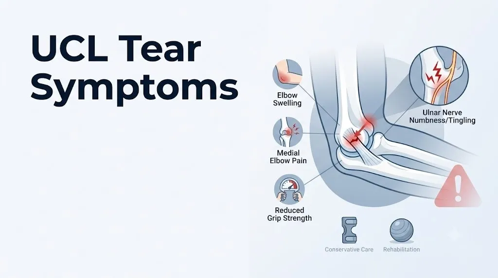
UCL tear symptoms typically begin with medial elbow pain during or immediately after a throwing motion or forceful overhead movement, and they can range from a mild aching discomfort that builds over a season to a sudden, sharp pop at the inner elbow that stops an athlete mid-pitch. The specific symptoms you experience depend on whether the tear is partial or complete, whether it happened acutely or developed through chronic overuse, and whether the adjacent ulnar nerve is involved.
The American Orthopaedic Society for Sports Medicine (AOSSM) reports that UCL reconstruction rates have increased dramatically over the past two decades, with estimates suggesting that one in three Major League Baseball pitchers has undergone the procedure. Research published in the American Journal of Sports Medicine documents that peak valgus torque during the baseball pitching motion can reach 64 Newton-meters at the medial elbow, a load that approaches or exceeds the tensile failure threshold of the UCL in some throwers.
This article covers the complete clinical picture: the anatomy that makes the UCL vulnerable, what each specific symptom means mechanistically, how partial and complete tears differ, how youth athletes present differently from adults, which symptoms point to nerve involvement, and which presentations require immediate medical attention rather than watchful waiting.
UCL Tear Symptoms: What Your Elbow Is Telling You
UCL tear symptoms are the body’s response to structural failure or progressive stress damage to the ulnar collateral ligament, the primary medial stabilizer of the elbow joint that runs from the medial epicondyle of the humerus to the sublime tubercle of the coronoid process of the ulna.
The UCL is not a single cord. It is a complex of three distinct anatomical bands: the anterior oblique band, the posterior oblique band, and the transverse band. The anterior oblique band carries roughly 54% of valgus resistance at 90 degrees of elbow flexion, the position at which peak throwing stress occurs, according to biomechanical cadaveric research published in the American Journal of Sports Medicine. When this specific band tears, the elbow loses its primary check against medial opening, and every subsequent throwing motion drives the joint toward abnormal gapping.
Think of the anterior oblique band like the main cable of a suspension bridge. The bridge can handle normal traffic with all cables intact. When the main cable frays, the structure doesn’t collapse immediately, but every vehicle that crosses adds stress to the remaining cables. That’s exactly how partial UCL tears work: throwing continues, compensation increases, symptoms worsen progressively, until the point of functional failure.
The resulting symptom pattern reflects which structures bear the abnormal load after the UCL fails to do its job.
Core UCL tear symptoms at a glance:
- Medial (inner) elbow pain during or immediately after throwing
- A sudden pop at the medial elbow at the moment of acute rupture (present in some but not all cases)
- Decreased throwing velocity or distance without obvious reason
- Loss of control or accuracy in overhead athletes
- Elbow stiffness, particularly in the morning or after rest
- Numbness or tingling in the ring and small fingers from ulnar nerve involvement
- Reduced grip strength on the affected side
- Swelling or tenderness directly over the medial epicondyle and the UCL’s path
What Is a UCL Tear and How Does It Happen
A UCL tear is a disruption of the fibers of the ulnar collateral ligament of the elbow, caused either by a single high-force valgus loading event or by the cumulative effect of repetitive submaximal valgus stress that exceeds the ligament’s repair capacity over time.
The anatomy is precise and worth understanding. The UCL originates on the anterior-inferior surface of the medial epicondyle of the humerus, the bony prominence on the inner side of the elbow that you can feel with your fingertip. It inserts on the sublime tubercle of the coronoid process of the ulna, approximately 18 to 20 millimeters distal to the joint line. The anterior oblique band, which is the clinically relevant portion in the vast majority of throwing athlete UCL injuries, is taut throughout elbow flexion from 30 to 120 degrees and provides the primary resistance against the valgus force generated during overhead throwing.
The late cocking and early acceleration phases of the baseball pitching motion generate the highest valgus torque at the medial elbow. During these phases, the shoulder is externally rotated and the elbow is near 90 degrees of flexion, placing maximum tension on the anterior oblique band simultaneously. Repeated exposure to these forces, across hundreds or thousands of pitches over a season or career, produces progressive microtearing and collagen fiber disruption that collectively attenuate the ligament before any single pitch causes visible failure.
Acute complete rupture without a chronic background is less common. It typically occurs in a non-throwing context: a fall on an outstretched or outstretched-and-abducted arm, a direct blow to the outer elbow that forces the joint into valgus, or a forceful twisting motion at the wrist that transmits valgus force up the forearm.
Common mechanisms of UCL tear by context:
| Mechanism | Population Affected | Typical Tear Pattern |
|---|---|---|
| Repetitive overhead throwing | Baseball, softball pitchers, javelin throwers | Chronic attenuation, partial or complete |
| Single high-force pitch | Any throwing athlete | Acute complete rupture with pop |
| Fall on outstretched hand | Recreational athletes, gymnasts | Acute valgus stress rupture |
| Direct elbow trauma | Contact sport athletes | Acute complete or partial tear |
| Volleyball overhead smash | Overhead non-throwing athletes | Chronic partial, less common acute |
Torn UCL Symptoms: The Full Clinical Picture
Torn UCL symptoms in a throwing athlete typically include a constellation of medial elbow pain, functional throwing deficit, and sometimes ulnar nerve involvement, and the full clinical picture is distinctly different from the isolated medial elbow tenderness seen in conditions like medial epicondylitis.
The most consistent symptom reported by athletes with confirmed UCL tears is pain at the medial elbow during the late cocking or early acceleration phase of throwing. This timing is not random. Research published in Clinics in Sports Medicine confirms that these phases correspond precisely to peak valgus torque at the elbow, meaning pain occurs when the damaged ligament is under maximum tensile load. Pain that occurs only during this specific phase, rather than throughout the entire throwing motion or at rest, is a clinically distinguishing feature of UCL insufficiency.
In acute complete ruptures, a sharp, audible or palpable pop at the inner elbow is present in approximately 35% to 50% of cases according to clinical series reviewed by the American Academy of Orthopaedic Surgeons (AAOS). The absence of a pop does not rule out a complete tear, particularly in the setting of chronic attenuation where the ligament fails gradually rather than suddenly.
According to the National Institute of Arthritis and Musculoskeletal and Skin Diseases (NIAMS), ligament injuries that disrupt more than 50% of the fiber cross-section are associated with measurable joint instability on physical examination, while partial tears involving less than 50% of fiber cross-section may produce pain and functional deficit without detectable laxity on a single static valgus stress test.
Full symptom inventory with clinical mechanism:
| Symptom | Anatomical Mechanism | Clinical Significance |
|---|---|---|
| Medial elbow pain during throwing | Anterior oblique band fiber disruption, nociceptor sensitization | Primary presenting symptom, phase-specific |
| Audible or felt pop | Acute complete fiber rupture | Indicates acute complete tear in ~35-50% of cases |
| Decreased velocity | Loss of valgus restraint, compensatory muscle guarding | Functional marker, often precedes pain in chronic cases |
| Elbow instability or looseness | Abnormal medial joint gapping (valgus instability) | Detected by valgus stress test at 30 degrees flexion |
| Ring and small finger numbness | Ulnar nerve traction in cubital tunnel | Indicates concurrent nerve involvement |
| Grip weakness | Ulnar nerve motor deficit or pain inhibition | Requires nerve assessment to distinguish causes |
| Medial elbow swelling | Inflammatory edema from capsule and ligament injury | Acute tear sign; less prominent in chronic attenuation |
Inner Elbow Pain From a UCL Tear
Inner elbow pain from a UCL tear originates at the medial epicondyle and travels along the course of the anterior oblique band toward the proximal ulna, producing a localized, deep aching pain that is distinctly different from the diffuse tenderness of a muscle strain.
The pain generator in a UCL tear is primarily the disrupted anterior oblique band itself and the surrounding joint capsule. Ligament tissue contains free nerve endings that respond to mechanical deformation and the local release of prostaglandin E2 and substance P from damaged collagen fibers. When the torn fibers are loaded during throwing, these nociceptors fire in proportion to the tensile force applied. This explains why the pain is phase-specific: it peaks during late cocking and early acceleration, eases between pitches, and may be absent entirely at rest in partial tears.
Pinpointing the location of tenderness helps differentiate UCL pathology from two conditions that produce overlapping medial elbow pain. Medial epicondylitis (golfer’s elbow) produces tenderness directly over the common flexor tendon origin at the anterior surface of the medial epicondyle, approximately 5 to 10 millimeters proximal and anterior to where the UCL inserts. A UCL tear produces maximum tenderness 5 to 10 millimeters distal to the medial epicondyle, along the course of the anterior oblique band toward the sublime tubercle. A trained examiner can reliably distinguish these two locations with a single-finger palpation test.
To accurately describe inner elbow pain to a sports medicine physician or orthopedic surgeon:
- Point to the exact location of maximum tenderness with one fingertip, starting at the medial epicondyle and moving distally.
- Report whether the pain occurs specifically during the late cocking and early acceleration phases or throughout the entire throwing motion.
- Note whether the pain began during a specific pitch or game or developed gradually over a season.
- Rate the pain on a scale of 0 to 10 at rest, during daily activities, and during throwing.
- Report any associated clicking, locking, or giving-way sensation at the elbow, which may indicate concurrent loose body formation or posteromedial impingement.
Athletes with concurrent valgus extension overload syndrome (posterior impingement at the posteromedial olecranon) may report pain at both the medial and posterior elbow, a pattern that indicates a more extensive injury complex requiring MRI evaluation to clarify.
Key Takeaway: UCL inner elbow pain is phase-specific during throwing, maximal 5 to 10 millimeters distal to the medial epicondyle along the anterior oblique band’s course, and distinctly different in location from golfer’s elbow, a distinction a sports medicine physician can confirm with targeted palpation.
UCL Tear Instability and Grip Weakness
UCL tear instability is the physical sensation that the elbow is about to give way under valgus load, and it reflects the loss of the anterior oblique band’s restraining function against medial joint gapping.
In an intact elbow, the anterior oblique band prevents the medial articular surface of the ulna from moving away from the medial trochlea of the humerus during valgus stress. When the band tears completely, the medial joint space opens abnormally under load, a phenomenon measurable on a valgus stress radiograph or musculoskeletal ultrasound stress test. Clinically, athletes describe this as a feeling that the elbow is “loose,” “wobbly,” or “going to give out” during the acceleration phase of throwing, even before severe pain develops.
Grip weakness in UCL tears has two distinct causes, and distinguishing between them is clinically important. The first cause is pain inhibition: the brain reduces voluntary motor output to the forearm and hand musculature to protect the injured medial elbow from further loading during gripping and wrist flexion, which engage the flexor-pronator mass and transmit force through the UCL attachment zone. The second cause is true motor weakness from ulnar nerve compromise, where the ulnar nerve’s traction or compression within the cubital tunnel impairs motor signaling to the intrinsic hand muscles (the hypothenar muscles, interossei, and medial two lumbricals), producing genuine muscle weakness independent of pain.
A clinician distinguishes between these two by testing grip strength with the elbow in extension (reducing UCL tension and pain) versus flexion (maximizing UCL and cubital tunnel stress). Pain-inhibited grip weakness improves substantially in extension. True ulnar motor weakness does not.
Signs of UCL instability worth reporting to a provider:
- A catching, clicking, or giving-way sensation at the inner elbow during overhead activity
- The sense that the throwing arm “doesn’t have the same power” without obvious pain as the reason
- Difficulty generating force during gripping tasks such as opening jars or carrying bags
- Progressive loss of throwing accuracy without any single identifiable injury event
Older recreational athletes with concurrent elbow osteoarthritis may have baseline grip weakness from osteophyte formation at the humeroradial joint. A UCL tear layered on top of pre-existing elbow arthritis can produce a more severe and difficult-to-interpret instability picture, requiring MRI arthrogram evaluation rather than standard MRI.
Swelling and Bruising After a UCL Tear
Swelling after a UCL tear is produced by the acute inflammatory response to collagen fiber disruption at the medial elbow, and its presence, location, and timing tell an examiner something specific about the nature of the injury.
Acute complete UCL ruptures cause rapid release of intracellular fluid, blood (hemarthrosis within the elbow joint or periligamentous hematoma in the soft tissues), and inflammatory mediators including TNF-alpha and IL-1 beta from the torn ligament fibers. This produces visible swelling at the medial elbow that typically develops within the first 30 to 60 minutes of an acute injury. The swelling concentrates directly over the UCL’s course, from the medial epicondyle to approximately 2 centimeters distally, and is associated with warmth and tenderness to light touch.
Bruising (ecchymosis) from acute UCL tears follows the path of least resistance through the subcutaneous tissue. It may appear at the medial elbow initially, then track distally along the forearm over 24 to 48 hours. Bruising visible below the elbow crease or extending toward the wrist after an acute elbow injury should prompt evaluation to rule out concurrent avulsion fracture at the medial epicondyle, where a bone fragment may have been pulled away by the intact UCL during the injury event.
Chronic UCL attenuation, by contrast, produces minimal or no acute swelling. The ligament fails gradually through repetitive microtrauma, and the inflammatory response is low-grade rather than acute. Athletes with chronic UCL injury often report no visible swelling but persistent diffuse medial elbow puffiness that is most noticeable in the morning.
Quick Tip:
- Ice applied to the medial elbow within 20 to 30 minutes of an acute injury can reduce early edema formation by causing vasoconstriction in the periligamentous capillaries. Apply for 15 to 20 minutes maximum, never directly on skin.
- Swelling that does not begin to decrease within 48 to 72 hours of rest and ice warrants evaluation by a primary care sports medicine physician.
- For skeletally immature athletes (under age 16), any acute medial elbow swelling after throwing requires same-day or next-day evaluation to rule out medial epicondyle apophyseal avulsion fracture, which can have a similar appearance to a UCL sprain clinically.
UCL Tear and Ulnar Nerve Symptoms
UCL tear ulnar nerve symptoms occur when the abnormal valgus instability produced by a torn UCL repeatedly stretches the ulnar nerve across the floor of the cubital tunnel, the fibro-osseous channel posterior to the medial epicondyle through which the ulnar nerve travels as it passes from the upper arm to the forearm.
The posterior oblique band of the UCL forms a portion of the cubital tunnel floor. When the anterior oblique band tears and the elbow becomes unstable in valgus, the posterior oblique band is subjected to abnormal tensile forces during each throwing cycle. This repeated mechanical disturbance of the cubital tunnel floor stretches the ulnar nerve, which carries sensory fibers to the ring and small fingers (fourth and fifth digits) and motor fibers to the intrinsic hand muscles including the hypothenar muscles, the interossei, and the medial two lumbricals.
Research published in the American Journal of Sports Medicine found that concurrent ulnar nerve symptoms are present in approximately 40% of athletes undergoing UCL reconstruction, indicating that ulnar nerve involvement is a common feature of UCL insufficiency rather than a rare complication.
Ulnar nerve symptoms in UCL tears follow a characteristic pattern. During the late cocking phase of throwing, when the elbow is maximally flexed and the valgus torque is highest, the athlete notices a transient electric shock or tingling sensation radiating from the inner elbow into the ring and small fingers. This symptom disappears when the throwing motion is completed and the elbow returns to extension, reducing cubital tunnel pressure.
With persistent UCL instability, this transient traction pattern can evolve into cubital tunnel syndrome: persistent numbness at the ring and small fingers, weakness of grip and pinch, and, in severe cases, wasting (atrophy) of the hypothenar muscles at the base of the small finger.
Ulnar nerve symptoms associated with UCL tears:
- Electric shock sensation (paresthesia) radiating from the inner elbow into the ring and small fingers during throwing
- Persistent numbness or tingling in the ring and small fingers at rest (indicates progressive cubital tunnel syndrome)
- Clumsiness or weakness when using the hand for fine tasks (buttoning, writing)
- Visible muscle wasting at the hypothenar eminence (base of small finger) in advanced cases
- A positive Froment’s sign: inability to hold a piece of paper between thumb and index finger using the flat thumb pad rather than the bent thumb tip, indicating weakness of the ulnar-innervated adductor pollicis
Key Takeaway: Ulnar nerve tingling in the ring and small fingers during throwing is present in roughly 40% of UCL tears and signals that the cubital tunnel floor is being disturbed by valgus instability, not that a separate nerve injury has occurred.
Partial UCL Tear Symptoms Versus Complete Tear
Partial UCL tear symptoms differ from complete tear symptoms primarily in their severity at rest, the presence or absence of a felt pop, and the degree of measurable instability on clinical examination, but the functional throwing deficit can be surprisingly similar between the two.
A partial UCL tear disrupts a portion of the anterior oblique band’s collagen fibers while leaving a measurable intact fiber population. The remaining intact fibers maintain some valgus restraint, meaning the medial elbow may not demonstrate clear laxity on a standard valgus stress test performed at rest. This is why partial tears are frequently missed on initial examination and misattributed to flexor-pronator muscle strain or medial epicondylitis. The pain during the phase of maximum valgus load during throwing is present, but the elbow “holds together” enough at rest that the instability is not obvious.
Complete UCL tears disrupt the entire cross-section of the anterior oblique band. Clinically, these produce a measurable increase in medial joint space opening (greater than 3 millimeters compared to the contralateral elbow) on a valgus stress radiograph or under musculoskeletal ultrasound dynamic assessment. The sensation of elbow looseness or giving way is more pronounced, and throwing is typically impossible at any competitive velocity.
The clinical examination tools that most reliably distinguish partial from complete tears include the moving valgus stress test (sensitivity approximately 100%, specificity 75% according to a study in the American Journal of Sports Medicine) and the milking maneuver. Both apply valgus stress dynamically rather than statically, which is why they detect partial tears that a static valgus stress test misses.
| Feature | Partial UCL Tear | Complete UCL Tear |
|---|---|---|
| Pop at time of injury | Uncommon | Present in 35-50% of acute cases |
| Pain at rest | Mild or absent | Moderate, increases in first 24-48 hours |
| Elbow laxity on static exam | Often absent | Usually measurable (greater than 3 mm opening) |
| Throwing ability | Reduced velocity and accuracy | Usually impossible at competitive level |
| Ulnar nerve symptoms | Present in some cases | Present in approximately 40% of cases |
| MRI finding | Partial thickness signal change in anterior oblique band | Full thickness tear with joint space widening |
Chronic UCL Tear Symptoms and Warning Signs
Chronic UCL tear symptoms develop gradually over a throwing season or career and are frequently dismissed as ordinary arm fatigue until the accumulated structural damage reaches a threshold that produces unmistakable functional failure.
The warning signs of progressive UCL attenuation precede complete tear by weeks to months in many athletes. The earliest reliable warning is a persistent decrease in throwing velocity that is not explained by fatigue, altered mechanics, or illness. Research published in Clinics in Sports Medicine documents that many pitchers who eventually undergo UCL reconstruction report a velocity drop of 3 to 5 miles per hour in the season before their acute injury event, suggesting that the structural failure was progressive rather than sudden. This velocity loss reflects the motor control system’s unconscious reduction of arm speed to protect a compromised ligament from loading at its tensile failure threshold.
Other early warning signs include a subtle change in the feel of the throwing motion, specifically a sense of “working harder” to achieve the same ball speed, increased medial elbow fatigue after normal throwing volumes, and a mild aching at the inner elbow that was not present earlier in the season and does not fully resolve between outings.
Advanced chronic UCL attenuation produces a pattern that clinicians call valgus extension overload syndrome when the posteromedial olecranon begins to impinge against the posterior trochlea during the extension phase of throwing. This adds a second pain generator: posterior elbow pain during ball release and follow-through, which is anatomically distinct from the medial pain of UCL origin and indicates that the instability has created secondary bony changes.
Warning signs that chronic UCL damage is progressing:
- Velocity decrease of 3 mph or more without a mechanical explanation
- Medial elbow aching that persists for 24 to 48 hours after a pitching outing (not resolved by the next day)
- The sense of “dead arm” or fatigue earlier in pitch counts than in previous seasons
- New posterior elbow pain during follow-through (suggests developing posteromedial osteophytes from valgus extension overload)
- Gradual loss of full elbow extension (a flexion contracture of even 5 to 10 degrees indicates chronic intra-articular changes)
Older athletes, particularly those over age 35 who continue throwing recreationally, may develop chronic UCL attenuation compounded by age-related collagen cross-linking changes that reduce the ligament’s elasticity and tensile strength, making them more susceptible to progressive fiber failure at lower cumulative loads than younger athletes.
UCL Tear Symptoms in Throwing Athletes and Pitchers
UCL tear symptoms in throwing athletes follow a pattern shaped by the specific biomechanics of the throwing motion, and understanding that pattern is what allows a sports medicine physician to distinguish UCL pathology from the many other causes of medial elbow pain in competitive athletes.
The pitching motion places the medial elbow under a valgus torque that peaks at approximately 64 Newton-meters during the late cocking and early acceleration phases, as documented in instrumented motion analysis studies referenced by the American Orthopaedic Society for Sports Medicine. This torque is applied approximately 1,500 to 3,000 times per year for a full-season starting pitcher, creating cumulative collagen fatigue that no other sport replicates in precisely the same way. Baseball pitchers, javelin throwers, and handball players share this biomechanical vulnerability.
Throwing athletes describe UCL symptoms in sport-specific terms that are diagnostically informative. A pitcher may report that the inner elbow pain begins during the late cocking phase (when the arm is cocked back and the shoulder is fully externally rotated), reaches its maximum during the early acceleration phase (when the arm begins to move forward and the elbow rapidly extends), and disappears almost completely during release and follow-through. This phase-specific pattern is one of the most reliable indicators of UCL involvement rather than flexor-pronator muscle strain, which produces pain throughout a broader range of the throwing cycle.
Decreased ball velocity is often the first objective sign. Athletes and coaches frequently notice it before pain becomes the dominant complaint. A study published in the American Journal of Sports Medicine found that pitchers undergoing UCL reconstruction reported statistically detectable velocity decreases beginning in the season before their operative intervention, with the average reduction approximately 3 to 4 miles per hour.
Throwing-specific UCL symptom patterns by sport:
| Sport | Mechanism | Characteristic Symptom Pattern |
|---|---|---|
| Baseball pitching | Repetitive valgus torque at peak load | Phase-specific medial elbow pain, velocity loss |
| Softball pitching (windmill) | Lower valgus torque; different timing | Less common UCL injury; more flexor-pronator strain pattern |
| Javelin throwing | High-load single-event or repetitive | Medial elbow pain at release; similar to baseball pattern |
| Volleyball (overhead) | Lower repetitive valgus; less common | Medial elbow aching with overhead contact, less instability |
| Tennis (serve) | Moderate valgus at ball contact | Medial elbow pain at contact phase; concurrent forearm involvement |
Key Takeaway: Phase-specific medial elbow pain that peaks during late cocking and early acceleration is the most reliable clinical indicator of UCL insufficiency in throwing athletes, often preceding detectable instability by weeks to months.
UCL Tear Symptoms in Youth and Adolescent Athletes
UCL tear symptoms in youth and adolescent athletes present differently from the adult pattern because the medial epicondyle apophysis (the growth plate at the medial epicondyle attachment of the UCL) is the structural weak point in skeletally immature elbows, not the ligament itself.
Before the medial epicondyle apophysis fuses, which typically occurs between ages 15 and 17 in boys and somewhat earlier in girls, the cartilaginous growth plate is weaker than the UCL ligament fibers. Repetitive valgus stress in this age group therefore tends to stress the apophysis rather than tear the ligament. The result is medial epicondyle apophysitis, also known as Little Leaguer’s elbow, a condition characterized by medial elbow pain, swelling directly over the medial epicondyle (rather than 5 to 10 millimeters distal to it as in adult UCL tears), and pain with any throwing activity.
In more severe cases, the valgus force can avulse the medial epicondyle apophysis, pulling a bone fragment away from the humerus with the attached UCL and flexor-pronator mass. A medial epicondyle avulsion fracture in a youth athlete is a clinical and imaging emergency: fragments displaced more than 5 to 15 millimeters (the exact threshold is debated in the orthopedic literature, with the AAOS noting that surgical fixation criteria remain an active area of clinical discussion) typically require surgical fixation. An avulsion fracture can also trap the avulsed bone fragment within the elbow joint, locking the joint and requiring urgent surgical intervention.
The AOSSM has documented a dramatic increase in youth elbow injuries over the past two decades, with overuse injuries in youth baseball players increasing in correlation with year-round specialization and elevated pitch counts.
Youth-specific UCL warning signs requiring same-day evaluation:
- Acute medial elbow pain during throwing with sudden inability to continue
- Medial elbow swelling that develops within 30 minutes of a throwing episode
- Loss of elbow range of motion following a specific throw
- A locking or inability to fully extend the elbow (possible intra-articular fragment)
- Medial elbow tenderness directly over the medial epicondyle tip in a player under age 16
A board-certified pediatric orthopedic surgeon or a sports medicine fellowship-trained orthopedic surgeon with youth athlete experience is the appropriate specialist for adolescent medial elbow injuries, given the distinct imaging criteria and surgical decision thresholds in this age group.
UCL Tear Symptoms in Female and Recreational Athletes
UCL tear symptoms in female athletes occur most commonly in overhead sports including volleyball, gymnastics, softball, and tennis, and the clinical presentation shares key features with the male thrower pattern while differing in injury mechanism frequency and anatomical context.
Female overhead athletes participating in volleyball and tennis experience medial elbow UCL stress, but the biomechanical demands of these sports generate lower peak valgus torques than baseball pitching. The AOSSM notes that UCL reconstruction rates in female athletes are substantially lower than in male baseball pitchers, reflecting both lower cumulative valgus loads and differences in sport-specific throwing mechanics. Softball windmill pitching generates valgus stress patterns that differ from overhand baseball pitching, making true UCL tears less common in fast-pitch softball pitchers than overhand baseball pitchers, though medial elbow pain from flexor-pronator overuse remains prevalent.
In female gymnasts and cheerleaders, UCL injury occurs through a different mechanism: upper extremity weight-bearing and tumbling skills that apply valgus stress to the elbow during floor and vault activities. These athletes present with medial elbow pain during skill execution, particularly during handspring and round-off landings where the elbow absorbs significant compressive and valgus force simultaneously.
Recreational adult athletes, regardless of sex, present with UCL injuries through fall mechanisms, contact sports, or the sudden resumption of throwing activity after extended periods of inactivity. Research suggests that the ligament’s tensile properties degrade with disuse and age, meaning a recreational adult who throws hard after months of inactivity may expose a deconditioned UCL to loads it cannot safely absorb.
Symptoms specific to female and recreational athlete UCL presentation:
- Medial elbow pain during overhead hitting or serving rather than during a specific throwing phase
- Pain during gymnastic weight-bearing skills, particularly during landing deceleration
- Acute onset medial elbow pain following a fall on an outstretched, abducted arm
- A first-time acute UCL rupture in a recreational adult who resumed throwing suddenly
A primary care sports medicine physician is the appropriate first contact for any athlete with medial elbow pain suspected to be UCL-related, with referral to a board-certified orthopedic surgeon as needed based on instability findings and imaging results.
How a UCL Tear Is Diagnosed
A UCL tear is diagnosed through a combination of patient history, physical examination using targeted stress tests, and imaging, with MRI arthrogram (MRI following intra-articular contrast injection) as the gold standard for confirming tear location and extent.
The physical examination begins with palpation along the UCL’s course. Maximum tenderness 5 to 10 millimeters distal to the medial epicondyle, at the anterior oblique band’s midsubstance, is a primary clinical indicator. The valgus stress test (applying a valgus force to the elbow at 30 degrees of flexion to unlock the olecranon from the olecranon fossa) assesses medial joint laxity but has limited sensitivity for partial tears because partial tears often maintain sufficient residual fiber integrity to prevent gross laxity at rest.
The moving valgus stress test, in which the examiner applies a constant valgus force while moving the elbow from full flexion to extension, reproduces the dynamic stress of the throwing motion and provokes pain specifically between 70 and 120 degrees of flexion in UCL-injured athletes. A 2005 study in the American Journal of Sports Medicine reported sensitivity of approximately 100% and specificity of 75% for this test in confirming UCL tears in throwing athletes.
Imaging begins with plain radiographs (X-rays) to rule out bony avulsion, calcification within the UCL, or posteromedial osteophyte formation. Standard MRI (without contrast) detects complete UCL tears reliably but may miss partial tears, particularly undersurface tears of the anterior oblique band. MRI arthrogram, in which gadolinium contrast is injected into the elbow joint under imaging guidance before MRI acquisition, improves sensitivity for partial tears to approximately 92% according to data cited by the AAOS.
The diagnostic process from first appointment to confirmed diagnosis:
- Sports medicine or orthopedic history: mechanism, timing, sport position, pitch count, symptom location, phase specificity, nerve symptoms.
- Physical examination: palpation, range of motion, valgus stress test, moving valgus stress test, milking maneuver, ulnar nerve assessment.
- Plain radiographs: assess for avulsion fracture, calcification, posteromedial osteophytes.
- Standard MRI: initial imaging for complete tears or when stress tests are clearly positive.
- MRI arthrogram: ordered when standard MRI is equivocal or when a partial undersurface tear is suspected based on history and examination.
- Musculoskeletal ultrasound with dynamic valgus stress: emerging tool, particularly useful for real-time assessment of medial joint gapping during applied stress.
Key Takeaway: MRI arthrogram is the most sensitive imaging study for confirming partial UCL tears (approximately 92% sensitivity), and the moving valgus stress test is the most sensitive physical examination maneuver for reproducing UCL-related pain during a throwing simulation.
When UCL Tear Symptoms Require Urgent Medical Evaluation
Most UCL tear symptoms warrant a scheduled sports medicine appointment within 48 to 72 hours, but certain presentations require same-day or emergency evaluation because they indicate neurovascular compromise or structural instability that worsens with delay.
The most serious acute scenario is a UCL injury accompanied by acute neurovascular compromise at the forearm or hand. This can occur when a medial epicondyle avulsion fracture traps the avulsed bone fragment within the elbow joint alongside the ulnar nerve, or when severe valgus instability acutely kinks or compresses the brachial artery or its branches. A hand that is pale, pulseless, or cold after a medial elbow injury, or an immediate complete loss of sensation in the ring and small fingers at the time of injury (rather than transient tingling), is a vascular or neurological emergency.
A locked elbow (inability to flex or extend beyond a fixed point) following an acute throwing injury indicates possible intra-articular fragment entrapment, which requires urgent orthopedic evaluation and may require surgical removal.
For youth athletes under age 16, any acute medial elbow pain with swelling, loss of range of motion, or tenderness directly over the medial epicondyle after a throwing incident warrants same-day evaluation to rule out medial epicondyle apophyseal avulsion fracture.
UCL-related symptoms requiring urgent or emergency evaluation:
| Symptom | Urgency | Provider and Action |
|---|---|---|
| Pale, cold, or pulseless hand after medial elbow injury | Emergency | Call 911 or go to the ER immediately |
| Complete sudden loss of hand sensation at time of injury | Emergency | Go to the nearest emergency room immediately |
| Locked elbow unable to move after throwing injury | Same day | Go to an urgent orthopedic clinic or ER |
| Medial epicondyle swelling in athlete under 16 after throwing | Same day to next day | Pediatric or sports orthopedic surgeon evaluation |
| Rapid progressive hand weakness or finger drop after elbow injury | Same day | Orthopedic sports medicine or ER evaluation |
| Medial elbow pain without neurovascular deficit in adult athlete | 48 to 72 hours | Primary care sports medicine or orthopedic surgeon |
Emergency Symptoms: When to Call 911 or Go to the ER
Certain symptoms associated with acute elbow injury, including UCL tears with concurrent structural complications, require immediate emergency evaluation. Do not wait to see if these resolve on their own.
Call 911 or go to the nearest emergency room immediately if you experience:
- A pale, cold, or pulseless hand following an acute elbow injury, which can indicate brachial artery disruption or vascular compromise from displaced bone or joint structures
- Complete sudden loss of sensation in the hand or fingers at the time of injury, indicating acute nerve compression or entrapment requiring immediate decompression
- A locked elbow that cannot be moved in any direction following acute medial elbow trauma, suggesting possible intra-articular bone fragment entrapment
- Severe deformity at the elbow following a high-force valgus injury, indicating possible elbow dislocation with or without fracture, which is a surgical emergency
- Medial elbow injury in a child or adolescent with inability to extend the arm and visible swelling at the inner elbow, requiring urgent imaging to rule out displaced avulsion fracture
These presentations can indicate acute neurovascular compromise, elbow dislocation, or intra-articular fracture and require emergency orthopedic or emergency medicine assessment, not an urgent care appointment or a call to schedule a sports medicine consultation for the following week.
UCL Tear Treatment: Surgery Versus Non-Surgical Options
UCL tear treatment depends on the degree of tear, the athlete’s throwing demands, the presence of ulnar nerve involvement, and the individual’s goals for return to throwing, with both non-surgical rehabilitation and surgical reconstruction supported by clinical evidence in different patient populations.
Non-surgical management is the appropriate first approach for partial UCL tears in athletes who do not require a return to high-level overhead throwing, and in some cases for partial tears in throwing athletes if instability is absent and neurological symptoms are not present. Non-surgical treatment consists of a structured rest period from throwing (typically 6 to 12 weeks), followed by a supervised physical therapy program targeting forearm and wrist flexor strengthening, elbow proprioception, scapular stabilization, and progressive return-to-throwing protocols.
Platelet-rich plasma (PRP) injection into the UCL has gained attention as an adjunct to non-surgical management. Research published in the American Journal of Sports Medicine in 2019 found that PRP injection combined with a structured rehabilitation protocol produced return-to-throwing rates of approximately 73% in baseball players with partial UCL tears, though the authors noted the study’s limitations including lack of a randomized control group. PRP is not a proven curative treatment based on current evidence, and patients should ask their orthopedic surgeon specifically about the evidence basis before pursuing this option.
Tommy John surgery, formally called ulnar collateral ligament reconstruction (UCLR), replaces the torn UCL with a tendon graft, most commonly the palmaris longus from the same forearm or the gracilis from the thigh. The AAOS notes that overall return-to-prior-level-of-play rates for professional pitchers following UCL reconstruction range from 83% in some series to lower rates in others depending on age, graft type, and position, with return-to-pitching timelines typically between 12 and 18 months.
The internal brace technique, a newer UCL repair approach (rather than reconstruction) that augments the torn native ligament with a suture tape, has shown early favorable outcomes for acute complete tears in preliminary studies, with faster return-to-sport timelines than traditional UCLR. However, the long-term data are still accumulating, and a board-certified orthopedic sports medicine surgeon is the appropriate specialist to discuss surgical options and their supporting evidence with a specific patient.
UCL Tear Recovery and Return-to-Sport Timeline
UCL tear recovery time varies based on whether treatment is non-surgical or surgical, the athlete’s age and baseline fitness, and the sport and position to which they are returning.
Non-surgical recovery for partial UCL tears in non-overhead athletes follows a predictable timeline. The initial rest and inflammation resolution phase typically lasts four to six weeks, during which ice, activity modification, and supervised physical therapy address swelling, elbow range of motion, and flexor-pronator muscle activation. The intermediate phase, from weeks six to twelve, progresses forearm strengthening and introduces elbow proprioception and scapular control exercises. Throwing athletes begin an interval throwing program (ITP) at weeks 10 to 14, starting with flat-ground short-toss and progressing through distance and intensity over an additional 6 to 12 weeks before return to competitive pitching.
After Tommy John surgery, the AOSSM documents a return-to-competitive-pitching timeline of 12 to 18 months for the majority of professional and collegiate athletes, with the graft maturation (ligamentization) process requiring a full 12 months before the reconstructed ligament achieves adequate tensile strength for competitive throwing loads. Return before 12 months is not recommended regardless of symptom resolution, because the remodeling graft has not yet achieved its final collagen architecture.
Youth athletes have a different recovery context. The AAOS recommends a minimum rest period of 6 to 8 weeks from all throwing for medial epicondyle apophysitis (Little Leaguer’s elbow), with a supervised return-to-throwing program thereafter. Athletes who return prematurely to throwing before full apophyseal healing are at risk of completing an avulsion fracture from a partially healed growth plate.
Recovery milestones by treatment approach:
| Treatment | Phase 1 (Rest/Inflammation) | Phase 2 (Strengthening) | Phase 3 (Throwing Program) | Return to Competition |
|---|---|---|---|---|
| Non-surgical, partial tear | 4 to 6 weeks | Weeks 6 to 12 | Weeks 10 to 24 | 4 to 6 months (non-thrower); 6 to 9 months (thrower) |
| Tommy John surgery (UCLR) | 6 to 8 weeks post-op | Weeks 8 to 16 | Months 6 to 12 | 12 to 18 months |
| Internal brace repair (selected cases) | 4 to 6 weeks | Weeks 6 to 12 | Months 4 to 7 | 7 to 12 months (preliminary data) |
| Apophysitis (youth, non-surgical) | 6 to 8 weeks | Weeks 6 to 12 | Weeks 12 to 20 | 4 to 6 months |
Pitch count guidelines from USA Baseball and the American Sports Medicine Institute remain the primary evidence-based tools for preventing UCL reinjury after return to throwing. Any throwing athlete who has recovered from UCL injury should have a written pitch count and rest day schedule reviewed by their sports medicine physician before returning to competitive play.
Frequently Asked Questions About UCL Tear Symptoms
What does a UCL tear feel like in the elbow?
A UCL tear typically feels like a sharp or deep aching pain at the inner elbow (medial side) that is worst during the late cocking and early acceleration phases of throwing, and it may be accompanied by a felt or heard pop at the moment of acute rupture.
Many athletes describe a sense that the elbow is “loose” or “about to give way” during overhead activity, along with a decrease in throwing speed or accuracy that precedes or accompanies the pain.
Some people also notice tingling or numbness in the ring and small fingers from concurrent ulnar nerve involvement in the cubital tunnel.
Can you still throw with a torn UCL?
Some athletes can continue throwing with a partial UCL tear, but velocity, accuracy, and endurance are typically reduced because the damaged anterior oblique band cannot fully resist the valgus torque generated during the throwing motion.
Continuing to throw through a partial tear risks completing the rupture and developing concurrent ulnar nerve dysfunction, both of which worsen treatment outcomes and extend recovery time.
A primary care sports medicine physician or board-certified orthopedic sports medicine surgeon should assess any throwing athlete with suspected UCL injury before they resume competitive throwing.
How do I know if my UCL is torn or just sprained?
A UCL sprain (stretching without significant fiber disruption) and a partial UCL tear can feel nearly identical clinically, with both producing medial elbow pain during throwing and tenderness along the UCL’s course.
The key distinctions are the presence of measurable valgus instability on a moving valgus stress test, a reduction in throwing velocity beyond 3 to 5 mph, and MRI arthrogram findings showing signal change within the anterior oblique band’s substance.
A board-certified orthopedic sports medicine surgeon or primary care sports medicine physician with access to MRI arthrogram is the appropriate resource to make this distinction definitively.
What are the symptoms of a UCL tear in the fingers?
UCL tear-related finger symptoms consist of numbness, tingling, or an electric shock sensation in the ring finger (fourth digit) and small finger (fifth digit), produced by traction on the ulnar nerve as it passes through the cubital tunnel during valgus stress at the elbow.
These symptoms typically occur specifically during the late cocking and early acceleration phases of throwing and may resolve between pitches in early nerve involvement, becoming more persistent as instability and cubital tunnel irritation worsen over time.
Continuous finger numbness at rest, weakness in the hand, or visible muscle wasting at the base of the small finger indicate progression to cubital tunnel syndrome and warrant evaluation by a sports medicine physician or orthopedic surgeon.
Can a UCL tear heal without surgery?
Partial UCL tears in athletes who are not required to return to high-level overhead throwing can heal without surgery through a structured rehabilitation program, with return-to-throwing success rates varying by the degree of tear and the individual’s sport demands.
A 2019 study in the American Journal of Sports Medicine found that PRP injection combined with rehabilitation produced return-to-throwing in approximately 73% of baseball players with partial UCL tears, though this was not a randomized controlled trial and results varied by grade of injury.
Complete UCL tears in competitive throwing athletes who require a return to full competitive throwing velocity almost always require UCL reconstruction (Tommy John surgery) based on the current clinical evidence.
How long does it take to recover from a UCL tear?
Non-surgical recovery from a partial UCL tear takes approximately 4 to 9 months for a throwing athlete to return to competitive play, depending on the severity of the tear and the demands of the sport.
Recovery after Tommy John surgery (UCL reconstruction) takes 12 to 18 months for the majority of competitive pitchers, with the American Orthopaedic Society for Sports Medicine noting that graft maturation requires a full 12 months before the reconstructed ligament has adequate tensile strength for competitive throwing loads.
Youth athletes recovering from medial epicondyle apophysitis without surgical intervention typically require 4 to 6 months before safe return to competitive throwing, with the exact timeline determined by pain resolution, radiographic healing, and a supervised interval throwing program.
Closing
The anatomy of the UCL explains everything. When the anterior oblique band fails, whether acutely or through the slow accumulation of thousands of valgus-loaded pitches, the elbow loses its primary medial stabilizer, and every structure nearby absorbs the consequences: the joint capsule strains, the ulnar nerve gets traction, the posteromedial olecranon begins to impinge, and the hand starts losing the grip and precision the athlete depends on.
The symptom pattern that results, phase-specific medial pain during late cocking, decreased velocity, and ring-and-small-finger tingling, is not vague. It is specific, anatomically traceable, and clinically distinguishable from other elbow problems with the right examination and imaging. If your elbow is telling you this story, a primary care sports medicine physician or board-certified orthopedic sports medicine surgeon is the first call, not the last resort.
The one thing that changes outcomes most is timing. Getting evaluated before a partial tear becomes complete, and before ulnar nerve irritation becomes permanent cubital tunnel syndrome, gives you the full menu of treatment options and the best possible return-to-sport result.