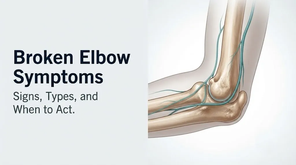
Broken Elbow Symptoms: Signs, Types, and When to Act
Broken elbow symptoms include immediate, severe pain at the elbow joint, rapid swelling, visible or felt deformity, bruising, and inability to fully straighten or bend the arm, all produced when one or more of the three bones forming the elbow joint fracture under impact or stress. What many people don’t realize is that some elbow fractures produce surprisingly mild pain in the first hour, particularly in children and in non-displaced radial head fractures, making symptom recognition more important than pain intensity alone.
According to the American Academy of Orthopaedic Surgeons (AAOS), elbow fractures account for a notable proportion of all upper extremity fractures evaluated in emergency departments, with supracondylar humeral fractures being the most common elbow fracture in children and radial head fractures being the most common type in adults. These are not minor injuries: the elbow joint houses the brachial artery, three major peripheral nerves, and a growth plate system in children that requires specific imaging expertise to evaluate correctly.
This article covers every major broken elbow symptom with the anatomical mechanism behind each one, explains how symptoms differ by fracture type and by patient age, identifies the neurovascular warning signs that make an elbow injury a genuine emergency, and tells you exactly which provider to contact and when.
Broken Elbow Symptoms: What They Feel Like
Broken elbow symptoms typically include immediate localized pain at or around the elbow joint, rapid swelling within minutes to hours of injury, limited or impossible elbow movement, and in some cases, visible deformity, a felt or heard crack at the time of injury, and numbness or tingling traveling into the forearm and hand.
The experience of a broken elbow varies considerably by fracture type and severity. A non-displaced radial head fracture, for example, may produce only moderate lateral elbow pain with mild swelling and minimal deformity. A displaced supracondylar fracture in a child can produce catastrophic swelling, gross deformity, and neurovascular compromise within the first 30 to 60 minutes.
Pain is never a reliable standalone indicator of fracture severity. Bone pain originates primarily from periosteal nociceptors, the dense network of pain receptors lining the outer bone surface. When the periosteum tears at a fracture site, these receptors fire acutely. But fractures through the cancellous (spongy) interior of a bone, such as impacted radial head fractures, cause less periosteal disruption and therefore less initial pain.
Core symptoms that suggest a broken elbow:
- Immediate pain at the elbow that worsens with any movement attempt
- Swelling beginning within minutes of injury, often rapid and pronounced
- Bruising (ecchymosis) appearing within hours, potentially spreading down the forearm by 24 to 48 hours
- Inability to fully straighten (extend) the elbow to 180 degrees
- Inability to fully bend (flex) the elbow toward the shoulder
- Point tenderness directly over a specific bone when pressed
- Numbness, tingling, or weakness in the hand or fingers
- Visible or palpable deformity of the elbow contour
- A crack, pop, or grinding sensation at the moment of injury
For older adults taking anticoagulant medications such as warfarin or direct oral anticoagulants: hematoma expansion is significantly accelerated after any fracture. Swelling and ecchymosis can develop faster and more extensively than in a person not on blood thinners, sometimes making the injury appear more severe than the fracture grade alone would suggest.
Elbow Anatomy and Fracture Types
The elbow is a complex hinge-and-pivot joint formed by three bones, each capable of fracturing at distinct anatomical locations that produce different symptom patterns. Understanding which bone broke and where determines the symptoms, the neurovascular risks, and the treatment approach.
The three bones are the distal humerus (the lower end of the upper arm bone), the radius (the forearm bone on the thumb side, whose rounded proximal end forms the radial head at the elbow), and the ulna (the forearm bone on the pinky side, whose prominent bony projection forms the olecranon at the tip of the elbow). The distal humerus itself has four named projections: the trochlea (which articulates with the ulna), the capitellum (which articulates with the radial head), the medial epicondyle (the inner bony bump), and the lateral epicondyle (the outer bony bump).
Think of the elbow like a door hinge with an extra rotating component. The hinge motion (flexion and extension) happens between the trochlea and ulna. The rotating motion (pronation and supination that turns your palm up and down) happens between the radial head and capitellum. A fracture anywhere in this system disrupts one or both motions.
| Fracture Type | Bone Involved | Most Common Cause | Population Most Affected |
|---|---|---|---|
| Radial head fracture | Proximal radius | Fall on outstretched hand | Adults, especially ages 20 to 60 |
| Olecranon fracture | Proximal ulna | Direct blow or forceful triceps contraction | Adults |
| Supracondylar fracture | Distal humerus (above condyles) | Fall on outstretched hand | Children ages 5 to 10 |
| Lateral condyle fracture | Distal humerus (lateral side) | Fall on outstretched hand | Children ages 5 to 10 |
| Medial epicondyle fracture | Medial humerus | Valgus stress or elbow dislocation | Children and adolescents |
| Monteggia fracture-dislocation | Proximal ulna plus radial head | High-energy forearm impact | Adults and children |
| Distal humerus fracture | Entire distal humerus | High-energy trauma or low-energy in osteoporosis | Adults, especially elderly women |
For women over age 65 with reduced bone mineral density from post-menopausal estrogen decline: a fall from standing height can produce a comminuted (shattered) distal humerus fracture where the same fall in a younger adult would produce only a soft tissue injury or a non-displaced fracture. The National Institute of Arthritis and Musculoskeletal and Skin Diseases (NIAMS) identifies osteoporosis as a primary risk factor for fragility fractures of the upper extremity.
Pain From a Broken Elbow
Pain from a broken elbow is produced by the tearing of the periosteum, stimulation of periosteal nociceptors (pain-sensing nerve endings), and the acute inflammatory response that floods the fracture site with prostaglandins, bradykinin, and histamine within seconds to minutes of injury. The pain is typically immediate, sharp, and localized precisely to the fracture site at first contact.
The location of pain points directly to the likely fracture site. Lateral elbow pain (pain on the thumb side of the elbow) following a fall on an outstretched hand strongly suggests a radial head fracture. Pain precisely at the olecranon tip (the bony point at the back of the elbow) after a direct blow or a fall onto a flexed elbow suggests an olecranon fracture. Pain above the elbow crease on the front of the arm in a child after a fall suggests a supracondylar fracture.
Pain in elbow fractures often worsens specifically with attempts to rotate the forearm (pronation and supination), not only with flexion and extension. This is because radial head fractures disrupt the pivot joint between the radial head and capitellum. Rotating the forearm grinds the fractured radial head against the capitellum, producing a sharp, localized pain on the outer elbow that is often described as worse than the resting pain.
What to tell your emergency room physician about elbow pain:
- State the exact location of maximum pain: outer elbow, inner elbow, tip of elbow, or above the joint
- Describe what movement makes it worse: bending, straightening, or rotating the palm up or down
- Report the mechanism of injury: fall on outstretched hand, direct blow, fall on a bent elbow
- State whether the pain started immediately at impact or developed over 30 to 60 minutes
- Note whether pain is spreading down the forearm or into specific fingers
For people with pre-existing elbow conditions such as rheumatoid arthritis or prior elbow surgery: baseline joint pain and limited range of motion can make it harder to assess whether a new injury has produced a fracture. A board-certified orthopedic surgeon or emergency medicine physician should be told about pre-existing joint disease so that imaging is interpreted relative to prior baseline anatomy.
Swelling and Bruising From a Broken Elbow
Swelling from a broken elbow begins within minutes of injury because fracture disrupts periosteal blood vessels and local capillaries, triggering immediate plasma extravasation into surrounding soft tissue and hematoma formation at the fracture site. Bruising (ecchymosis) typically appears later, tracking along fascial planes as blood from the hematoma travels superficially through soft tissue.
The timeline of swelling and bruising is clinically predictable. Swelling begins within 15 to 30 minutes of injury as the inflammatory response drives vasodilation and increased capillary permeability via histamine and prostaglandin release. By 2 to 6 hours, swelling is often pronounced enough to obscure the normal bony contours of the elbow. Bruising typically becomes visible at 12 to 24 hours and may travel distally down the forearm and even into the palm by 48 to 72 hours, following the path of gravity and fascial plane drainage.
Swelling in the elbow carries a specific clinical concern that distinguishes it from swelling at other joints. The elbow joint capsule is an enclosed space. When a fracture fills the joint capsule with blood (hemarthrosis), the pressure inside the joint rises rapidly. This elevated intra-articular pressure is part of what prevents elbow extension in the first hours after a fracture, before the surrounding muscles even go into spasm.
Quick Tip:
- Pronounced elbow swelling that develops within 30 minutes of injury warrants emergency evaluation, not a wait-and-see approach
- Swelling that extends circumferentially around the entire forearm (rather than just the elbow) raises concern for compartment syndrome and requires immediate emergency evaluation
- For children: forearm swelling disproportionate to the elbow injury is a red flag for a Monteggia fracture-dislocation where the radial head dislocates alongside an ulna fracture
For patients on corticosteroid medications (oral prednisone, long-term inhaled steroids): chronic corticosteroid use reduces tissue integrity and may cause swelling to appear more florid and bruising to develop faster and more extensively than expected from the fracture grade, because corticosteroids impair the capillary fragility regulatory mechanisms.
Key Takeaway: If the elbow swells visibly within the first 30 minutes after an injury, treat it as a probable fracture until imaging proves otherwise, regardless of how much pain the person reports.
Loss of Range of Motion From a Broken Elbow
Loss of elbow range of motion after a fracture occurs through two distinct mechanisms: mechanical blockage from displaced bone fragments obstructing joint movement, and protective muscle guarding driven by pain signals from periosteal nociceptors that cause the surrounding muscles to contract and resist any movement that stresses the fracture site.
Normal elbow range of motion spans from 0 degrees (full extension, arm fully straight) to approximately 145 degrees of flexion (forearm nearly touching the upper arm), with 80 to 90 degrees of pronation and supination. A broken elbow typically limits extension most severely in the acute phase. Attempting to straighten a fractured elbow stretches the inflamed, blood-filled joint capsule anteriorly, producing sharp pain that reflexively triggers protective muscle guarding.
The specific pattern of motion loss helps localize the fracture. Inability to rotate the forearm (supination and pronation) with relatively preserved flexion and extension points to the radial head, because radial head integrity is required for the pivot joint function. Inability to extend the elbow at all with preserved rotation points more toward the ulna or distal humerus. A locked elbow that cannot move in any direction after trauma suggests a dislocation component or a large displaced fragment obstructing the joint.
According to the AAOS OrthoInfo clinical guidelines on elbow fractures, loss of motion that persists beyond the acute injury phase, particularly a flexion contracture of more than 30 degrees at rest, is a recognized early sign that the fracture needs prompt orthopedic evaluation to prevent permanent stiffness.
For elderly patients: pre-existing elbow stiffness from degenerative joint disease makes it harder to determine whether loss of motion is new from a fracture or chronic from arthritis. A baseline comparison by the patient (“I normally can straighten my arm to this point”) is the most reliable guide for the evaluating emergency medicine physician.
Numbness and Tingling From a Broken Elbow
Numbness and tingling from a broken elbow indicate that one or more peripheral nerves traveling through or adjacent to the elbow joint has been compressed, stretched, or contused by the fracture, displaced bone fragment, or expanding hematoma. The specific nerve affected determines precisely where in the hand numbness or tingling occurs.
Three major nerves pass through the elbow region, each with a distinct anatomical course that makes it vulnerable to specific fracture types. The radial nerve (specifically its posterior interosseous branch) runs near the radial head and is at risk in radial head and lateral epicondyle fractures, producing numbness on the back of the thumb and first two fingers, plus weakness in wrist and finger extension. The ulnar nerve runs through the cubital tunnel on the medial (inner) side of the elbow, making it vulnerable in medial epicondyle fractures and olecranon fractures, producing the classic “funny bone” sensation: numbness and tingling in the ring and little finger, with hand grip weakness. The anterior interosseous nerve, a branch of the median nerve, runs anteriorly near the supracondylar region and is the nerve most commonly injured in supracondylar fractures in children, producing inability to make an “OK” sign with the thumb and index finger (flexion of the distal joints is lost).
| Nerve | Location at Elbow | Fracture Types That Risk It | Sensory Loss Location | Motor Loss |
|---|---|---|---|---|
| Radial nerve / posterior interosseous branch | Lateral, near radial head | Radial head fracture, lateral condyle | Dorsal thumb and first two fingers | Wrist drop, finger extension loss |
| Ulnar nerve | Medial, cubital tunnel | Medial epicondyle fracture, olecranon fracture | Ring and little finger | Grip weakness, intrinsic hand muscle weakness |
| Anterior interosseous nerve (AIN) | Anterior, near supracondylar region | Supracondylar fracture | Minimal sensory loss | Loss of distal thumb and index finger flexion (cannot make “OK” sign) |
| Median nerve | Anterior | Elbow dislocation, supracondylar fracture | Palm, thumb, first two fingers | Thumb opposition weakness |
For children specifically: anterior interosseous nerve injury occurs in approximately 7 to 10 percent of supracondylar humeral fractures, according to published research in the Journal of Bone and Joint Surgery. Because children may not reliably report sensory changes, checking motor function (asking the child to make an “OK” sign or pinch their fingers) is more diagnostically reliable than asking about numbness alone.
Visible Deformity and Sounds at Time of Injury
Visible deformity at the elbow after trauma indicates that a bone has fractured and either the bone ends have displaced from their normal alignment or the joint has dislocated, shifting the entire anatomical relationship between the humerus, radius, and ulna. An audible crack, pop, or crunch at the moment of injury is produced by the rapid fracture of cortical bone or by the sudden separation of bone surfaces at the fracture line.
The normal elbow has identifiable surface landmarks: the olecranon is the most prominent posterior point, and the two epicondyles (medial and lateral) form a triangle with the olecranon when the elbow is flexed to 90 degrees. This triangle is called the cubital triangle or, in clinical practice, the Hueter triangle. Disruption of this triangle, where one of the three bony points is no longer in its expected position relative to the other two, indicates either a fracture with displacement or a dislocation.
The sound at the time of injury is not always a dramatic crack. Some fractures, particularly impacted or non-displaced fractures, produce only a muffled thud or a sensation of something giving way rather than an audible sound. Others, particularly olecranon avulsion fractures where the triceps tendon pulls the olecranon tip off the bone, produce a sharp snap that is felt and heard simultaneously.
The presence of visible deformity should never be used to rule a fracture in or out. According to Mayo Clinic clinical guidance on elbow injuries, some severely displaced fractures in children produce dramatic deformity, while some complete radial head fractures in adults produce no visible deformity whatsoever. The decision to obtain imaging should not hinge on whether deformity is visible.
For athletes in contact sports: deformity from elbow dislocation can look very similar to deformity from a severely displaced distal humerus fracture. Both require emergency evaluation; attempting to reduce a dislocated elbow before ruling out a fracture risks displacing fracture fragments into neurovascular structures.
Radial Head Fracture Symptoms
Radial head fracture symptoms are centered on the lateral (outer) elbow and include localized tenderness directly over the radial head (the bony structure just below the lateral epicondyle on the outer forearm), pain that is specifically worsened by rotating the forearm palm-up or palm-down, and swelling confined to the outer elbow. This is the most common elbow fracture in adults, typically produced by falling on an outstretched hand with the elbow slightly flexed.
The mechanism of a radial head fracture is a transmitted axial load. When the hand hits the ground during a fall, force travels up the radius and concentrates at the radial head as it impacts the capitellum of the humerus. The radial head, which is rounded and relatively small, absorbs the impact and cracks. The fracture ranges from a hairline crack that barely interrupts the bone surface (Mason Type I) to a severely comminuted fracture with multiple fragments (Mason Type III) where the joint surface is destroyed.
A key clinical feature: radial head fractures frequently cause a block to forearm rotation rather than to elbow flexion. If a patient cannot rotate their palm fully upward (supination) after a fall but can still flex and extend the elbow reasonably well, a radial head fracture should be the primary consideration until imaging confirms otherwise.
Characteristic symptoms of a radial head fracture:
- Point tenderness directly over the radial head (press the outer elbow just below the lateral epicondyle)
- Pain with forearm rotation (especially supination, turning the palm up)
- Lateral elbow swelling, often with hemarthrosis producing a firm, tense swelling
- Limited but partially preserved elbow flexion and extension
- No visible deformity in non-displaced or minimally displaced fractures (Mason Type I)
- Possible visible lateral elbow fullness from joint effusion
For women between ages 30 and 55: radial head fractures are among the most frequently missed elbow injuries in this age group, according to clinical observation reported in orthopedic emergency literature, because the initial pain may be moderate and swelling subtle enough to be attributed to a sprain. Any lateral elbow pain with forearm rotation restriction after a fall warrants X-ray evaluation, not an assumed sprain diagnosis.
Key Takeaway: Pain specifically worsened by rotating the forearm palm-up or palm-down after a fall on an outstretched hand is the defining feature of a radial head fracture and warrants imaging regardless of how mild the pain feels initially.
Olecranon Fracture Symptoms
Olecranon fracture symptoms are centered at the tip and posterior aspect of the elbow, with point tenderness directly over the olecranon process, a palpable gap or step in the bone where the fracture has separated, and inability to extend the elbow against resistance because the triceps brachii muscle attaches via its tendon directly to the olecranon. When the olecranon fractures and displaces, triceps-driven elbow extension fails.
The olecranon is the bony projection that you feel when you put your elbow on a table. It forms the posterior prominence of the elbow and is covered only by a thin layer of skin and the olecranon bursa, with almost no muscle padding. This means a direct blow to the posterior elbow, such as falling directly onto the tip of a bent elbow or receiving a direct hit during a sports contact, transmits full force to the olecranon with no soft tissue buffer.
One distinguishing feature of an olecranon fracture is whether the patient can extend the elbow against gravity. If the fracture is displaced, the pull of the triceps muscle separates the fracture fragment proximally, creating a gap in the bone. The patient can flex the elbow passively (gravity-assisted) but cannot straighten it actively against any resistance. This extension weakness is a clinical sign that is not present in simple elbow contusions or bursitis.
The American Academy of Orthopaedic Surgeons states that olecranon fractures represent approximately 10 percent of all elbow fractures in adults and are more common in elderly patients who fall directly onto a flexed elbow. Displaced olecranon fractures typically require surgical repair with open reduction and internal fixation (ORIF) using a tension band wiring or plate-and-screw construct to restore the extensor mechanism.
For individuals with olecranon bursitis (a separate, non-fracture condition involving fluid in the bursal sac at the elbow tip): distinguishing bursal swelling from post-fracture swelling requires palpation. A fractured olecranon produces a palpable bony discontinuity and point tenderness on the bone itself, whereas isolated bursitis produces a soft, fluctuant fluid pocket with normal bone contour underneath.
Supracondylar Elbow Fracture Symptoms in Children
Supracondylar humeral fracture symptoms in children include immediate pain above the elbow crease on the front and medial side of the arm, rapid and pronounced swelling that can distort the elbow’s normal shape within 30 to 60 minutes, refusal to move the arm, and in more severe cases, dimpling of the skin at the anterior elbow, pallor or blueness of the hand, or coldness of the fingers indicating vascular compromise.
The supracondylar fracture occurs just above the two condyles of the distal humerus, in the narrowest and most structurally vulnerable portion of the bone. In children, this area is mechanically weak because the cortical bone is thin relative to the bony diameter at this point. A fall on an outstretched hand with the elbow hyperextended drives the olecranon into this thin area like a wedge, snapping the distal humerus posteriorly. The result is a fracture that displaces the elbow’s distal fragment backward relative to the shaft.
This posterior displacement brings the proximal fracture fragment into contact with the brachial artery, which runs directly anterior to the supracondylar region. This is the anatomical basis for why supracondylar fractures carry a vascular injury risk that no other elbow fracture type matches. According to research published in Pediatrics (the journal of the American Academy of Pediatrics), vascular injury occurs in approximately 10 to 20 percent of displaced supracondylar fractures, ranging from arterial spasm to complete brachial artery laceration.
Quick Tip:
- Check the child’s hand immediately: if the fingers are pale, blue, cold, or pulseless, this is a vascular emergency
- Ask the child to touch their thumb to each finger to screen for anterior interosseous nerve function
- Do not attempt to straighten or splint the arm if the elbow is severely deformed; support it in the position of comfort and go to the emergency room immediately
- The Gartland classification (Type I, II, III) guides surgical decision-making, with Type III displaced fractures requiring urgent surgical pinning
For children under age 8: the distal humerus ossification centers appear at predictable ages following the CRITOE sequence (Capitellum appearing on X-ray at approximately age 1, Radial head at age 3, Internal/medial epicondyle at age 5, Trochlea at age 7, Olecranon at age 9, External/lateral epicondyle at age 11). Any ossification center that appears out of sequence or in an unexpected position on pediatric elbow X-ray indicates a fracture or avulsion at that center.
Broken Elbow Symptoms in Adults vs. Children
Broken elbow symptoms differ between adults and children because children’s bones contain an active physis (growth plate) composed of cartilage that is mechanically weaker than surrounding bone but invisible on standard X-ray, meaning fractures in children can look deceptively normal on plain radiographs while the child has a genuine bony injury at a growth plate location.
In adults, elbow fractures more commonly occur through cortical bone that is clearly visible on X-ray. The fracture line is usually detectable on a properly positioned lateral and anteroposterior elbow X-ray. When no fracture line is visible in an adult but swelling and pain are present after trauma, the posterior fat pad sign on a lateral X-ray is the key finding: the posterior fat pad, normally hidden within the olecranon fossa, is displaced upward by joint effusion (hemarthrosis), appearing as a triangular shadow posterior to the distal humerus. This “sail sign” indicates intra-articular pathology even when no fracture line is visible.
| Feature | Adults | Children |
|---|---|---|
| Most common fracture type | Radial head fracture | Supracondylar fracture |
| Growth plate involvement | No (physis fused) | Yes (physis open, varies by age) |
| X-ray visibility of fracture | Usually detectable | May be invisible (Salter-Harris Type I) |
| Primary ligament sprain vs. fracture | Both occur | Fracture more likely than ligament sprain |
| Neurovascular complication risk | Present but lower | Higher (especially brachial artery in supracondylar) |
| Key imaging sign | Posterior fat pad sign (sail sign) | CRITOE sequence disruption, fat pad sign |
| Deformity visibility | Variable | Often pronounced in supracondylar |
In children, the clinical guideline from the AAOS specifies that a child who refuses to use an arm after trauma, even without obvious deformity and even with a normal-appearing X-ray, should be treated as having a probable elbow fracture and referred to a pediatric orthopedic surgeon for further evaluation. The absence of a visible fracture line on a child’s X-ray does not rule out a physeal injury.
For adolescent athletes ages 13 to 17: the growth plate at the medial epicondyle is the last to fuse in the elbow (typically ages 15 to 17). Throwing athletes in this age group are at specific risk for medial epicondyle apophysitis (Little Leaguer’s Elbow) or avulsion fracture from the repetitive valgus stress of overhead throwing, producing medial elbow pain, swelling, and weakness with throwing that requires evaluation by a pediatric orthopedic surgeon or sports medicine physician experienced in pediatric throwing injuries.
Neurovascular Injury Signs With a Broken Elbow
Neurovascular injury signs with a broken elbow are the most urgent symptoms to recognize because they indicate that the fracture has damaged the brachial artery, one of the peripheral nerves, or has produced rising pressure in the forearm compartment (compartment syndrome), all of which can cause permanent limb damage or tissue death within hours if not treated surgically.
The “5 P’s” of acute limb ischemia provide the clinical framework for neurovascular assessment after any elbow fracture: Pain (out of proportion to the injury, or pain that increases despite immobilization), Pallor (pale or white skin color in the hand or fingers), Pulselessness (absent or diminished radial pulse at the wrist), Paresthesia (numbness or tingling in the hand or fingers), and Paralysis (inability to move the fingers or hand). Any one of these signs after an elbow fracture, particularly after a supracondylar fracture or elbow dislocation, is a surgical emergency.
Compartment syndrome of the anterior forearm compartment is the most feared complication of elbow fractures with significant swelling. The anterior forearm compartment contains the flexor muscles of the fingers and wrist, the median and anterior interosseous nerves, and the radial and ulnar arteries. When swelling raises pressure within this closed fascial compartment above approximately 30 mmHg (or within 30 mmHg of diastolic blood pressure), blood flow to the compartment’s contents ceases. Untreated compartment syndrome progresses to Volkmann ischemic contracture, a permanent contracture of the forearm and hand muscles from ischemic death of muscle and nerve tissue, within 6 to 8 hours of onset.
According to the Journal of Bone and Joint Surgery, compartment syndrome following pediatric supracondylar fracture, while not common, represents one of the most devastating complications in pediatric orthopedic trauma, with permanent functional deficits occurring in cases where compartment pressure was not recognized and released within the critical window.
For patients who have received a plaster or fiberglass splint or cast in an emergency setting: increasing pain under the cast, tingling that was not present at the time of casting, or a feeling of the cast being too tight are warning signs that compartment pressure may be rising inside the immobilized limb. Return to the emergency room immediately if any of these symptoms develop after an elbow has been casted or splinted.
Key Takeaway: Pallor, pulselessness, or pain out of proportion to the injury in a hand or forearm after any elbow fracture are a surgical emergency: go to the nearest emergency room immediately without waiting for a scheduled orthopedic appointment.
Hairline and Stress Fracture of the Elbow
Hairline and stress fractures of the elbow produce a distinctly different symptom pattern from traumatic complete fractures: the onset is gradual rather than sudden, the pain is activity-related rather than constant, there is typically no dramatic swelling or deformity, and the injury is often mistaken for tendinitis, bursitis, or overuse strain for weeks before the correct diagnosis is made.
A hairline fracture (also called an occult fracture or incomplete fracture) occurs when a single traumatic force partially cracks a bone without fully separating the bone ends. At the elbow, hairline fractures of the radial head are particularly common after a FOOSH (fall on outstretched hand) mechanism where the impact force was insufficient to produce a complete fracture but still cracked the cortical surface. The pain is moderate, rotation of the forearm is restricted and painful, but the elbow appears almost normal and the patient may initially dismiss the injury.
A stress fracture results from repetitive sub-threshold loading that accumulates microtrauma in bone faster than the bone’s remodeling capacity can repair it. At the elbow, stress fractures occur most commonly in overhead-throwing athletes (particularly baseball pitchers and javelin throwers) and in gymnasts who load the elbow in extension repeatedly. The olecranon is the most common stress fracture site at the elbow in throwers, producing a posterior elbow ache that initially appears only during throwing and progressively becomes constant.
The National Institutes of Health (NIH) NIAMS division notes that stress fractures are more common in female athletes compared to male athletes at the same training level, a pattern associated with lower bone mineral density in some women at reproductive age, particularly those with relative energy deficiency in sport (RED-S), where caloric restriction impairs bone formation relative to resorption.
For military recruits and endurance athletes: elbow stress fractures, while less common than lower extremity stress fractures in these populations, should be suspected when lateral or posterior elbow pain develops progressively over several weeks of high-volume training without a single traumatic event. Magnetic resonance imaging (MRI) detects stress fractures at the elbow with far greater sensitivity than plain X-ray in the early weeks of injury.
Broken Elbow vs. Sprained Elbow Symptoms
Distinguishing a broken elbow from a sprained elbow by symptoms alone is not reliably possible, and no amount of online symptom-checking substitutes for clinical examination and X-ray imaging, because the symptom overlap between an elbow fracture and a significant elbow ligament sprain is extensive and clinically meaningful.
Both conditions produce pain, swelling, tenderness to touch, and limited range of motion after trauma. The physiological reason for the overlap is that both fractures and significant ligament tears trigger the same inflammatory cascade: histamine and prostaglandin release, vasodilation, plasma extravasation producing edema, and periosteal or ligamentous nociceptor activation producing pain. The inflammatory response itself does not distinguish its cause.
| Feature | Fracture More Likely | Sprain More Likely |
|---|---|---|
| Mechanism | High-energy fall or direct blow | Twisting, valgus force without direct impact |
| Pain onset | Immediate and severe | May build over minutes |
| Swelling onset | Within minutes, often rapid and large | Within hours, often more gradual |
| Point tenderness | Over bone (radial head, olecranon, condyles) | Over ligament (UCL on inner elbow, RCL on outer) |
| Deformity | May be present | Absent (unless dislocation) |
| Range of motion | Severely limited in all planes | Limited but may retain some arc |
| Sound at injury | Crack or pop | Pop or tearing sensation (high-grade tears) |
| Neurovascular symptoms | Possible | Absent |
The most reliable clinical differentiator is point tenderness location. Tenderness directly over bone (pressing the radial head, olecranon tip, or condyles) strongly suggests fracture. Tenderness directly over the ligament origin and insertion sites (medial ulnar collateral ligament, lateral radial collateral ligament) with relatively preserved point tenderness over bone suggests ligamentous injury.
However, according to the Cleveland Clinic clinical guidance on elbow injuries, a definitive distinction between fracture and ligament sprain requires plain X-ray imaging as a minimum standard, because even experienced clinicians examining an acutely swollen elbow cannot reliably exclude fracture by physical examination alone. Any significant elbow injury that limits normal daily function warrants imaging.
How a Broken Elbow Is Diagnosed
A broken elbow is diagnosed through a clinical examination combined with plain X-ray imaging as the first-line investigation, followed by computed tomography (CT scan) or magnetic resonance imaging (MRI) for complex fractures, occult injuries, or cases where X-ray findings are inconclusive.
The clinical examination begins with a neurovascular assessment: radial pulse at the wrist, capillary refill in the fingertips, grip strength, and sensory testing of the hand to check for nerve or vascular injury before any manipulation or imaging is performed. This sequence is non-negotiable because an undetected vascular injury can worsen during transport or positioning for X-ray.
Plain X-ray of the elbow uses at minimum two views: an anteroposterior (AP) view and a true lateral view with the elbow at exactly 90 degrees of flexion. The lateral view is the most diagnostically important for detecting the fat pad signs. On a normal lateral X-ray, the anterior fat pad is barely visible as a faint shadow anterior to the distal humerus. The posterior fat pad is not normally visible at all. Joint effusion from a fracture (hemarthrosis) pushes both fat pads out of their recesses, making the anterior fat pad more prominent and making the posterior fat pad visible as the characteristic “sail sign.” A visible posterior fat pad sign on a lateral elbow X-ray is treated as evidence of a fracture until proven otherwise, even if no fracture line is seen.
Diagnostic imaging modalities for elbow fractures:
- Plain X-ray (AP and lateral views): First-line; detects most displaced fractures; may miss non-displaced radial head fractures and pediatric physeal injuries
- CT scan (computed tomography): Best for complex fractures, comminuted fractures, distal humerus fractures requiring surgical planning; provides three-dimensional fracture mapping
- MRI (magnetic resonance imaging): Best for stress fractures, occult non-displaced fractures, and ligamentous injury assessment; highest sensitivity for early stress fracture
- Ultrasound: Useful for assessing joint effusion and ligament continuity in some clinical settings; operator-dependent
For pregnant women who sustain an elbow fracture: plain X-ray with appropriate abdominal shielding is generally considered acceptable by the American College of Obstetricians and Gynecologists (ACOG) when imaging is medically necessary, as the radiation dose to the fetus from a focused extremity X-ray is negligible. MRI without gadolinium contrast is the preferred alternative when CT would be used for surgical planning, as MRI avoids ionizing radiation entirely.
Emergency Signs of a Broken Elbow
Certain broken elbow symptoms require immediate emergency evaluation. Do not wait to see if these resolve on their own.
Go to the nearest emergency room immediately or Call 911 if the following are present after an elbow injury:
- Pallor, bluish discoloration (cyanosis), or cold skin in the hand or fingers: These indicate brachial artery injury or acute compartment syndrome, both of which cause limb-threatening ischemia within hours
- Absent or diminished radial pulse at the wrist: A pulseless hand after elbow fracture is a surgical emergency requiring vascular repair
- Severe pain in the forearm that is worsening despite rest and immobilization: Pain out of proportion to the visible injury, or pain that increases under a cast or splint, is the most important early sign of compartment syndrome
- Rapid, circumferential swelling of the entire forearm: This indicates rising pressure in the forearm compartments, not just soft tissue swelling at the fracture site
- Complete inability to move any finger: Paralysis of the hand after elbow fracture indicates severe nerve injury or ischemia to the anterior forearm compartment muscles
- Open wound over the elbow with visible bone: An open (compound) fracture carries immediate infection risk and requires emergency surgical debridement and irrigation within hours
- Elbow deformity in a child with any signs of vascular compromise: Displaced supracondylar fractures with vascular involvement require emergency surgical reduction and pinning, not a morning orthopedic appointment
- Progressively worsening symptoms after the elbow has already been evaluated and splinted: Increasing pain, tightness, or numbness under a splint or cast requires immediate return to the emergency room for cast removal and pressure assessment
These presentations can indicate brachial artery laceration, acute compartment syndrome, Volkmann ischemic contracture in evolution, or open fracture infection risk, all of which require emergency surgical evaluation, not an urgent care appointment or a next-day phone call.
Frequently Asked Questions About Broken Elbow Symptoms
How do I know if my elbow is broken or just sprained?
The only reliable way to distinguish a broken elbow from a sprained elbow is plain X-ray imaging, because both conditions produce pain, swelling, and limited motion that cannot be differentiated by symptoms alone.
Point tenderness directly over a specific bone (such as the radial head on the outer elbow or the olecranon at the tip) suggests fracture more than sprain, but this is not definitive without imaging.
Any elbow injury that prevents normal daily function, produces rapid swelling within the first 30 minutes, or causes numbness or tingling in the hand should be evaluated by an emergency medicine physician with X-ray the same day.
Can you move your arm with a broken elbow?
Some movement is often possible with a broken elbow, particularly with non-displaced or hairline fractures where the bone ends remain in alignment and muscle guarding is incomplete.
A radial head fracture, for example, may allow moderate elbow flexion and extension while blocking forearm rotation (turning the palm up and down) due to the radial head’s role in the pivot joint.
The ability to move the arm does not rule out a fracture, and any attempt to force range of motion testing on a suspected fracture before imaging is obtained can displace fracture fragments into neurovascular structures.
What does a broken elbow feel like?
A broken elbow typically feels like immediate, sharp, localized pain at the elbow that is worse with any attempt at movement, accompanied by a sensation of swelling building rapidly within the first 15 to 30 minutes.
Many people describe the acute pain as distinctly different from a sprain: deeper, more precisely located at a single bony point, and associated with a feeling that something is mechanically wrong inside the joint rather than a surface ache.
In some fractures, particularly non-displaced radial head fractures, the initial pain may be moderate enough that the person initially believes they have sprained the elbow, with the severity of the injury only becoming apparent when forearm rotation is attempted.
Can a broken elbow heal on its own without treatment?
Non-displaced, stable fractures such as Mason Type I radial head fractures can often heal with non-surgical management (sling immobilization for a brief period followed by early mobilization), but this still requires clinical evaluation and imaging to confirm the fracture is truly non-displaced.
No elbow fracture should be assumed to be self-managing without X-ray confirmation, because a fracture that appears minor can have displacement or an associated ligament injury that changes the treatment plan entirely.
Displaced fractures, fractures with ligamentous instability, fractures with neurovascular compromise, and open fractures all require medical or surgical treatment under the care of a board-certified orthopedic surgeon.
How long does a broken elbow take to heal?
Healing time for a broken elbow ranges from 3 to 4 weeks for minor non-displaced radial head fractures managed without surgery to 3 to 6 months for complex distal humerus fractures requiring open reduction and internal fixation, followed by structured physical and occupational therapy to restore range of motion.
Pediatric elbow fractures that do not involve growth plate disruption generally heal faster than adult fractures, with callus formation visible on X-ray within 3 to 4 weeks in most children.
Patients with osteoporosis, diabetes, or who smoke heal fractures more slowly, with available clinical evidence indicating that smoking impairs bone healing by reducing periosteal blood flow and osteoblast activity.
What are the signs of nerve damage from a broken elbow?
Signs of nerve damage from a broken elbow include numbness or tingling in specific fingers (ring and little finger for ulnar nerve injury, thumb and first two fingers for median or radial nerve injury), weakness in hand grip, wrist drop, or inability to extend the fingers for radial nerve involvement.
The anterior interosseous nerve, a branch of the median nerve, is the nerve most commonly injured in supracondylar fractures; its injury is detected by asking the patient to make an “OK” sign, because distal flexion of the thumb and index finger is lost when this nerve is damaged.
Any numbness, tingling, or new weakness in the hand after an elbow fracture should be reported to an emergency medicine physician or orthopedic surgeon immediately, as nerve injuries that are identified early have better outcomes than those detected late.
Taking the Next Step After a Suspected Broken Elbow
The symptoms described in this article, taken together, give you a clinically grounded picture of what a broken elbow produces at each anatomical location and in each patient population. The most important thing to act on is this: swelling that begins within 30 minutes, point tenderness over bone, restricted forearm rotation, or any sign of numbness in the hand after an elbow injury all warrant same-day X-ray evaluation by an emergency medicine physician. These are not symptoms to monitor at home.
If the injury is your own, immobilize the arm in the position of comfort, apply ice wrapped in a cloth for 15 to 20 minutes, and go to the emergency room rather than urgent care, because urgent care facilities may lack the imaging or orthopedic consultation capacity to manage complex fractures or neurovascular complications. If the injured person is a child, be especially alert to hand and finger circulation, because supracondylar fractures in children carry a vascular injury risk that requires immediate orthopedic evaluation.
A board-certified orthopedic surgeon, or a pediatric orthopedic surgeon for anyone under age 15, is the appropriate specialist for any confirmed elbow fracture. You have every piece of information you need to recognize this injury accurately and act on it without delay.