Broken Hand Symptoms: What Your Injury Is Telling You
Broken hand symptoms typically include sharp, localized pain at the injury site, swelling that develops within minutes, bruising, reduced grip strength, and sometimes visible deformity or a sensation of pins and needles in the fingers. The specific pattern depends entirely on which of the hand’s 27 bones are involved and how the fracture has displaced.
Hand fractures are far more common than most people expect. According to the National Institute of Arthritis and Musculoskeletal and Skin Diseases (NIAMS), fractures of the hand and wrist collectively account for roughly one-sixth of all fractures seen in emergency departments in the United States, with metacarpal fractures representing the most frequently fractured group among young adults.
This article covers the full symptom picture of a broken hand, explains why each symptom happens at the bone, nerve, and tissue level, maps symptoms to specific fracture types, and identifies the presentations that require immediate emergency evaluation rather than a wait-and-see approach.
Broken Hand Symptoms: The Clinical Overview
Broken hand symptoms include pain that worsens with movement or pressure, swelling that can develop rapidly, ecchymosis (bruising), stiffness, reduced range of motion, and in some cases numbness or tingling in the fingers. These symptoms exist on a spectrum from subtle, in the case of a nondisplaced scaphoid fracture, to immediately obvious, in the case of a displaced metacarpal fracture with visible angular deformity.
Not every broken hand looks dramatic. Some fractures produce no visible deformity at all, which is one of the leading reasons people delay seeking care. Pain that feels “not that bad” an hour after a hand injury can reflect a crack through the cortical bone of the scaphoid that will look completely normal on an initial X-ray and cause serious long-term damage if missed.
The general rule that orthopedic surgeons and emergency physicians apply is straightforward: any hand injury that produces pain with direct pressure on bone, swelling localized to a specific area, or a noticeable loss of grip function warrants imaging. Self-assessment cannot confirm or rule out a fracture. Only radiographic evaluation can do that.
Key symptoms at a glance:
- Immediate or rapidly developing pain at the injury site
- Localized tenderness directly over bone when touched
- Swelling appearing within 15 to 30 minutes of injury
- Bruising that may appear hours later and can spread beyond the fracture site
- Reduced ability to make a full fist or grip objects
- Stiffness and reluctance to move the affected finger or hand
- Visible or palpable deformity in some fracture types
- Numbness, tingling, or burning in one or more fingers (nerve involvement)
- A “pop” or “crack” sensation heard or felt at the moment of injury
Symptoms of a Broken Hand: What Actually Happens Inside the Hand
The symptoms of a broken hand are not random. Each one traces directly to a specific tissue disruption occurring at the moment of fracture and in the hours immediately following. Understanding this makes it much easier to interpret what your body is reporting.
When a bone breaks, the impact disrupts the periosteum, the fibrous membrane that wraps around bone and is densely populated with nociceptive nerve fibers (specifically substance P-releasing C-fibers and A-delta fibers). These fibers generate the immediate, sharp pain signal sent to the brain within milliseconds of the break. This periosteal pain is why even a nondisplaced fracture that has not moved bone fragments apart hurts so sharply when pressed.
Simultaneously, the fracture tears intraosseous blood vessels within the cancellous bone and ruptures small periosteal vessels. Blood leaks into the fracture site and surrounding soft tissue, forming a hematoma. Within minutes, damaged mast cells release histamine and the inflammatory cascade activates, increasing capillary permeability through prostaglandin E2 production. Plasma and blood cells flood the interstitial space. That flood is visible swelling.
Think of the hand’s anatomy like a tightly organized filing cabinet. The bones, tendons, nerves, and blood vessels are stacked in precise compartments with very little spare room. When fluid rushes into that system after a fracture, there’s nowhere for it to go. Pressure builds fast. That’s why hand swelling after a fracture can feel tight and throbbing, unlike swelling in a more loosely organized body region like the thigh.
According to the American Academy of Orthopaedic Surgeons (AAOS), fractures that produce significant swelling within the first 30 minutes are more likely to involve displaced bone fragments or associated soft tissue injury, and warrant the same day evaluation rather than monitoring at home.
Hand Anatomy and Why Fractures Produce the Symptoms They Do
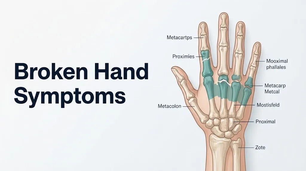
The hand contains 27 bones: 8 carpal bones in the wrist (scaphoid, lunate, triquetrum, pisiform, trapezium, trapezoid, capitate, and hamate), 5 metacarpal bones forming the palm, and 14 phalangeal bones making up the fingers (two in the thumb, three in each of the remaining four fingers). Each group of bones has a different structural role, a different fracture mechanism, and a different symptom signature.
The carpal bones form the proximal foundation of the hand. Because they are arranged in two rows and bound tightly by ligaments, fractures here (especially the scaphoid) often produce minimal swelling but deep, aching pain. The metacarpals are the structural columns of the palm. Fractures here, particularly at the neck of the fourth or fifth metacarpal, frequently produce the visible “knuckle depression” and angular deformity associated with a boxer’s fracture. The phalanges are the most commonly fractured hand bones overall, largely because the fingers are exposed to direct trauma.
Each bone is surrounded by a richly innervated periosteum, which is why point tenderness, meaning pain that is sharply reproduced by pressing directly over a specific bone, is one of the most clinically reliable signs of fracture rather than soft tissue injury.
The hand also has multiple fascial compartments: the thenar compartment (housing the thenar muscles serving the thumb), the hypothenar compartment (housing muscles serving the fifth finger), four dorsal interosseous compartments, three palmar interosseous compartments, and the adductor pollicis compartment. These tight compartments become critically relevant when post-fracture swelling is severe enough to cause compartment syndrome.
| Bone Group | Location | Common Fracture Types | Primary Symptom Location |
|---|---|---|---|
| Scaphoid | Wrist (proximal carpal row) | Waist, proximal pole, distal pole | Anatomical snuffbox (base of thumb, dorsal wrist) |
| Metacarpals | Palm | Neck (boxer’s), base (Bennett’s), shaft | Dorsal hand, knuckle region |
| Proximal phalanges | Finger base segments | Shaft, condylar, base | Along individual finger |
| Middle phalanges | Finger mid-segments | Shaft, collateral ligament avulsion | Middle portion of finger |
| Distal phalanges | Fingertips | Tuft, shaft | Fingertip, subungual hematoma |
Key Takeaway: The hand contains 27 distinct bones organized in tight fascial compartments, which is why fractures produce swift swelling, sharp point tenderness, and occasionally the cascading pressure that leads to compartment syndrome.
Hand Fracture Types and Their Specific Symptom Patterns
Different hand fracture types produce distinctly different symptom patterns, which is why a detailed description of where the pain is located, what the injury mechanism was, and what the hand looks like are all clinically meaningful to an examining physician.
A research review published in the Journal of Hand Surgery (American Volume) found that metacarpal fractures (fractures of the five palm bones) are the most common hand fractures presenting to emergency departments, accounting for roughly 40% of all hand fractures. Phalangeal fractures (finger bone fractures) are the second most frequent, and carpal fractures, primarily the scaphoid, are the third.
Within these groups, the symptom fingerprint differs meaningfully:
| Fracture Type | Common Mechanism | Key Symptom | Distinguishing Feature |
|---|---|---|---|
| Fifth metacarpal neck (boxer’s) | Punch to a hard surface | Dorsal hand pain and swelling, depressed fourth or fifth knuckle | Knuckle that appears lower than its neighbors |
| First metacarpal base (Bennett’s) | Axial load on the thumb | Pain at the base of the thumb, difficulty pinching | Swelling near the carpometacarpal joint of the thumb |
| Scaphoid waist | Fall on outstretched hand (FOOSH) | Deep aching pain in the anatomical snuffbox | Minimal swelling, easy to miss on initial X-ray |
| Proximal phalanx shaft | Direct crush or lateral force | Swelling and pain along the finger shaft | Rotational deformity when making a fist |
| Distal phalanx tuft | Crush injury (door, tool) | Intense fingertip pain, subungual hematoma | Blood collecting under the nail |
People with osteoporosis or those taking long-term corticosteroids may sustain any of these fracture types from lower-energy mechanisms, making the injury mechanism history less reliable as a severity indicator in these populations.
Boxer’s Fracture Symptoms: What the Fifth Metacarpal Feels Like When It Breaks
Boxer’s fracture refers to a fracture of the neck of the fifth metacarpal bone, the slender column of bone connecting the base of the palm to the base of the small (fifth) finger. It is called a boxer’s fracture because it typically results from striking a hard surface with a closed fist, though it also occurs in non-athletic settings including fist-wall impacts during moments of anger or accidental falls.
The symptom presentation is usually unmistakable to anyone who knows what to look for. The most visually distinctive sign is a depressed or absent knuckle: the normally prominent bony knuckle of the small finger appears lower than its neighbors because the fractured metacarpal neck has angulated, pushing the metacarpal head downward toward the palm. Not all boxer’s fractures produce obvious deformity; some lower-grade angulations (under 30 to 40 degrees, depending on the metacarpal involved) may be detectable only on X-ray.
Pain is sharp and concentrated over the dorsum (back) of the hand at the fourth and fifth metacarpal region. Swelling develops rapidly and can extend across much of the dorsal hand surface within the first hour. Bruising follows over the subsequent 12 to 24 hours and may spread toward the wrist or along the finger.
A specific finding that distinguishes boxer’s fracture from a knuckle contusion is rotational malalignment: when the patient attempts to make a fist, the small finger may cross over (rotate toward) the ring finger instead of pointing straight ahead toward the wrist. This scissoring deformity indicates malrotation at the fracture site and requires orthopedic evaluation because malrotated fractures that heal without correction produce lasting functional impairment.
The American Society for Surgery of the Hand (ASSH) states that fourth and fifth metacarpal neck fractures with acceptable angulation can often be managed conservatively with an ulnar gutter splint, but displaced or malrotated fractures typically require orthopedic consultation for potential surgical fixation.
Scaphoid Fracture Symptoms: The Most Frequently Missed Hand Fracture
The scaphoid fracture is the single most frequently missed fracture in the hand and wrist, and understanding its deceptively subtle symptoms can prevent a long-term complication that has serious functional consequences. The scaphoid bone sits at the base of the thumb side of the wrist, bridging the two rows of carpal bones, and it has a notoriously tenuous blood supply that enters primarily through its distal end, meaning proximal pole fractures can cut off circulation to a segment of the bone.
The hallmark symptom of a scaphoid fracture is pain and tenderness in the anatomical snuffbox, the small depression visible on the dorsum of the wrist just proximal to the base of the thumb when the thumb is extended. Pressing into this area with a fingertip in a person with a scaphoid fracture produces sharp, reproducible pain. This is called the snuffbox tenderness test and it is one of the most sensitive clinical signs for scaphoid fracture.
What makes this fracture clinically treacherous is what it often lacks: dramatic swelling, obvious deformity, or severe pain that forces the person to seek care immediately. A 2022 review published in the Journal of Bone and Joint Surgery found that a substantial proportion of scaphoid fractures present with pain mild enough that the patient initially attributes it to a wrist sprain and delays evaluation by days to weeks.
This delay matters. When a scaphoid fracture goes untreated, the already-limited blood supply to the proximal pole can be completely interrupted by the fracture line, leading to avascular necrosis: the death of bone tissue from lack of blood flow. Avascular necrosis of the scaphoid is a serious condition requiring complex surgical management and producing lasting wrist dysfunction.
Anyone who falls on an outstretched hand (the “FOOSH” mechanism) and has pain in the snuffbox region, especially pain that persists beyond 48 to 72 hours without improvement, should be evaluated by an emergency medicine physician or orthopedic surgeon. A normal initial X-ray does not rule out a scaphoid fracture. MRI or CT scanning is required for definitive exclusion.
Key Takeaway: Scaphoid fractures frequently feel like minor wrist sprains, but pain in the anatomical snuffbox after a fall on an outstretched hand warrants imaging beyond a plain X-ray, because a missed scaphoid fracture can progress to avascular necrosis and permanent wrist dysfunction.
Hand Injury Symptoms vs. Sprained Hand: How to Tell the Difference
Distinguishing between hand injury symptoms from a fracture and symptoms from a sprain (ligament injury) is one of the most common clinical challenges in emergency and primary care settings, and the honest answer is that reliable differentiation requires imaging in most cases.
That said, there are clinical patterns that shift the probability strongly toward fracture:
Patterns that suggest fracture over sprain:
- Pain that is precisely located over bone rather than over the joint space or surrounding soft tissue
- Point tenderness that can be reproduced by pressing directly on a specific bone
- Visible deformity or angulation at a joint or along a bone segment
- Rotational malalignment of a finger when making a fist
- A felt or heard “crack” at the moment of injury (though sprains can also produce a pop)
- Swelling that is dense and firm rather than boggy and diffuse
- Inability to move the finger or hand at all within the first 15 minutes (extreme early stiffness suggests significant structural disruption)
Patterns that suggest sprain over fracture (without ruling fracture out):
- Pain located directly over a joint rather than over the bone shaft
- Some preserved range of motion, though painful
- Swelling that is softer and more diffuse across the joint
- Injury mechanism involving twisting or hyperextension rather than direct impact
A 2021 study published in the Annals of Emergency Medicine confirmed that clinical examination alone, even by experienced emergency physicians, correctly distinguishes fracture from sprain in hand injuries only about 65 to 70% of the time without imaging. The conclusion is not that clinical assessment is useless; it is that clinical assessment identifies who needs imaging, not who has a fracture.
For anyone with a hand injury producing moderate to severe pain, swelling, and functional limitation, plain radiographs are the appropriate next step. Waiting to see whether symptoms improve before getting an X-ray is a pattern that leads to missed fractures, delayed treatment, and avoidable complications.
Swelling and Bruising as Hand Fracture Symptoms
Swelling is the most universally present of all hand fracture symptoms, and it begins at the moment of injury through a cascade that unfolds in two distinct phases.
The first phase, occurring within the first 15 to 30 minutes, is vascular. Fractured bone tears intraosseous blood vessels, and that blood immediately begins to collect at the fracture site, forming a hematoma. Simultaneously, damaged cells release histamine and bradykinin, which cause local capillary walls to dilate and become permeable. Plasma leaks out of the bloodstream into the surrounding tissue. This is the early, firm swelling that appears so rapidly after a hand injury.
The second phase, occurring over the following 6 to 24 hours, is the full inflammatory response. Prostaglandin E2 production increases, amplifying capillary permeability further. White blood cells migrate to the injury site. More fluid accumulates. Swelling typically peaks between 24 and 72 hours post-injury and can be substantial enough to make the hand appear twice its normal size in severe fractures.
Bruising, technically called ecchymosis, appears later because it takes time for the blood from the fracture hematoma to track along fascial planes toward the skin surface. Bruising that appears immediately is almost certainly from subcutaneous skin injury rather than the fracture itself. Bruising from a deeper fracture typically appears 12 to 48 hours after injury and may spread significantly beyond the fracture site, sometimes appearing at the wrist or partway up the forearm in a hand fracture, which surprises people but reflects normal hematoma tracking.
Quick Tip:
- Apply ice wrapped in a cloth for 15 to 20 minutes at a time in the first 48 hours to reduce swelling, but do not apply pressure wrapping that could increase compartment pressure
- Elevate the injured hand above heart level by resting it on pillows or a sling to reduce fluid accumulation from gravity
- People taking blood thinners (warfarin, apixaban, rivaroxaban, clopidogrel) should be aware that bruising will spread more extensively and develop faster than in a person not on anticoagulation therapy, which does not by itself indicate a more severe fracture
Grip Weakness and Stiffness From a Hand Fracture
Grip weakness after a hand fracture is not simply a consequence of pain avoidance. It reflects a specific disruption of the mechanical system that generates grip force, and understanding why it happens helps explain why it persists even after the initial pain has partially settled.
Normal grip force is generated by a coordinated system involving the flexor digitorum profundus and flexor digitorum superficialis tendons (which flex the fingers), the intrinsic interosseous and lumbrical muscles (which control fine finger positioning), and the thenar muscle group (which drives thumb opposition, the key to most functional grip). A fracture anywhere along the metacarpals or phalanges disrupts the precise geometry of this system. A fractured metacarpal, for example, shortens or angulates the structural column that the flexor tendons wrap around, reducing the mechanical efficiency of every finger movement and generating pain with any attempt to generate force.
Stiffness in a hand fracture has two causes. The first is protective: the nervous system reflexively reduces motor commands to muscles surrounding a painful injury site, limiting movement to protect the area. The second is structural: edema within the tightly packed fascial compartments restricts tendon gliding, because swollen tissue literally does not let the tendon move through its normal excursion.
A 2020 study published in the Journal of Hand Surgery (American Volume) found that patients who begin supervised hand therapy (directed finger and wrist range-of-motion exercises) within the first two weeks of a hand fracture, once the fracture is appropriately immobilized, have shorter recovery times and better long-term grip strength than those who remain completely immobilized without any movement protocol. This is why early involvement of a certified hand therapist (an occupational therapist with hand therapy specialization) is part of the standard of care in most hand fracture treatment protocols.
Athletes in throwing, racquet, or contact sports should know that return to grip-demanding activity before confirmed fracture union on imaging significantly increases the risk of refracture and malunion.
Key Takeaway: Grip weakness after a hand fracture reflects both pain-protective motor inhibition and mechanical disruption of the hand’s flexor-tendon-bone geometry, not simple pain avoidance, which is why normal grip function can take weeks to fully return even after the fracture is healing well.
Broken Hand Numbness and Nerve Involvement
Numbness in a broken hand is a symptom that requires careful attention because it indicates involvement of one or more peripheral nerves, either through direct trauma, compression from swelling, or impingement from displaced bone fragments.
The three major nerves of the hand each supply distinct regions of skin sensation. The median nerve supplies the palm side of the thumb, index, middle, and half of the ring finger. The ulnar nerve supplies the palm side of the small finger and the other half of the ring finger, plus most of the dorsal (back) surface of the hand. The radial nerve’s superficial sensory branch supplies the dorsal surface of the thumb and radial side of the index finger.
A fracture that produces numbness in a specific finger or region of the hand indicates which nerve is affected:
| Numbness Location | Likely Nerve Involved | Clinical Significance |
|---|---|---|
| Thumb, index, middle finger (palm side) | Median nerve | Possible carpal tunnel pressure from edema or displaced carpal bone fragment |
| Small finger, ring finger (palm side), and dorsal ulnar hand | Ulnar nerve | Hamate fracture, severe medial hand swelling, ulnar nerve stretch |
| Dorsal thumb and index finger | Radial sensory nerve | Distal radius or first metacarpal fracture with radial nerve compression |
| Single finger tip only | Digital nerve | Direct contusion or laceration of the digital nerve at the fracture site |
Numbness that develops gradually over the first few hours post-injury and is mild may reflect simple swelling-related nerve compression. Numbness that is severe, appears immediately with the injury, or is accompanied by weakness in finger movement (not just pain-related weakness) suggests direct nerve injury or compartment syndrome and requires emergency evaluation.
Older adults with pre-existing diabetic peripheral neuropathy or carpal tunnel syndrome may have baseline numbness that makes it harder to interpret new numbness after a hand injury, and should communicate both their baseline sensation status and their current symptoms explicitly to the evaluating physician.
Hand Fracture Symptoms in Children and Growth Plate Injuries
Hand fracture symptoms in children follow the same general pattern as in adults, with one critical difference that alters both diagnosis and management: children’s bones contain growth plates, also called the physis, which are regions of actively dividing cartilage at the ends of the long bones where bone elongation occurs. These growth plates are the weakest point in a child’s bone-ligament-tendon system, which means that forces sufficient to sprain a ligament in an adult may instead fracture through the growth plate in a child.
Growth plate fractures in the hand are classified by the Salter-Harris system on a scale from Type I (fracture only through the growth plate, no bone fragment, often invisible on plain X-ray) to Type V (compression of the growth plate itself). Type I and II are by far the most common in finger fractures.
The symptom picture of a growth plate injury can be deceiving. Because the physis is made of cartilage (which does not appear on plain X-ray), and because Type I fractures produce no visible bone displacement, a child can have a true fracture that shows a completely normal X-ray. The clinical diagnosis relies heavily on the combination of:
- Point tenderness directly over the growth plate (at the base of the finger bone in most pediatric hand fractures, specifically over the proximal phalangeal base)
- Swelling localized to the base of the affected finger
- Pain with gentle axial loading (pressing along the long axis of the finger)
- A mechanism consistent with fracture (a fall, crush, or lateral force)
The American Academy of Pediatrics recommends that pediatric hand injuries with point tenderness over a growth plate be treated as probable fractures even when X-rays are normal, typically with buddy taping or splint immobilization and follow-up imaging in 10 to 14 days if symptoms persist.
Parents should bring children with hand injuries that have point tenderness over the bone base to a pediatrician or emergency medicine physician for evaluation the same day, not the following day or after a weekend.
Hand Fracture Symptoms in Older Adults With Osteoporosis
Hand fracture symptoms in older adults present a distinct clinical picture because the underlying bone quality is often compromised by osteoporosis (defined as bone mineral density more than 2.5 standard deviations below the young adult mean on DXA scanning) or its precursor, osteopenia. Lower bone density means fractures occur from lower-energy mechanisms, such as a simple fall from standing height or even a forceful grip on a surface during a stumble, mechanisms that would not produce fractures in a younger adult with normal bone density.
According to the National Institute of Arthritis and Musculoskeletal and Skin Diseases (NIAMS), postmenopausal women have a significantly elevated fracture risk compared to age-matched men, primarily due to estrogen loss after menopause, which removes estrogen’s protective effect on osteoblast activity and bone remodeling. After menopause, bone resorption outpaces bone formation, progressively weakening cortical and cancellous bone structure.
The symptom pattern in an older adult with osteoporosis-related hand fracture may be:
- Pain that seems disproportionately severe relative to a minor mechanism
- Swelling that develops relatively slowly (reduced inflammatory response in older adults due to decreased prostaglandin sensitivity)
- More pronounced bruising extending over a wider area (atrophied skin with less collagen allows blood to spread more easily)
- Greater functional limitation early in the course due to muscle atrophy and reduced hand strength
- Higher likelihood of comminuted fractures (bone shattering into multiple fragments rather than a clean break) requiring surgical management
Older adults on anticoagulant medications (warfarin, apixaban, rivaroxaban) will have more extensive bruising and a higher risk of significant hematoma formation. Those on long-term corticosteroids (prednisone, prednisolone) for conditions such as rheumatoid arthritis, COPD, or inflammatory bowel disease have accelerated bone loss that further compounds fracture risk and may slow healing.
Any older adult who sustains a hand fracture from a low-energy mechanism should be referred to their primary care physician or a bone health specialist (endocrinologist or rheumatologist) for bone mineral density evaluation if not already on osteoporosis treatment, because a hand fracture is a recognized fragility fracture that substantially increases the likelihood of future hip, vertebral, and wrist fractures.
Key Takeaway: In older adults, particularly postmenopausal women, a hand fracture from a minor mechanism is a fragility fracture that signals underlying bone density loss and warrants evaluation for osteoporosis management, not just fracture treatment.
Compartment Syndrome of the Hand: Symptoms and Emergency Threshold
Compartment syndrome of the hand is the most serious acute complication of a hand fracture, and it is a surgical emergency. Understanding its specific symptoms and how they differ from normal post-fracture swelling can be the difference between a hand that heals fully and one that sustains permanent muscle and nerve damage.
The hand has ten named fascial compartments, including the thenar compartment, the hypothenar compartment, the adductor pollicis compartment, four dorsal interosseous compartments, and three palmar interosseous compartments. These are enclosed spaces bounded by inelastic fascia. When post-fracture edema and hematoma cause pressure within any of these compartments to rise, the pressure can reach a level that collapses capillaries and cuts off blood flow to the muscles and nerves inside.
The clinical threshold established by the Whitesides compartment pressure criterion, published in the Journal of Bone and Joint Surgery, identifies a compartment as at risk for ischemic injury when the tissue pressure comes within 30 mmHg of the patient’s diastolic blood pressure. In practice, clinical signs of impending compartment syndrome include:
The “6 Ps” of compartment syndrome in the hand:
- Pain out of proportion to what the fracture mechanism would predict, and pain that is worsening rather than stabilizing
- Pressure producing a tensely swollen, rock-hard feeling hand rather than the softer, pitting edema of normal post-fracture swelling
- Paralysis or weakness of finger movement beyond what pain alone would explain
- Paresthesia (numbness and tingling) in the fingers, particularly in the web spaces between fingers (the area supplied by digital nerve branches running through the interosseous compartments)
- Pallor of the fingers (blanched skin color suggesting reduced arterial inflow)
- Pulselessness in the digital arteries (absent capillary refill in the fingertips, or absent pulse at the radial artery for wrist-level involvement)
Pain with passive stretch of the fingers (pain when someone else gently straightens the fingers) is one of the earliest and most reliable signs of compartment syndrome in the hand, because it stretches the ischemic muscle within the compartment.
Compartment syndrome in the hand most often results from crush injuries, high-velocity fractures, reperfusion after arterial injury, or circumferential bandaging applied too tightly after a fracture. Surgical fasciotomy (cutting open the fascial compartments to release the pressure) must be performed within 6 hours of compartment pressure reaching critical levels to prevent irreversible muscle and nerve death, which can result in a permanent contracture called Volkmann’s contracture.
Emergency Symptoms: When to Call 911 or Go to the ER for a Broken Hand
Certain symptoms associated with hand fractures require immediate emergency evaluation. Do not wait to see if these resolve on their own.
Call 911 or go to the nearest emergency room immediately if you experience:
- Pain that is worsening rapidly rather than plateauing after a hand injury, especially if the hand is becoming rock-hard and tensely swollen
- Numbness, tingling, or complete loss of sensation in one or more fingers developing after the initial injury
- Inability to move the fingers at all, beyond normal pain-related reluctance
- Fingers that look pale, grey, or bluish compared to the uninjured hand
- Fingertips that do not pink up within 2 seconds when you press them and release (poor capillary refill)
- A visible open wound at or near the fracture site with bone visible or suspected to be exposed (open fracture)
- A hand injury in which the skin is broken and the fracture mechanism involved soil, animal contact, or contaminated surfaces (high infection risk)
- Any hand fracture with progressive swelling after splinting or casting that is causing increasing tightness or pain (possible cast-related compartment syndrome)
- Severe deformity or rotation of a finger that was not present immediately after the injury
These presentations can indicate acute compartment syndrome, open fracture with infection risk, or neurovascular compromise, all of which require emergency surgical assessment, not an urgent care appointment or a phone call to a nurse line.
Call 911 immediately if transportation to the emergency room is not immediately available, or if the person with the injury is experiencing confusion, extreme distress, or signs of shock (pale skin, rapid weak pulse, faintness) alongside the hand injury.
How a Hand Fracture Is Diagnosed
Diagnosing a hand fracture accurately begins with a clinical examination and proceeds to imaging, with the specific imaging choice determined by which fracture is suspected.
The physical examination performed by an emergency medicine physician or orthopedic surgeon includes:
- Inspection: Observing the hand for visible deformity, swelling distribution, skin changes, and whether fingers align correctly when partially flexed
- Palpation: Pressing systematically over each bone to identify the specific location of maximal point tenderness, which identifies the likely fracture site
- Range of motion assessment: Testing active and passive movement of each finger joint to identify which joints are restricted and whether restriction reflects pain, mechanical block, or tendon involvement
- Neurovascular check: Testing sensation in the finger pulp of each digit, checking capillary refill time, and assessing intrinsic muscle strength to identify nerve or vascular compromise
- Rotational alignment: Asking the patient to partially flex the fingers and assessing whether all four fingers point toward the scaphoid (normal) or whether one crosses over its neighbor (rotational malalignment)
Imaging is the confirmatory step:
| Imaging Type | What It Shows | When It Is Ordered |
|---|---|---|
| Plain X-ray (3 views: PA, lateral, oblique) | Cortical bone breaks, displacement, angulation, gross alignment | First-line for all suspected hand fractures |
| CT scan | Fracture pattern detail, intra-articular involvement, comminution | Complex fractures, pre-surgical planning, joint-involving fractures |
| MRI | Soft tissue injury, bone marrow edema, suspected scaphoid fracture with normal X-ray | Suspected scaphoid fracture, ligament injury, avascular necrosis assessment |
| Bone scan (nuclear scintigraphy) | Hot spot of increased metabolic activity at fracture site | Suspected fracture with negative X-ray and MRI not available |
The American College of Radiology recommends MRI as the preferred imaging modality for suspected scaphoid fractures when plain radiographs are negative, due to its high sensitivity for occult (hidden) fractures that would otherwise be missed.
A physician evaluating a hand injury at an urgent care without imaging capability should refer the patient for same-day X-ray at a facility that can obtain them, not tell them to wait and see.
Key Takeaway: A normal plain X-ray does not rule out a hand fracture, particularly a scaphoid fracture, and persistent snuffbox pain after a normal X-ray warrants MRI within 72 hours for definitive exclusion.
Hand Fracture Healing Symptoms and Recovery Timeline
The symptoms of a healing hand fracture are distinct from the symptoms of the acute injury, and knowing what normal healing feels like helps patients distinguish expected recovery from signs that something has gone wrong.
In the first 1 to 2 weeks (the inflammatory and early repair phase), the primary symptoms are swelling, bruising, aching pain at rest that worsens with movement, and stiffness. This is the period when the fracture hematoma is being organized and the periosteum begins generating new bone-forming cells called osteoblasts. Pain should be gradually reducing over this period but will not disappear.
Between 2 and 6 weeks (the reparative phase), a soft callus forms at the fracture site, made initially of cartilage tissue that bridges the fracture gap. During this phase, the fracture site itself may feel less acutely painful but remain tender to firm pressure. The hand will still feel stiff, and swelling will be reducing but not gone. Some patients experience a paradoxical increase in discomfort when they begin hand therapy exercises, which is normal as structures that have been immobilized begin to mobilize.
Between 6 and 12 weeks (the remodeling phase), the soft callus is progressively replaced by woven bone and then by lamellar bone, the same dense bone as the original structure. Symptoms during this phase include residual stiffness, mild aching with prolonged use, and gradually improving grip strength. Swelling should be nearly resolved by 8 weeks in most fractures.
Signs that healing may not be progressing normally:
- Pain that is not reducing over the first 3 to 4 weeks
- Return of significant swelling or warmth after an initial improvement
- A sensation of the fracture site “giving way” or crepitus with movement
- Persistent numbness that is not improving
- Fever or increasing redness at the site (possible infection, especially in post-surgical or open fracture cases)
The AAOS recommends that any patient whose hand fracture symptoms are not improving as expected at the 4-week mark be evaluated with repeat imaging to assess fracture union and rule out non-union or malunion.
Recovery time varies substantially: finger phalangeal fractures in young adults without displacement typically heal in 3 to 4 weeks. Metacarpal fractures require 4 to 6 weeks. Scaphoid fractures, due to their limited blood supply, require 6 to 12 weeks for nondisplaced fractures and longer after surgery for displaced ones. Full return of hand strength and fine motor function after any hand fracture can take 3 to 6 months with appropriate therapy.
Frequently Asked Questions About Broken Hand Symptoms
Can you move a broken hand?
Yes, in many hand fractures, some movement remains possible, which is why the ability to move the hand or fingers does not rule out a fracture.
Movement ability depends on the specific bone fractured, the degree of displacement, and pain tolerance; nondisplaced fractures of the metacarpals or phalanges frequently allow partial range of motion.
The inability to move the hand or fingers is a sign of either severe fracture, tendon injury, or neurovascular compromise and warrants emergency evaluation by an emergency medicine physician.
What does a broken hand feel like compared to a sprain?
A broken hand typically produces sharp, precisely localized pain directly over bone when pressed, whereas a sprain produces pain centered over the joint space and surrounding ligaments.
Fractures more often produce immediate firm swelling, possible deformity, and sometimes a crack or pop sensation at the moment of injury.
However, distinguishing a fracture from a sprain by symptoms alone is unreliable; a 2021 study in the Annals of Emergency Medicine found that clinical examination without imaging correctly identifies fractures only about 65 to 70% of the time.
How do I know if I need an X-ray for a hand injury?
Any hand injury producing point tenderness directly over bone, significant swelling localized to one region, visible deformity, rotational malalignment of a finger, or numbness warrants same-day X-ray evaluation.
The American Academy of Orthopaedic Surgeons recommends imaging for hand injuries that do not follow the expected pattern of a simple soft tissue contusion.
When in doubt, get the X-ray; the cost and inconvenience of imaging is substantially lower than the cost of managing a missed fracture.
Can a hand fracture heal on its own without treatment?
Some nondisplaced hand fractures, particularly minor phalangeal fractures, can heal without formal medical treatment, but this should not be assumed without imaging to confirm the fracture is nondisplaced and properly aligned.
Fractures that heal in poor alignment (malunion) can produce permanent functional problems including limited range of motion, chronic pain, and reduced grip strength.
Any suspected hand fracture should be evaluated by an emergency medicine physician or primary care physician to confirm alignment and determine whether immobilization, buddy taping, or surgical intervention is needed.
How long does swelling last after a hand fracture?
Swelling after a hand fracture typically peaks between 24 and 72 hours post-injury and begins reducing over the following 1 to 2 weeks with appropriate elevation and immobilization.
Residual mild swelling can persist for 4 to 8 weeks in metacarpal and phalangeal fractures and up to 3 months in scaphoid fractures.
Swelling that returns or increases after an initial improvement, especially accompanied by increased warmth or pain, should prompt same-day evaluation by the treating physician.
What are the signs that a broken hand is getting worse?
Signs of worsening include pain that is increasing rather than gradually improving, swelling that is becoming tighter and more rock-hard (especially if accompanied by finger numbness or pallor), and any new or increasing inability to move the fingers.
These patterns can indicate developing compartment syndrome, fracture displacement, or neurovascular compromise.
Go to the nearest emergency room immediately if any of these signs appear, because compartment syndrome of the hand requires surgical intervention within hours to prevent permanent damage.
What You Should Take Away From This
Broken hand symptoms range from dramatic and immediately obvious to subtle enough that people walk around for days with an undiagnosed fracture. The location and character of the pain, the pattern of swelling, and the integrity of finger alignment and rotation all carry diagnostic information that an examining physician will use to determine whether you need imaging, what type of imaging, and what treatment is required.
The symptom that most often leads to serious complications is not the dramatic one but the subtle one: the scaphoid fracture that feels like a “minor sprain,” the growth plate injury in a child whose X-ray looks normal, the early compartment syndrome that seems like ordinary post-injury tightness. Knowing what these presentations look and feel like, specifically rather than vaguely, is what allows you to make the right call about when to go in.
If you’ve injured your hand and you have point tenderness over bone, swelling that is worsening rather than stabilizing, finger numbness, or any doubt about the mechanism, see an emergency medicine physician or go to the emergency room the same day. A plain X-ray is a low-barrier, low-cost test that confirms or rules out fracture within minutes. The consequences of missing a hand fracture are measurable and preventable