Eye Anemia Symptoms: Signs, Causes, and When to Act
Eye anemia symptoms range from pale inner eyelids and mild visual fatigue to retinal hemorrhage, optic nerve damage, and sudden vision loss, with the specific pattern depending on the type of anemia, its severity, and the blood vessels involved. The eyes are one of the few places in the body where the effects of reduced hemoglobin are directly visible, making ocular examination a genuinely useful clinical window into a person’s blood status.
The National Heart, Lung, and Blood Institute defines anemia as a hemoglobin concentration below 13.5 g/dL in adult men and below 12.0 g/dL in adult women. At severe levels, particularly below 8 g/dL, the retina, which has the highest oxygen consumption per unit of tissue weight of any organ in the human body, begins to show measurable functional and structural effects. Recognizing these effects early is clinically relevant because some anemia-related eye complications carry narrow treatment windows.
This article covers every clinically recognized ocular manifestation of anemia with the physiological mechanism behind each one. It distinguishes between the eye findings specific to iron deficiency, sickle cell, pernicious, and aplastic anemia, covers the populations at highest ocular risk, explains how these signs are detected clinically, and specifies precisely which eye symptoms require emergency evaluation rather than a routine appointment.
Eye Anemia Symptoms: Overview and Clinical Picture
Eye anemia symptoms encompass a spectrum of ocular signs and visual disturbances produced when reduced hemoglobin concentration impairs oxygen delivery to the eye’s highly metabolically active tissues, including the retina, optic nerve, and conjunctiva.
The clinical picture ranges from subtle and easily overlooked to vision-threatening, depending on both hemoglobin level and the specific type of anemia. Mild anemia produces primarily cosmetic changes visible in the inner eyelid. Moderate to severe anemia produces visual symptoms and fundoscopic findings. Certain anemia subtypes produce severe, potentially irreversible ocular complications even at hemoglobin levels that would be considered only moderately low by standard laboratory thresholds.
According to the Mayo Clinic, the most recognizable ocular sign of anemia is pallor of the inner eyelid lining, a finding that experienced clinicians use as a rapid bedside screening tool. However, this represents only the most superficial layer of the ocular anemia picture.
| Ocular Manifestation | Anemia Type | Severity Threshold | Clinical Urgency |
|---|---|---|---|
| Palpebral conjunctival pallor | All types, especially IDA | Moderate to severe anemia | Non-urgent, schedule evaluation |
| Blurry vision or visual fatigue | All types | Moderate to severe | Schedule evaluation within days |
| Retinal hemorrhage | Aplastic, severe anemia | Severe with thrombocytopenia | Urgent, same day |
| Optic disc pallor | Pernicious anemia, chronic severe | Chronic severe | Evaluate within days |
| Floaters, flashes, visual field defects | Sickle cell, severe | Any in sickle cell | Urgent to emergency |
| Sudden vision loss | Sickle cell, ischemic optic neuropathy | Any | Emergency: Call 911 |
| Scleral icterus (yellow tinge) | Hemolytic anemia | Any level of hemolysis | Non-urgent, but requires diagnosis |
People with underlying cardiovascular disease, elevated intraocular pressure, or diabetes mellitus develop clinically meaningful ocular symptoms at hemoglobin levels higher than the thresholds listed above, because their retinal and optic nerve vascular autoregulation is already compromised before anemia adds its additional hemoglobin-reduction burden.
How Anemia Affects the Eyes and Visual System
Anemia affects the eyes by reducing the oxygen-carrying capacity of the blood supplying the retina, optic nerve head, choroid, and conjunctiva, triggering a cascade of compensatory and pathological vascular responses that produce the characteristic eye signs and symptoms of the condition.
The retina is extraordinary in its metabolic demands. The photoreceptor layer, particularly the cone photoreceptors of the fovea, consumes oxygen at a rate comparable to cardiac muscle during peak activity. This oxygen is supplied by two separate vascular systems: the central retinal artery, which branches from the ophthalmic artery (itself a branch of the internal carotid artery) and supplies the inner retinal layers, and the posterior ciliary arteries, which supply the outer retinal layers and choriocapillaris through the choroid.
When hemoglobin drops, the body compensates by increasing retinal blood flow through vasodilation of the central retinal artery and its branches. This autoregulatory response, governed by nitric oxide released from retinal vascular endothelial cells, can maintain adequate oxygen delivery down to a hemoglobin of approximately 8 g/dL in healthy adults. Below that threshold, or at higher hemoglobin levels in patients with concurrent vascular disease, autoregulation cannot fully compensate. The retina and optic nerve begin to experience hypoxia.
Think of the retinal blood supply like a city’s water pressure system with two independent mains. When water pressure drops, the system can increase pump speed to maintain flow for a while. But if pressure drops far enough, even maximum pump output cannot keep the distant neighborhoods supplied. The retina is the most distant, highest-demand neighborhood in this system. It’s the first to notice when supply drops critically low.
Blood viscosity changes in anemia add a second layer of complexity. Severely anemic blood is less viscous than normal blood, increasing flow velocity through retinal vessels but also increasing turbulent flow and vessel wall shear stress. This contributes to the retinal hemorrhages seen in severe anemia independent of the presence of thrombocytopenia.
People with glaucoma or elevated intraocular pressure deserve specific mention here. Intraocular pressure reduces optic nerve perfusion pressure (the difference between mean arterial pressure and intraocular pressure). When anemia lowers mean arterial pressure through reduced blood viscosity and cardiac output changes, optic nerve perfusion pressure falls further in glaucoma patients, dramatically amplifying their risk of ischemic optic neuropathy at hemoglobin levels that would be tolerated without visual consequence in normotensive individuals.
What Causes Ocular Symptoms in Anemia
Ocular symptoms in anemia are caused by three distinct pathological mechanisms: generalized tissue hypoxia from reduced hemoglobin, anemia subtype-specific vascular complications, and secondary thrombocytopenia-driven hemorrhagic events.
The first mechanism, generalized hypoxia, is common to all anemia types and severity-dependent. It produces the symptoms most people recognize: pallor, visual fatigue, and mild blurring. These symptoms are reversible with treatment of the underlying anemia and do not cause structural ocular damage in most cases.
The second mechanism is subtype-specific. In sickle cell anemia, hemoglobin S (HbS) polymerization under deoxygenated conditions distorts red blood cells into a crescent shape that physically occludes small caliber retinal arterioles and choroidal capillaries. This produces retinal infarction, not just hypoxia. Downstream of the occlusion, ischemic retinal tissue releases vascular endothelial growth factor (VEGF), driving neovascularization that leads to proliferative sickle cell retinopathy. In pernicious anemia, vitamin B12 deficiency impairs myelin synthesis in the optic nerve axons, producing a progressive demyelinating optic neuropathy through a mechanism entirely unrelated to hemoglobin levels.
The third mechanism involves thrombocytopenia, the reduction in platelet count that accompanies aplastic anemia and some hemolytic conditions. When platelet counts fall below 50,000 cells per microliter, spontaneous retinal hemorrhages become clinically likely. Below 10,000 cells per microliter, the risk of severe, vision-threatening intraocular hemorrhage rises sharply, according to clinical data referenced in the Survey of Ophthalmology.
Dietary deficiencies driving anemia, particularly iron deficiency in menstruating women and vitamin B12 deficiency in older adults following strict plant-based diets, account for a large proportion of the mild-to-moderate eye anemia symptom presentations seen in primary care settings. These are reversible causes. Genetic causes such as sickle cell disease and thalassemia produce progressive ocular complications that require longitudinal specialty management.
Key Takeaway: The eye symptoms of anemia are not one condition. They represent at least three different pathological pathways, and the ocular risk profile of sickle cell anemia is fundamentally different from that of iron deficiency anemia.
Pale Conjunctiva: The Primary Eye Sign of Anemia
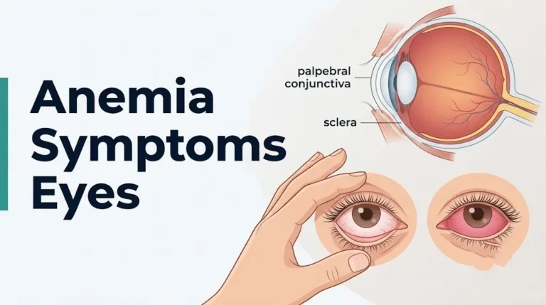
Pale conjunctiva, specifically pallor of the palpebral conjunctiva (the inner surface of the lower eyelid), is the most clinically established ocular sign of anemia and can be assessed by any person at home by gently pulling down the lower eyelid and examining its inner lining.
The palpebral conjunctiva is normally a vivid salmon-pink or deep red color from the network of capillaries visible through its transparent epithelium. When hemoglobin concentration falls, the blood in those capillaries carries less oxyhemoglobin, which is the bright red pigment responsible for the normal pink color. The capillaries become visible but pale, producing the characteristic washed-out appearance clinicians describe as conjunctival pallor.
A study published in the British Journal of Ophthalmology and referenced in clinical assessment guides found that conjunctival pallor, when assessed by trained clinicians, has a sensitivity of approximately 70 to 80 percent for detecting anemia with hemoglobin below 9 g/dL. Its specificity is higher, meaning pallor is a more reliable confirmatory than screening sign. The hemoglobin threshold at which pallor becomes visible to the naked eye in most adults is approximately 9 to 10 g/dL.
To assess your own palpebral conjunctiva for pallor:
- Stand in front of a mirror in good natural or bright artificial light.
- Wash your hands before touching your eyelid.
- Place your index finger on your lower eyelid and gently pull downward to expose the inner lining.
- Compare the color to a photo of normal salmon-pink conjunctiva: if your inner lid appears pale pink, white, or washed out, this is abnormal.
- Repeat for the other eye. Asymmetric pallor may indicate a local rather than systemic cause.
- Do not use this observation to self-diagnose anemia. Use it as a reason to schedule an appointment with your primary care physician for a complete blood count.
Children with nutritional iron deficiency anemia may show conjunctival pallor at hemoglobin levels above the adult threshold because their smaller blood volume makes hemoglobin changes more physiologically concentrated. Any child with pale inner eyelids warrants evaluation by a pediatrician.
Anemia and Blurry Vision or Visual Disturbance
Anemia causes blurry vision and visual disturbance through retinal and optic nerve hypoxia when hemoglobin falls below the threshold at which retinal autoregulation can maintain adequate oxygen delivery to the photoreceptor layer and retinal ganglion cells.
The visual disturbance of anemia is often described as a generalized dimming or haziness rather than a focal blind spot or sudden loss. This diffuse character reflects the widespread nature of retinal hypoxia when it is caused by low hemoglobin rather than a discrete vascular occlusion. Patients frequently describe it as looking through frosted glass or as difficulty reading in low-light conditions that they previously managed without difficulty.
Fatigue-related visual disturbance is a specific and clinically distinct pattern: visual acuity is normal at rest but degrades after sustained visual effort such as reading, driving, or computer work. The mechanism is analogous to exercising a muscle with inadequate oxygen supply. The photoreceptors and supporting cells of the retina metabolize rapidly during sustained visual attention, and when hemoglobin is reduced, this metabolic demand cannot be fully met. The result is visual fatigue that worsens with use.
According to the National Heart, Lung, and Blood Institute, anemia-related symptoms including fatigue and weakness are the result of tissues receiving inadequate oxygenation. The eye, as the highest per-gram oxygen consumer in the body, translates this insufficiency into visual symptoms before many other tissues show functional impairment.
People with type 2 diabetes who develop anemia face a compounded visual risk. Diabetic retinopathy already impairs retinal capillary function and reduces the oxygen extraction efficiency of the retinal microvasculature. Adding anemia-related hemoglobin reduction onto a retinal microvasculature that is already structurally compromised accelerates visual deterioration substantially compared to non-diabetic individuals at the same hemoglobin level. A board-certified ophthalmologist should evaluate any diabetic patient who develops new visual symptoms alongside a confirmed hemoglobin reduction.
Anemia and Eye Floaters, Flashes, and Field Defects
New onset eye floaters, flashing lights, or a shadow across the visual field in an anemic patient is a clinical red flag that may indicate retinal hemorrhage, retinal vascular occlusion, or sickle cell retinal vaso-occlusion, rather than the benign posterior vitreous detachment floaters that affect millions of adults.
Floaters in anemia arise through a specific mechanism. In sickle cell retinopathy, sickling events in the peripheral retinal vasculature produce retinal infarcts. These can lead to vitreous hemorrhage when new, fragile neovascular vessels bleed into the vitreous cavity. Blood in the vitreous appears to the patient as sudden, numerous dark floaters, often described as a shower of black dots or a spider web appearing suddenly. This is a distinct clinical emergency.
Flashing lights (photopsia) in anemic patients can indicate traction on the retina from neovascular membranes in proliferative sickle cell retinopathy, or from retinal ischemia directly stimulating retinal ganglion cells without adequate inhibitory signaling. The physiology differs from the harmless photopsia of posterior vitreous detachment in that anemia-associated photopsia from retinal ischemia carries a higher risk of progressing to retinal detachment or vascular occlusion.
Visual field defects, areas of missing or reduced vision in part of the visual field, in anemic patients may indicate:
- Retinal infarction from sickle cell vaso-occlusion in the peripheral retina
- Branch retinal artery occlusion in a patient with severe anemia reducing perfusion reserve
- Ischemic optic neuropathy affecting a segment of the optic nerve head
Any new visual field defect in an anemic patient, whether described as a dark spot, a missing patch, or a curtain effect, warrants same-day evaluation by a board-certified ophthalmologist or retinal specialist. Do not wait to see if it resolves.
Iron Deficiency Anemia Eye Symptoms
Iron deficiency anemia produces the most recognizable and most clinically common pattern of eye anemia symptoms: palpebral conjunctival pallor, visual fatigue, and in some cases, subconjunctival hemorrhage from altered blood fragility, without the severe retinal vascular complications seen in other anemia types.
Iron deficiency anemia is the most prevalent anemia type globally. The World Health Organization estimates it affects over 1.2 billion people. In the eye, it produces symptoms primarily through the hemoglobin-reduction pathway rather than through subtype-specific vascular mechanisms. The palpebral conjunctiva becomes pale as oxyhemoglobin concentrations fall. Visual fatigue and difficulty with sustained near vision develop as retinal metabolic demand begins to exceed oxygen supply at moderate to severe levels of hemoglobin reduction.
A specific finding in iron deficiency anemia that is absent in acid reflux and many other conditions is koilonychia (spoon-shaped nails) and glossitis alongside the ocular pallor, providing a clinical pattern that distinguishes iron deficiency from other causes of anemia. Clinicians who see pale conjunctiva combined with these systemic findings have strong clinical grounds for ordering a complete blood count with a serum ferritin level.
Iron deficiency anemia eye symptoms, in order of clinical frequency:
- Pale palpebral conjunctiva (inner lower eyelid), often the first visible sign
- Generalized visual fatigue and difficulty sustaining visual attention on tasks
- Mild blurring with prolonged reading or screen use that resolves with rest
- Subconjunctival hemorrhage in rare cases of severe anemia with altered platelet function
- Dark circles under the eyes from periorbital skin pallor (vasoconstrictive response to reduced cardiac output)
Menstruating women between the ages of 15 and 50 account for the largest demographic group with iron deficiency anemia-related eye symptoms due to monthly iron losses from menstruation. Women with heavy menstrual bleeding (menorrhagia) are at particular risk and should have serum ferritin levels checked by their primary care physician or obstetrician-gynecologist whenever they report progressive visual fatigue or conjunctival pallor.
Key Takeaway: Iron deficiency anemia produces eye symptoms through the general hemoglobin reduction pathway and does not typically cause retinal vascular damage, making it more reversible than sickle cell or aplastic anemia-related ocular complications if treated before severe deficiency develops.
Sickle Cell Anemia Eye Symptoms
Sickle cell anemia produces the most serious and potentially vision-threatening eye anemia symptoms of any anemia subtype, driven by mechanical occlusion of retinal and choroidal blood vessels by sickled red blood cells rather than by simple hemoglobin reduction.
The American Academy of Ophthalmology classifies sickle cell retinopathy into nonproliferative and proliferative stages. Nonproliferative sickle cell retinopathy includes peripheral retinal infarcts (visible as silver or white streaks on fundoscopy), salmon patch hemorrhages (intraretinal hemorrhages in the peripheral fundus), and black sunburst lesions from old hemorrhage sites. These findings may cause no noticeable visual symptoms if they remain in the peripheral retina away from the macula.
Proliferative sickle cell retinopathy is more dangerous. When peripheral retinal ischemia releases VEGF, neovascular fronds called sea fan neovascularization grow on the retinal surface and into the vitreous cavity. These fragile vessels bleed spontaneously, producing vitreous hemorrhage that appears as a sudden shower of dark floaters or a red tint to the visual field. Without treatment, neovascular traction can produce tractional retinal detachment, leading to permanent vision loss.
The eye symptoms that should prompt immediate evaluation in any patient with known sickle cell anemia include:
- Sudden onset of multiple new floaters or a sudden change in floater pattern
- Flashing lights, especially in the peripheral visual field
- Any shadow, curtain, or area of missing vision in part of the visual field
- Sudden blurring or loss of vision in one eye
- A sudden red tint or haziness to vision in one eye
Sickle cell hemoglobin C disease (HbSC) carries a higher retinopathy risk than homozygous sickle cell (HbSS), despite the somewhat milder systemic anemia profile of HbSC, because the higher hematocrit in HbSC produces more viscous blood that increases the risk of vascular stasis in the peripheral retinal circulation. All patients with any sickle cell genotype should have annual dilated fundus examination by a board-certified ophthalmologist or retinal specialist, beginning in childhood.
Pernicious Anemia and Optic Nerve Symptoms
Pernicious anemia produces optic nerve symptoms through a mechanism entirely distinct from hemoglobin reduction: vitamin B12 deficiency impairs myelin sheath synthesis in the optic nerve axons, producing a progressive demyelinating optic neuropathy that can cause permanent visual impairment if B12 deficiency remains uncorrected.
Pernicious anemia occurs when autoimmune destruction of gastric parietal cells eliminates production of intrinsic factor, the glycoprotein required for vitamin B12 absorption in the terminal ileum. Without intrinsic factor, B12 cannot be absorbed regardless of dietary intake. The resulting B12 deficiency disrupts the synthesis of myelin, the lipid-protein sheath that insulates the axons of the optic nerve (cranial nerve II), optic chiasm, and optic tracts.
Think of the optic nerve axons like electrical wires. Myelin is the insulation. When B12 deficiency degrades the insulation, the electrical signal traveling from the retina to the visual cortex becomes degraded, slow, and unreliable. The patient perceives this as a gradual, bilateral dimming or fogging of vision, color desaturation, and difficulty distinguishing contrast, rather than the sudden or focal vision loss associated with vascular occlusions.
The optic findings in pernicious anemia include:
- Optic disc pallor: a pale, washed-out appearance of the optic disc on fundoscopy, indicating loss of axonal tissue in the optic nerve head
- Reduced color vision: particularly along the red-green axis, detectable with Ishihara color plates
- Central or cecocentral scotoma: a visual field defect centered on or near the fixation point, often noticed as difficulty reading fine print
- Reduced visual acuity: typically bilateral and gradually progressive rather than sudden
Older adults over 60 following strict plant-based diets, those with prior gastric bypass surgery (which reduces intrinsic factor production), and those on long-term metformin therapy (which reduces ileal B12 absorption) are at highest risk for pernicious anemia-related optic neuropathy. A board-certified ophthalmologist noting optic disc pallor in an older adult should prompt referral to a primary care physician for serum B12 level measurement.
Aplastic Anemia and Retinal Hemorrhage Symptoms
Aplastic anemia causes retinal hemorrhage through thrombocytopenia rather than through hemoglobin-mediated hypoxia: the failure of the bone marrow to produce adequate platelets removes the primary barrier to spontaneous ocular bleeding, and retinal hemorrhage can occur even when hemoglobin levels are not critically low.
In aplastic anemia, autoimmune destruction of hematopoietic stem cells in the bone marrow reduces production of all blood cell lines simultaneously: red blood cells (producing anemia), white blood cells (producing immunosuppression), and platelets (producing thrombocytopenia). The retinal hemorrhages in aplastic anemia arise when platelet counts fall below 50,000 cells per microliter, at which point the normal platelet-mediated sealing of microvessel wall defects in the retinal capillary network becomes insufficient.
Research published in JAMA Ophthalmology has documented retinal hemorrhage as a presenting or early complication in aplastic anemia, sometimes occurring before the diagnosis is formally established. These hemorrhages appear on fundoscopy as flame hemorrhages (following the nerve fiber layer pattern of the inner retina), dot-and-blot hemorrhages (in the mid-retinal layers), and occasionally Roth spots (white-centered hemorrhages from fibrin or platelet aggregates), a pattern seen in other hematologic disorders including leukemia.
The visual impact of aplastic anemia retinal hemorrhages depends on location. Hemorrhages in the peripheral retina may produce no noticeable symptoms. Hemorrhages at or near the macula, which contains the fovea centralis responsible for central high-acuity vision, produce sudden central visual blurring, distortion, or a central blind spot. Hemorrhage into the vitreous from severe aplastic anemia-related thrombocytopenia produces the same floater-and-red-tint presentation as vitreous hemorrhage from other causes.
People with aplastic anemia who develop any sudden change in vision, new floaters, central visual distortion, or red tint to vision should be evaluated as an emergency by a board-certified ophthalmologist or retinal specialist on the same day, not at a routine appointment.
Key Takeaway: Aplastic anemia retinal hemorrhage is driven by platelet deficiency rather than hemoglobin level, meaning that a patient with aplastic anemia can have a dangerously low platelet count and severe retinal hemorrhage risk even at a hemoglobin level that does not yet produce visual fatigue or conjunctival pallor.
Chronic Anemia Eye Symptoms and Long-Term Ocular Complications
Chronic anemia produces a distinct ocular presentation from acute anemia: gradual structural changes in the optic nerve, retinal nerve fiber layer, and retinal vasculature accumulate over years, sometimes without causing noticeable visual symptoms until the damage is advanced.
The most clinically important chronic ocular complication of long-standing anemia is optic disc pallor, a sign that the optic nerve has lost axonal mass from sustained hypoxia. Unlike the acute optic disc pallor of ischemic optic neuropathy, which produces sudden vision loss, the optic disc pallor of chronic anemia develops insidiously and may be noticed by an ophthalmologist on a routine examination before the patient reports any visual complaint.
Chronic severe anemia also produces measurable thinning of the retinal nerve fiber layer (RNFL), the innermost retinal layer containing the axons of retinal ganglion cells traveling to the optic nerve. Research using optical coherence tomography (OCT), published in clinical ophthalmology literature, has documented RNFL thinning in patients with long-standing severe anemia from various causes, consistent with cumulative retinal ganglion cell loss from chronic hypoxic stress. This finding has clinical significance because RNFL thinning is irreversible once established.
The acute versus chronic distinction in anemia-related ocular symptoms:
| Feature | Acute Anemia | Chronic Anemia |
|---|---|---|
| Symptom onset | Rapid, days to weeks | Gradual, months to years |
| Conjunctival pallor | Present if severe | Present, may be adapted to by patient |
| Visual fatigue | Prominent early symptom | May normalize through adaptation |
| Optic disc pallor | Absent unless vascular event | May develop gradually |
| RNFL thinning | Absent acutely | Possible with prolonged severe disease |
| Retinal hemorrhage | If thrombocytopenia present | Can occur in any severe chronic anemia |
| Patient awareness | Often notices symptoms promptly | May not recognize gradual vision changes |
Patients with chronic kidney disease-related anemia deserve particular clinical attention. Renal anemia from reduced erythropoietin production coexists with hypertensive retinopathy and diabetic retinopathy in many of these patients, creating a compounded ocular risk environment that requires annual dilated fundus examination by a board-certified ophthalmologist regardless of subjective visual complaint.
Eye Anemia Symptoms in Specific Populations
The ocular manifestations of anemia differ substantially across specific population groups, and the standard clinical picture does not apply uniformly to pregnant women, children, older adults, or people with concurrent ocular disease.
Pregnant women experience physiological anemia of pregnancy from plasma volume expansion diluting red blood cell concentration. The American College of Obstetricians and Gynecologists defines anemia in pregnancy as hemoglobin below 11 g/dL in the first and third trimesters. Mild physiological anemia of pregnancy rarely produces significant ocular symptoms. However, women who develop iron deficiency anemia during pregnancy on top of the physiological hemodilution can reach hemoglobin levels of 7 to 8 g/dL, which, combined with the elevated cardiac output demands of pregnancy, can produce more prominent visual fatigue and occasionally conjunctival pallor. Any pregnant woman reporting new visual disturbance should be evaluated promptly by her obstetrician-gynecologist because visual symptoms in pregnancy can also indicate preeclampsia, an entirely different and potentially emergency-level condition that requires immediate differentiation.
Children with nutritional anemia from iron or vitamin B12 deficiency may show conjunctival pallor at hemoglobin levels that fall within what would be considered the mild-to-moderate range for adults, because the higher basal metabolic rate and rapid growth-related oxygen demands of childhood mean that retinal and systemic tissues reach the functional limit of reduced hemoglobin supply at higher thresholds. Parents who observe pale inner eyelids in their child alongside fatigue, reduced activity tolerance, or developmental concerns should arrange evaluation by the child’s pediatrician within days rather than weeks.
Older adults over 65 have reduced retinal vascular autoregulatory reserve from age-related vascular stiffness and reduced endothelial nitric oxide production. This means they develop clinically meaningful visual symptoms and optic nerve perfusion compromise at hemoglobin levels that would be tolerated without ocular consequence in younger adults. The American Society of Hematology notes that anemia in older adults is associated with worse functional outcomes than in younger populations.
People with pre-existing glaucoma face a specific and underappreciated risk. Any reduction in mean arterial pressure from anemia-related cardiovascular changes further reduces optic nerve perfusion pressure in an already pressure-compromised optic nerve head, increasing the risk of ischemic optic nerve damage at hemoglobin levels that would not threaten a non-glaucomatous eye.
How Anemia Eye Signs Are Diagnosed Clinically
Anemia eye signs are diagnosed through a combination of bedside clinical examination, fundoscopy, optical coherence tomography, and laboratory hematological testing, with the specific tests ordered depending on the ocular finding and the suspected anemia type.
The clinical examination begins with assessment of the palpebral conjunctiva: a clinician asks the patient to look up and gently everts the lower eyelid to expose the inner surface. Pallor is assessed and graded. The bulbar conjunctiva and sclera are also examined: yellow discoloration (scleral icterus) indicates elevated bilirubin from hemolytic anemia. The cornea may show Kayser-Fleischer rings in Wilson’s disease causing hemolytic anemia, though this is a rare specialized finding.
Fundoscopy (ophthalmoscopy) is the gold standard examination for detecting retinal anemia-related findings. A dilated fundus examination with an indirect ophthalmoscope or a slit-lamp with a condensing lens allows visualization of:
- Retinal hemorrhages and their morphological pattern (flame, dot-blot, Roth spots, preretinal)
- Optic disc appearance and pallor grading
- Retinal vasculature caliber and tortuosity
- Neovascularization in sickle cell retinopathy
- Peripheral retinal infarcts and pigmented lesions in sickle cell disease
Optical coherence tomography (OCT) provides a quantitative cross-sectional image of the retinal layers and measures retinal nerve fiber layer (RNFL) thickness, which can document subclinical ganglion cell loss in chronic anemia before it becomes visible on standard ophthalmoscopy.
The laboratory tests that complete the ocular anemia workup include:
- Complete blood count (CBC) with differential: hemoglobin, hematocrit, mean corpuscular volume (MCV), platelet count, white cell count
- Peripheral blood smear: to identify sickle cells, target cells, fragmented cells, or hypersegmented neutrophils (B12 deficiency)
- Serum ferritin and transferrin saturation: for iron deficiency anemia
- Serum vitamin B12 and folate: for pernicious or nutritional megaloblastic anemia
- Reticulocyte count: to assess bone marrow response
- Hemoglobin electrophoresis: to identify sickle cell genotype and thalassemia variants
Patients presenting with ocular findings suspected to be from anemia should be co-managed by both a primary care physician (for the hematological workup) and a board-certified ophthalmologist (for the ocular assessment and management). Sickle cell retinopathy specifically requires a retinal specialist (vitreoretinal surgeon) for evaluation and laser treatment of proliferative disease.
Anemia-Related Eye Symptoms vs. Other Eye Conditions
Anemia-related eye symptoms overlap with several other common eye conditions, and distinguishing them is clinically important because the treatments differ entirely and delaying the correct diagnosis can worsen outcomes.
The most important distinction is between anemia-related visual fatigue and early cataracts or refractive error. All three produce difficulty with sustained visual tasks and a sense that vision is not as clear as it once was. Cataracts produce a more consistent dimming that does not improve with rest, while anemia-related visual fatigue characteristically fluctuates with systemic energy level and improves after sleep or rest. Refractive error is correctable with a glasses prescription, while anemia-related blur is not correctable by lenses. The clinical test is a complete blood count combined with a refraction assessment and anterior segment examination.
Optic disc pallor from anemia versus glaucoma is a second clinically important distinction. Glaucoma produces optic disc cupping with pallor, while anemia-related optic atrophy produces diffuse pallor without the characteristic cup-to-disc ratio enlargement of glaucoma. OCT RNFL mapping differentiates the two patterns in most cases.
The scleral icterus of hemolytic anemia must be distinguished from hepatic jaundice (from liver disease), viral hepatitis, and Gilbert’s syndrome. All produce yellow discoloration of the sclera from elevated bilirubin, but the underlying cause, urgency, and management differ completely. Any new-onset yellow discoloration of the sclera requires blood testing including liver function tests, complete blood count, and bilirubin fractionation, ordered by a primary care physician.
| Eye Symptom | Anemia Cause | Alternative Diagnosis | Distinguishing Feature |
|---|---|---|---|
| Visual fatigue | Retinal hypoxia from low Hgb | Refractive error, early cataract | Anemia fatigue fluctuates with rest; not correctable by lenses |
| Pale inner eyelid | Low oxyhemoglobin in conjunctival capillaries | Normal variation in fair-skinned individuals | Confirmed by CBC showing low Hgb |
| Optic disc pallor | Axonal loss from chronic hypoxia or B12 deficiency | Glaucoma, previous optic neuritis | OCT cup-to-disc ratio distinguishes glaucoma; history distinguishes optic neuritis |
| Yellow sclera | Hemolysis-induced unconjugated hyperbilirubinemia | Liver disease, hepatitis, Gilbert’s | LFTs and bilirubin fractionation required |
| Retinal hemorrhage | Thrombocytopenia in aplastic anemia | Diabetic retinopathy, hypertensive retinopathy | CBC platelet count, blood glucose, blood pressure assessment |
People taking warfarin, heparin, or direct oral anticoagulants who also develop anemia face a compounded retinal hemorrhage risk, because anticoagulation plus thrombocytopenia (if aplastic anemia is the cause) produces a hemorrhagic risk that is substantially greater than either factor alone. This combination warrants urgent evaluation by a primary care physician or hematologist.
Key Takeaway: Anemia-related eye symptoms mimic several common eye conditions, and a complete blood count is the single most important test to add to any routine eye examination in a patient reporting visual fatigue, pallor of the inner eyelid, or unexplained retinal hemorrhage.
Emergency Eye Symptoms: When to Call 911 or Go to the ER
Certain eye symptoms associated with anemia require immediate emergency evaluation and cannot safely wait for a routine ophthalmology appointment.
Call 911 or go to the nearest emergency room immediately if you experience:
- Sudden vision loss in one or both eyes, which can indicate central retinal artery occlusion from anemia-related reduced perfusion pressure or sickle cell vascular occlusion; the treatment window for central retinal artery occlusion is approximately 4 to 6 hours from symptom onset
- A sudden curtain, shadow, or dark area appearing across part of your visual field, which can indicate retinal detachment from traction by sickle cell neovascular membranes or from severe retinal hemorrhage
- A sudden shower of new floaters, especially if accompanied by flashing lights, which in an anemic patient can indicate vitreous hemorrhage from sickle cell sea-fan neovascularization rupturing, requiring same-day retinal specialist evaluation
- Sudden onset of double vision (diplopia) combined with systemic symptoms of severe anemia (fainting, rapid heart rate, pallor, chest pain), which may indicate cerebrovascular ischemia from severe anemia-related reduced cerebral perfusion
- Sudden severe eye pain with redness and visual clouding, which can indicate acute angle-closure glaucoma triggered by anemia-related autonomic dysregulation, an ocular emergency requiring intraocular pressure reduction within hours
- Any new visual symptom in a patient with sickle cell anemia, regardless of how mild it seems, because sickle cell retinal vascular occlusion can progress rapidly and the treatment window for laser photocoagulation to prevent permanent vision loss is narrow
These presentations can indicate central retinal artery occlusion, ischemic optic neuropathy, vitreous hemorrhage, tractional retinal detachment, or sickle cell retinal vaso-occlusion, all of which require emergency medical assessment and not a phone call to an eye doctor’s answering service.
Frequently Asked Questions About Eye Anemia Symptoms
What do eyes look like with anemia?
The most visible sign of anemia in the eyes is pallor of the inner lower eyelid lining (the palpebral conjunctiva), which appears washed-out, pale pink, or nearly white instead of the normal vivid salmon-pink color.
The sclera (white of the eye) may appear slightly yellow in hemolytic anemia due to elevated bilirubin from red blood cell breakdown.
These changes are observable on physical examination, but confirmation requires a complete blood count ordered by a primary care physician.
Can anemia cause permanent eye damage?
Yes, anemia can cause permanent eye damage in specific circumstances, including ischemic optic neuropathy from severe anemia, proliferative sickle cell retinopathy with vitreous hemorrhage or tractional retinal detachment, and optic nerve axonal loss from vitamin B12 deficiency in pernicious anemia.
Damage from iron deficiency anemia-related visual fatigue is typically reversible with treatment of the underlying deficiency.
Sickle cell and pernicious anemia-related ocular complications carry the highest risk of permanent structural damage and require proactive monitoring by a board-certified ophthalmologist.
How do I check my own eyes for signs of anemia?
Pull your lower eyelid gently downward to expose the inner lining and compare its color to a normal salmon-pink reference: if it appears pale, whitish, or washed out, this is a reason to schedule a complete blood count with your primary care physician.
Dark circles under the eyes and a pale or grayish appearance to the skin around the eyes can accompany the conjunctival pallor of anemia, though these signs are less specific.
Self-examination can prompt you to seek evaluation but cannot confirm anemia or its severity.
Can anemia cause sudden vision loss?
Yes, anemia can cause sudden vision loss, and this represents a medical emergency.
Severe anemia can precipitate central retinal artery occlusion by reducing optic nerve perfusion pressure, and sickle cell anemia can cause sudden vision loss through vascular occlusion of the central retinal artery or vitreous hemorrhage from proliferative retinopathy.
Call 911 or go to the nearest emergency room immediately if you experience sudden vision loss in one or both eyes, as the treatment window for central retinal artery occlusion is approximately 4 to 6 hours from onset.
What type of anemia is most dangerous for eye health?
Sickle cell anemia poses the greatest risk to long-term eye health because it can produce proliferative retinopathy, vitreous hemorrhage, and tractional retinal detachment through a vascular occlusion mechanism that operates independently of the overall hemoglobin level.
Aplastic anemia carries severe short-term ocular hemorrhage risk from thrombocytopenia, and pernicious anemia carries irreversible optic nerve damage risk from B12 deficiency-related demyelination.
Iron deficiency anemia produces the mildest and most reversible ocular effects of the major anemia types, typically limited to conjunctival pallor and visual fatigue.
What doctor should I see for eye symptoms caused by anemia?
Start with your primary care physician for a complete blood count to confirm anemia and identify its type and severity.
Once anemia is confirmed, any visual symptoms beyond mild fatigue warrant referral to a board-certified ophthalmologist for a dilated fundus examination and OCT assessment of the retina and optic nerve.
Sickle cell patients with any new ocular symptoms should be evaluated urgently by a retinal specialist (vitreoretinal surgeon), and any patient experiencing sudden vision loss should go to the nearest emergency room immediately without waiting for an outpatient appointment.
The single most actionable thing you can do after reading this article is look at your inner lower eyelid in good light. If it looks pale, washed-out, or white rather than a vivid salmon-pink, schedule an appointment with your primary care physician for a complete blood count. Do not try to treat suspected anemia with dietary changes alone before confirming the type, because the treatments for iron deficiency, B12 deficiency, and sickle cell-related anemia are entirely different.
If you have already been diagnosed with anemia and develop any new visual symptom beyond mild eye fatigue, ask your primary care physician for a referral to a board-certified ophthalmologist. If you have sickle cell disease and notice new floaters, flashes, or any area of missing vision, go to an emergency room the same day.