Anemia Symptoms Eyes: Every Eye Sign Explained 2026
Anemia symptoms in the eyes include pale inner eyelids, yellowing of the whites, blurred vision, retinal hemorrhage, floaters, and dark circles, and each of these signs has a distinct anatomical and physiological explanation tied to the specific type and severity of anemia present. The eyes are not a secondary symptom location in anemia: for clinicians assessing a patient without access to immediate bloodwork, the inner eyelid is one of the fastest, most reliable physical exam sites for detecting low hemoglobin.
The World Health Organization defines anemia as a hemoglobin concentration below 13 g/dL in adult males, below 12 g/dL in non-pregnant adult females, and below 11 g/dL in pregnant women. According to the National Heart, Lung, and Blood Institute, anemia affects approximately 3 million Americans, with iron-deficiency anemia representing the most common subtype worldwide. Yet the eye-specific presentation of different anemia subtypes, including which signs point to hemolytic causes versus iron deficiency versus sickle cell disease, is almost never explained in depth in public-facing health content.
This article covers every clinically recognized eye sign of anemia, explains exactly what is happening in the ocular anatomy and vasculature to produce each sign, differentiates findings by anemia subtype, and tells you precisely when each sign warrants a routine appointment versus same-day evaluation versus an emergency room visit.
Anemia Symptoms Eyes: What the Eyes Reveal About Low Blood Counts
The eyes reveal anemia through changes in the color of the inner eyelids, the whites of the eyes, and the retinal blood vessels at the back of the eye, each reflecting a different physiological consequence of reduced hemoglobin or altered red blood cell structure.
Not all eye signs of anemia appear together. A patient with iron-deficiency anemia typically shows pale inner eyelids without any yellowing of the sclera. A patient with hemolytic anemia, where red blood cells are being destroyed faster than they are made, typically shows yellow sclera (scleral icterus) as the primary visible eye sign. A patient with severe aplastic anemia may show retinal hemorrhage visible only on fundoscopic examination by a clinician. Understanding which sign accompanies which subtype allows for more targeted clinical suspicion before laboratory results are available.
Key eye signs of anemia by clinical category:
| Eye Sign | Anemia Type Most Associated | Clinical Significance |
|---|---|---|
| Pale inner eyelids (conjunctival pallor) | Iron-deficiency, aplastic, chronic disease anemia | Reflects low hemoglobin in conjunctival capillaries |
| Yellow sclera (scleral icterus) | Hemolytic anemia, sickle cell disease | Reflects unconjugated bilirubin deposition in scleral elastin |
| Blurred vision | Moderate-to-severe anemia (any type) | Retinal hypoxia reducing photoreceptor function |
| Retinal hemorrhage | Severe anemia (Hgb below 8 g/dL), aplastic anemia | Capillary breakdown under hypoxic stress |
| Eye floaters | Severe anemia, pre-retinal hemorrhage | Vitreous involvement, requires urgent evaluation |
| Dark circles under eyes | Chronic iron-deficiency, nutritional anemia | Dermal hypopigmentation and vascular dilation |
The eyes are particularly useful for clinical assessment in individuals with darker skin tones, where skin pallor is less visible due to melanin concentration in the epidermis. The palpebral conjunctiva (inner eyelid lining) contains a very thin, non-pigmented epithelium overlying a dense capillary network, making it visible regardless of skin tone. Clinicians have used conjunctival pallor as a bedside anemia screening tool in resource-limited settings where complete blood count testing is not immediately available.
How Anemia Affects the Eyes: The Physiological Mechanism
Anemia affects the eyes through three primary physiological mechanisms: reduced hemoglobin concentration in ocular microvasculature, elevated unconjugated bilirubin from hemolysis depositing in scleral tissue, and retinal hypoxia caused by inadequate oxygen delivery.
Each red blood cell (erythrocyte) carries four molecules of hemoglobin, and each hemoglobin molecule binds four oxygen molecules at the iron-containing heme groups. The vivid red color of oxygenated blood comes from oxyhemoglobin. When hemoglobin concentration falls below normal thresholds, blood carries less oxygen per unit volume, and the visual appearance of blood in superficial capillaries shifts from deep red toward a pale pinkish-white.
The posterior conjunctival arteries, branches of the ophthalmic artery (itself the first branch of the internal carotid artery), supply a dense subepithelial capillary plexus within the palpebral conjunctiva. This capillary bed lies directly beneath a translucent epithelial layer only a few cell layers thick. The color seen through this thin covering reflects the hemoglobin saturation and concentration of the blood flowing through these capillaries in real time. Think of it like a screen printed over a color-changing liquid: when the liquid is richly red, the screen looks pink-red; when the liquid is pale and dilute, the screen looks white or pale salmon.
For the retina, the mechanism is different. The central retinal artery enters the eye through the optic disc, branching into four main quadrants of retinal arterioles. The retina is one of the most metabolically active tissues in the human body per unit weight, consuming oxygen at a rate comparable to brain tissue. According to research published in JAMA Ophthalmology, severe anemia (hemoglobin below approximately 8 g/dL) produces measurable retinal hypoxia that triggers compensatory vascular dilation, capillary microaneurysm formation, and eventually capillary rupture producing retinal hemorrhage.
Pregnant women with anemia may notice eye symptoms at slightly higher hemoglobin levels than non-pregnant adults. Pregnancy causes a physiological plasma volume expansion of approximately 45 to 50 percent that dilutes hemoglobin, but it also increases retinal and conjunctival vascular flow. This means that the hemoglobin threshold at which ocular symptoms appear may differ from non-pregnant adults, and assessment should use pregnancy-specific normal ranges throughout gestation.
Pale Conjunctiva: The Most Reliable Eye Sign of Anemia
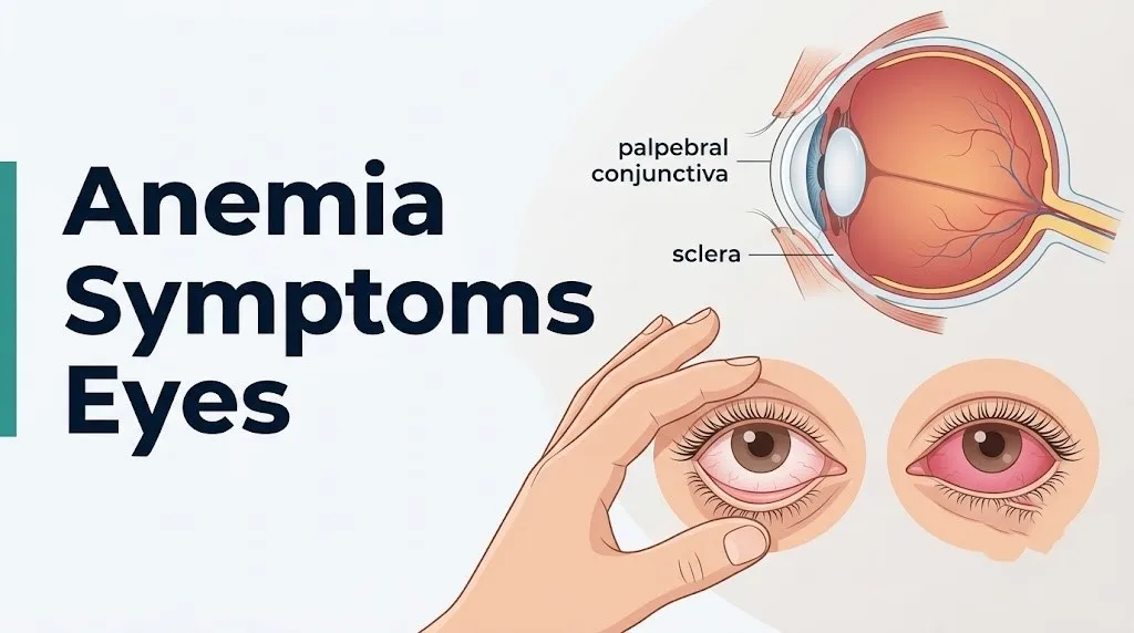
Pale conjunctiva, specifically pallor of the palpebral conjunctiva (the moist inner surface of the lower eyelid), is the most clinically reliable eye sign of anemia and can be assessed without any equipment in under 10 seconds.
The palpebral conjunctiva is preferred over the bulbar conjunctiva (the thin covering over the white of the eye) for pallor assessment because it is more richly vascularized and its capillary plexus sits closer to the surface. A published clinical accuracy study in Annals of Internal Medicine found that palpebral conjunctival pallor assessed by clinicians showed a specificity of approximately 82 percent and sensitivity of approximately 49 to 75 percent for detecting hemoglobin levels below 11 g/dL, depending on the experience of the examiner. This makes it a useful screening tool, not a diagnostic one, but it is substantially more informative than skin pallor assessment.
In healthy adults with normal hemoglobin levels, the inner surface of the lower eyelid appears deep pink to red. As hemoglobin concentration falls, the color transitions:
| Conjunctival Color | Approximate Hemoglobin Range | Clinical Interpretation |
|---|---|---|
| Deep pink to red | Greater than 11 g/dL | Normal in most adults |
| Light pink | Approximately 8 to 11 g/dL | Mild to moderate anemia possible |
| Pale pink to salmon | Approximately 6 to 8 g/dL | Moderate anemia, clinical evaluation needed |
| White or very pale | Below approximately 6 g/dL | Severe anemia, urgent evaluation |
It is worth noting that conjunctival color is not perfectly linear with hemoglobin levels. Vasomotor tone, ambient temperature, and hydration status all affect capillary filling in the palpebral conjunctiva. A well-hydrated, warm individual may show a deeper color than their hemoglobin level would predict. For this reason, conjunctival pallor is a screening signal, not a diagnostic endpoint.
Children have a higher compensatory response to anemia through increased cardiac output before symptoms appear, which means conjunctival pallor in a child may appear at lower hemoglobin levels than in older adults. In elderly patients, baseline conjunctival vascularity may be reduced, making color assessment less reliable.
How to Check for Anemia in the Eyes at Home
You can perform a basic conjunctival pallor self-check at home by gently pulling down the lower eyelid to expose the inner surface and comparing its color to a reference.
To examine the inner eyelid for signs of anemia:
- Stand in front of a mirror in good natural daylight or under a bright white LED light (avoid warm-toned or yellow incandescent lighting, which skews color perception)
- Wash your hands thoroughly before touching your face or eyelid
- Using a clean index finger, gently pull the lower eyelid downward to expose the entire inner surface (palpebral conjunctiva)
- Look at the color of the inner surface: it should appear deep pink to red in a person with normal hemoglobin levels
- Compare both eyes: color should be symmetric; asymmetric pallor in one eye suggests a local eye condition rather than systemic anemia
- Note whether the color is uniformly pale across the whole inner surface or patchy: uniform pallor is more consistent with systemic anemia; patchy paleness may reflect local vascular changes
Quick Tip:
- Photograph the inner eyelid in good lighting to share with your primary care physician if you are concerned: color documentation helps the clinician assess change over time.
- Do not use this check as a substitute for blood testing. A complete blood count (CBC) is the only way to confirm anemia and measure hemoglobin accurately.
- Individuals taking beta-blockers or other vasoconstrictors may have paler conjunctiva than their hemoglobin level warrants, because these medications reduce peripheral capillary filling.
The home check is a directional signal only. If you observe pale inner eyelids alongside fatigue, shortness of breath, rapid heartbeat, dizziness, or unusual cold in the hands and feet, these combined findings warrant a call to your primary care physician for a same-week appointment and a CBC.
Key Takeaway: The inner surface of the lower eyelid (palpebral conjunctiva) is the most reliable eye site for spotting anemia pallor, and it works regardless of skin tone, but it screens rather than diagnoses: a complete blood count is always required for confirmation.
Anemia and Yellow Eyes: Scleral Icterus Explained
Yellow coloration of the whites of the eyes in anemia is called scleral icterus and occurs specifically in hemolytic anemia subtypes when the accelerated destruction of red blood cells overloads the liver’s capacity to process bilirubin.
The mechanism is precise. When red blood cells are destroyed, the heme component of hemoglobin is broken down by macrophages in the spleen and liver through the enzyme heme oxygenase, producing biliverdin, which is then reduced to unconjugated (indirect) bilirubin. The liver normally conjugates bilirubin (attaches glucuronic acid via UDP-glucuronosyltransferase) to make it water-soluble for biliary excretion. In hemolytic anemia, the rate of red cell destruction outpaces the liver’s conjugating capacity. Unconjugated bilirubin accumulates in the bloodstream and deposits preferentially in elastin-rich connective tissue, which is abundant in the sclera, because unconjugated bilirubin has a high affinity for elastin.
The sclera turns yellow before the skin shows jaundice because scleral elastin binds bilirubin at lower serum concentrations. Clinically, scleral icterus becomes visible when the serum bilirubin level exceeds approximately 2 to 3 mg/dL, while skin jaundice typically requires levels above 5 mg/dL, according to data referenced by the Mayo Clinic in its clinical education materials.
Yellow eyes in anemia do not mean liver disease, though liver disease also causes yellow eyes through a related but distinct mechanism (hepatocellular conjugation failure rather than hemolytic overproduction). Distinguishing hemolytic anemia from hepatic jaundice requires laboratory testing including a fractionated bilirubin panel, reticulocyte count, peripheral blood smear, and liver function tests.
Individuals with glucose-6-phosphate dehydrogenase (G6PD) deficiency, the most common enzyme deficiency in the world affecting an estimated 400 million people according to the World Health Organization, are at particular risk for episodic hemolytic crises triggered by oxidative stress (from certain medications, infections, or fava bean ingestion). During these episodes, scleral icterus can appear within hours and may be the first visible sign of a hemolytic crisis.
Anemia and Blurred Vision
Blurred vision in anemia results from retinal tissue hypoxia caused by inadequate oxygen delivery to the photoreceptors and retinal ganglion cells when hemoglobin concentration falls below the level needed to meet the retina’s high metabolic demands.
The retina is one of the most oxygen-dependent tissues in the body. Its photoreceptors (rods and cones) and the retinal ganglion cells whose axons form the optic nerve require continuous, high-volume oxygen delivery. The central retinal artery supplies the inner retinal layers. The choriocapillaris (a dense capillary layer in the choroid behind the retinal pigment epithelium) supplies the outer retinal layers including the photoreceptors. When hemoglobin drops severely, both supply pathways deliver less oxygen per unit volume, and retinal cell function begins to deteriorate.
The result is visual blurring, typically bilateral and gradual in onset with chronic anemia. It is not the sharp, sudden-onset blurring of a retinal artery occlusion: it tends to be described as a general haziness or difficulty focusing, worse in low-light conditions (reflecting preferential rod photoreceptor impairment under hypoxia).
According to the National Eye Institute, any new or worsening blurred vision warrants ophthalmic evaluation, and blurred vision occurring alongside known anemia, particularly severe anemia with hemoglobin below 8 g/dL, should prompt same-day evaluation rather than a scheduled appointment weeks out.
Blurred vision in anemia can affect individuals across age groups, but older adults are at higher risk for compounding factors. Age-related reduction in retinal vascular reserve (reduced choroidal blood flow capacity) means that even moderate anemia may produce more pronounced visual symptoms in a person over 65 than in a younger adult with the same hemoglobin level. Clinicians managing anemia in older patients should ask specifically about visual symptoms at each assessment.
Anemia Retinal Hemorrhage and Vision Changes
Retinal hemorrhage in anemia occurs when severe hemoglobin deficiency pushes the retinal microvasculature past its compensatory capacity, causing capillary wall breakdown and bleeding into the layers of the retina or the vitreous cavity.
The retinal microvasculature compensates for reduced hemoglobin-carried oxygen by vasodilating (widening the caliber of arterioles) to increase blood flow volume. This is an effective compensatory mechanism down to a threshold. Below hemoglobin levels of approximately 8 g/dL, and particularly below 6 g/dL, the combination of hypoxia, increased vascular wall stress from dilated vessels, and reduced platelet function (which often co-occurs with severe bone marrow failure anemia) leads to capillary rupture. Blood escapes into the surrounding retinal tissue.
Retinal hemorrhages in anemia take several forms depending on the depth and location of the bleed:
- Flame hemorrhages: occur in the superficial nerve fiber layer, producing a feathered, flame-shaped appearance on fundoscopy
- Dot-blot hemorrhages: occur in the deeper retinal layers, appearing as round, well-demarcated red dots
- Pre-retinal (subhyaloid) hemorrhages: occur between the retina and vitreous, can be large, may produce a visible fluid level on fundoscopy
- Vitreous hemorrhage: blood enters the vitreous cavity, producing sudden floaters, blurring, or complete loss of red reflex
Research published in JAMA Ophthalmology has documented retinal hemorrhage as a recognized complication of severe anemia across subtypes including aplastic anemia, hemolytic anemia, and severe iron-deficiency anemia in the setting of concurrent thrombocytopenia (low platelet count). Aplastic anemia is particularly associated with retinal hemorrhage because it simultaneously depresses hemoglobin and platelet production.
Individuals with aplastic anemia being managed with immunosuppressive therapy or awaiting bone marrow transplantation should have scheduled dilated fundus examinations to screen for subclinical retinal hemorrhage, as small bleeds near the macula can threaten central vision without producing dramatic symptoms.
Key Takeaway: Retinal hemorrhage in anemia is a complication of severe hemoglobin deficiency (typically below 8 g/dL), particularly when low platelet counts co-occur, and requires dilated fundus examination by an ophthalmologist to detect before it threatens central vision.
Anemia Eye Floaters and Visual Disturbances
Eye floaters in anemia represent a symptom that demands careful clinical attention because, depending on their characteristics, they may indicate pre-retinal or vitreous hemorrhage rather than benign posterior vitreous detachment.
Floaters are perceived when particles or debris within the vitreous humor (the gel-filled cavity that makes up approximately 80 percent of the eye’s volume) cast shadows on the retina. In anemia-related ocular disease, floaters can arise from two distinct mechanisms. The first is benign posterior vitreous detachment (PVD), which is more common in the general population and is not directly caused by anemia. The second, more concerning mechanism is pre-retinal or vitreous hemorrhage: blood entering the vitreous from ruptured retinal capillaries produces red blood cells that the patient perceives as sudden, numerous, dark floaters, sometimes described as “a shower of dots” or “a dark curtain.”
The clinical distinction matters enormously. A few stable floaters of gradual onset that have been present for months suggest benign vitreous changes. Sudden onset of multiple floaters, especially when accompanied by flashes of light (photopsia from mechanical retinal traction) or a dark shadow at the edge of vision (peripheral field defect), suggests hemorrhage or retinal tear and requires same-day emergency ophthalmic evaluation.
Floaters appearing in a patient with known severe anemia or known sickle cell disease should be assumed to represent vitreous or pre-retinal hemorrhage until proven otherwise by a dilated fundus examination by an ophthalmologist or retinal specialist.
Older adults are at greater baseline risk for posterior vitreous detachment, meaning that floaters in an older adult with anemia may reflect either condition or both simultaneously. A board-certified ophthalmologist, not a primary care physician, should evaluate new floaters in this demographic given the compounding risk factors.
Anemia Eye Twitching: Is It Related to Anemia?
Eye twitching (myokymia) in anemia is most likely related to the electrolyte imbalances, magnesium depletion, and generalized neuromuscular fatigue that often accompany nutritional deficiency anemias rather than being a direct consequence of low hemoglobin itself.
Myokymia is an involuntary, repetitive contraction of the orbicularis oculi muscle (the ring-shaped muscle surrounding the eye that controls eyelid closure), typically affecting the lower eyelid. It is produced by spontaneous discharge from the facial nerve (cranial nerve VII) motor branches supplying the orbicularis oculi. The most common triggers in the general population are fatigue, caffeine excess, and magnesium deficiency.
Iron-deficiency anemia frequently co-occurs with magnesium deficiency and other micronutrient gaps, particularly in individuals with restrictive diets, malabsorption syndromes (celiac disease, inflammatory bowel disease), or chronic blood loss. Magnesium plays a direct role in stabilizing neuromuscular membrane potential: when intracellular magnesium falls, spontaneous motor nerve discharge becomes more likely, producing the twitching pattern.
Anemia-related fatigue may independently worsen eye twitching severity by increasing the overall excitability of an already fatigued neuromuscular system. This is consistent with the clinical observation that twitching is more common and more prolonged in periods of physical exhaustion and poor nutritional intake.
Eyelid twitching alone is not a red flag sign of anemia. If twitching is persistent (lasting more than two weeks continuously), affects both eyelids simultaneously, spreads beyond the eyelid to the face or cheek, or is accompanied by other involuntary movements, a neurological evaluation by a primary care physician or neurologist is warranted to rule out benign essential blepharospasm or hemifacial spasm, which are unrelated to anemia.
Women of reproductive age with iron-deficiency anemia frequently report muscle cramps, spasms, and twitching as part of their symptom picture, possibly reflecting concurrent magnesium and calcium absorption impairment in the iron-deficient intestinal mucosa.
Key Takeaway: Eye twitching in anemia reflects neuromuscular irritability from co-occurring nutritional deficiencies (particularly magnesium) rather than low hemoglobin directly, but persistent or spreading twitching needs neurological assessment regardless of anemia status.
Anemia Dry Eyes and Eye Discomfort
Dry eyes in anemia are associated with reduced tear film quality and quantity, particularly in iron-deficiency and vitamin B12-deficiency anemia subtypes, through mechanisms affecting the glandular tissue responsible for tear production.
The lacrimal gland (located at the superolateral orbit) and the meibomian glands (sebaceous glands embedded in the tarsal plates of the eyelids, secreting the lipid layer of the tear film) both require adequate oxygenation and nutritional support to maintain secretory function. The tear film that covers the cornea has three layers: an inner mucin layer (secreted by conjunctival goblet cells), a middle aqueous layer (secreted by the lacrimal gland), and an outer lipid layer (secreted by the meibomian glands). Disruption of any layer produces dry eye symptoms.
In vitamin B12-deficiency anemia (pernicious anemia), peripheral neuropathy affecting the autonomic fibers of the facial nerve (cranial nerve VII) that stimulate lacrimal gland secretion may reduce basal tear production. Research published in the British Journal of Ophthalmology has found an association between vitamin B12 deficiency and reduced Schirmer test scores (a clinical measure of tear production), though the evidence remains observational rather than from controlled trials.
In iron-deficiency anemia, hypoxic stress on conjunctival goblet cells (which produce mucin, the foundation layer of the tear film) may reduce goblet cell density over time, impairing tear film stability. A reduced tear film breakup time (the interval before a dry patch appears on the cornea after blinking) has been reported in some iron-deficient patients.
Dry eye symptoms include:
- Gritty or sandy sensation in the eyes
- Burning or stinging, particularly in air-conditioned or dry environments
- Excessive tearing (paradoxically: the eye tries to compensate for chronic dryness with reflex tearing)
- Blurred vision that temporarily clears with blinking
- Sensitivity to light (photophobia)
Individuals with autoimmune conditions associated with anemia, particularly systemic lupus erythematosus (SLE) or Sjögren’s syndrome, may have co-occurring dry eye disease as a primary feature of the autoimmune process itself, separate from the anemia-related mechanism.
Anemia Dark Circles Under Eyes
Dark circles under the eyes in anemia are produced by a combination of dermal vascular dilation, reduced hemoglobin-driven skin color change, and in some cases thinning of the periorbital skin from nutritional deficiency.
The periorbital skin (skin immediately surrounding the eyes) is among the thinnest skin on the body, measuring approximately 0.5 mm in thickness. The orbicularis oculi muscle and the underlying periosteum of the orbital rim are visible through this thin layer, and the venous plexus in this region produces the bluish-purple undertone that most people recognize as “dark circles.” When hemoglobin is low and the blood’s oxygen-carrying capacity is reduced, venous blood (deoxygenated, darker in color) may contribute more visibly to the periorbital appearance.
In iron-deficiency anemia specifically, reduced iron availability impairs the synthesis of collagen (iron is a cofactor for prolyl hydroxylase and lysyl hydroxylase, the enzymes required for collagen crosslinking). Thinner, less structurally supported periorbital skin allows the underlying vascular and muscular structures to show through more prominently.
Anemia-related dark circles tend to be bilateral and are typically accompanied by other systemic anemia symptoms such as fatigue, pallor, and cold extremities. They are not a specific clinical marker for anemia on their own, as dark circles have multiple causes including genetics, chronic sleep deprivation, allergic shiners (venous congestion from nasal allergy), and photoaging.
Dark circles associated with anemia in children, particularly in combination with pale inner eyelids and fatigue, should prompt a primary care pediatrician evaluation including a CBC, especially if dietary iron intake is poor or if the child has a history of pica (craving non-food substances, a recognized sign of iron-deficiency anemia).
Sickle Cell Anemia Eye Symptoms
Sickle cell disease (SCD) produces ocular complications through a mechanism entirely distinct from iron-deficiency or nutritional anemia: the polymerization of abnormal hemoglobin S (HbS) under low-oxygen conditions causes red blood cells to deform into a sickle shape, obstructing the small-caliber microvasculature of the peripheral retina.
The retinal vasculature in sickle cell disease develops a pattern of progressive ischemic damage called proliferative sickle retinopathy (PSR). The peripheral retina (particularly in the superotemporal quadrant) receives blood through the most distal, smallest arterioles: these are the first vessels to be occluded by sickled erythrocytes during hypoxic episodes. Chronic vaso-occlusion produces peripheral retinal ischemia, which stimulates upregulation of vascular endothelial growth factor (VEGF). VEGF drives abnormal new blood vessel growth (neovascularization) from the retinal surface into the vitreous. These fragile new vessels can rupture, producing vitreous hemorrhage, or contract and pull on the retina, causing tractional retinal detachment.
PSR staging, described by the Goldberg classification:
- Stage I: peripheral arteriolar occlusions (no symptoms)
- Stage II: peripheral arteriovenous anastomoses (no symptoms)
- Stage III: neovascularization (“sea-fan” formations, often asymptomatic)
- Stage IV: vitreous hemorrhage (sudden floaters, vision loss)
- Stage V: tractional retinal detachment (severe, potentially permanent vision loss)
The American Academy of Ophthalmology recommends annual dilated fundus examination beginning at age 10 for all patients with sickle cell disease, specifically to detect PSR in the earlier, asymptomatic stages before vitreous hemorrhage or detachment occurs.
Patients with hemoglobin SC disease (a compound heterozygous form) have a paradoxically higher rate of PSR than patients with homozygous HbSS disease, because higher hematocrit in SC disease produces greater blood viscosity and more complete microvascular occlusion, according to research published in the British Journal of Haematology.
Key Takeaway: Sickle cell disease produces proliferative retinopathy through peripheral retinal vaso-occlusion and VEGF-driven neovascularization, a mechanism entirely different from simple hemoglobin deficiency, and annual dilated eye exams starting at age 10 are the AAO-recommended standard for all SCD patients.
Iron Deficiency Anemia Eye Symptoms
Iron-deficiency anemia produces eye symptoms primarily through reduced hemoglobin concentration (causing pallor and hypoxic effects) and through iron’s direct roles in ocular tissue metabolism beyond erythropoiesis.
Iron is required for the function of multiple enzymes throughout the body. In the eye, iron-dependent enzymes include cytochrome c oxidase (a key enzyme in the mitochondrial electron transport chain, present in retinal photoreceptors and the retinal pigment epithelium), ribonucleotide reductase (required for DNA synthesis in rapidly dividing conjunctival and corneal epithelial cells), and the collagen-synthesis hydroxylases described previously. When iron stores are depleted, these enzymatic processes operate below capacity.
Specific eye symptoms most associated with iron-deficiency anemia include:
- Pale palpebral conjunctiva (the most reliable physical sign)
- Dry, irritated eyes from reduced goblet cell mucin secretion
- Fatigue-related blurring (difficulty maintaining visual focus during sustained tasks)
- Dark periorbital circles from dermal thinning and vascular visibility
- In severe deficiency: blurred vision and, rarely, papilledema (optic disc swelling) from intracranial hypertension associated with severe iron-deficiency anemia in children
The association between iron-deficiency anemia and idiopathic intracranial hypertension (IIH, pseudotumor cerebri) is supported by case series and observational data, though the mechanism is not fully established. One hypothesis involves altered cerebrospinal fluid absorption via iron-dependent metabolic processes. IIH produces papilledema (optic disc swelling visible on fundoscopy) and can threaten optic nerve function if untreated.
Women of reproductive age represent the highest-risk demographic for iron-deficiency anemia, with the National Heart, Lung, and Blood Institute estimating that approximately 12 percent of US women aged 12 to 49 have iron-deficiency anemia. For this group, the combination of heavy menstrual bleeding and inadequate dietary iron intake creates a cumulative deficit that may take months to produce symptomatic anemia and visible eye changes.
Anemia Eye Symptoms During Pregnancy
Anemia during pregnancy produces eye symptoms at hemoglobin thresholds different from the non-pregnant adult because physiological plasma volume expansion alters the relationship between circulating hemoglobin concentration and actual oxygen delivery capacity.
During pregnancy, plasma volume increases by approximately 45 to 50 percent beginning in the first trimester and peaking around 28 to 32 weeks gestation. Red blood cell mass also increases, but by only approximately 18 to 30 percent (without iron supplementation). This relative dilution produces a physiological fall in hemoglobin that is distinct from true anemia. The WHO and the American College of Obstetricians and Gynecologists (ACOG) define anemia in pregnancy as hemoglobin below 11 g/dL in the first and third trimesters, and below 10.5 g/dL in the second trimester.
Eye symptoms during pregnancy-associated anemia:
- Pale inner eyelids: appear at hemoglobin levels proportionally similar to non-pregnant ranges, though plasma dilution can make clinical assessment less reliable
- Blurred vision: must be carefully evaluated in pregnancy because it can also indicate preeclampsia (a hypertensive disorder of pregnancy), which carries its own retinal pathology including cortical visual disturbances, retinal artery spasm, and rarely, hypertensive retinopathy
- Visual disturbances including scotomas (blind spots), flashes of light, or sudden vision changes in a pregnant woman should be treated as a potential preeclampsia emergency regardless of whether anemia is present
The co-occurrence of anemia and hypertensive disorders in pregnancy makes symptom interpretation more complex. A pregnant woman who reports blurred vision or other eye changes should contact her obstetrician or obstetric emergency triage line immediately rather than attributing the symptom to anemia alone.
Gestational vitamin B12 deficiency, more common in vegan and vegetarian pregnancies, can also produce anemia (megaloblastic) with associated optic neuropathy risk if left untreated. Any pregnant woman with a plant-based diet and visual symptoms alongside anemia symptoms should have B12 levels tested as part of a prenatal anemia workup, as managed by her obstetrician-gynecologist.
When to See a Doctor for Anemia Eye Symptoms
Any eye sign of anemia warrants a medical appointment, but the urgency of that appointment depends on which symptom is present, how rapidly it developed, and whether it is accompanied by systemic anemia symptoms or underlying conditions that increase ocular risk.
To determine the appropriate level of care, organize symptoms by urgency:
| Symptom | Urgency Level | Who to Contact |
|---|---|---|
| Pale inner eyelids without other symptoms | Routine appointment this week | Primary care physician, request CBC |
| Pale eyelids with fatigue, shortness of breath, palpitations | Same week, sooner if symptoms worsen | Primary care physician |
| Yellow sclera (new onset) | Within 24 to 48 hours | Primary care physician (bilirubin panel needed) |
| Blurred vision (gradual, bilateral) with known anemia | Within days | Primary care physician or ophthalmologist |
| New-onset floaters or flashes of light | Same day | Ophthalmologist (dilated fundus exam) |
| Sudden vision loss (any degree, any eye) | Emergency: go immediately | Emergency room or call 911 |
| Visual field defect or dark shadow in vision | Emergency | Emergency room or call 911 |
| Eye symptoms in pregnancy with headache or high blood pressure | Emergency | Obstetric emergency triage or 911 |
What to tell your primary care physician at the appointment:
- Describe when you first noticed the eye change and whether it has progressed
- List all associated symptoms: fatigue, shortness of breath, heart racing, pallor, cold extremities, dizziness on standing
- Report your diet (vegetarian, vegan, restricted intake) and any recent blood loss (heavy periods, gastrointestinal bleeding, recent surgery)
- Name all current medications including NSAIDs (which increase GI blood loss risk), proton pump inhibitors (which reduce iron absorption), and metformin (associated with B12 malabsorption)
- Note any family history of sickle cell disease, G6PD deficiency, or hereditary hemolytic anemia
- Bring a photograph of your inner eyelid taken in good light if you observed pallor at home
Emergency Symptoms: When to Call 911 or Go to the ER
Certain eye symptoms associated with anemia require immediate emergency evaluation. Do not wait to see if these resolve on their own.
Call 911 or go to the nearest emergency room immediately if you experience:
- Sudden, partial, or complete vision loss in one or both eyes, regardless of whether anemia is already known, because this can indicate central retinal artery occlusion, retinal detachment, or severe vitreous hemorrhage requiring same-hour intervention
- A sudden shower of floaters combined with flashing lights, which signals possible retinal tear or detachment with associated vitreous hemorrhage, a time-sensitive surgical emergency
- A dark curtain or shadow spreading across your visual field, indicating retinal detachment that can become permanent without immediate surgical repair
- Any visual change in a pregnant woman accompanied by headache, swollen hands or face, or blood pressure readings above 140/90 mmHg, as this constellation indicates possible preeclampsia with severe features, not anemia
- Profound pallor, fainting, and severe shortness of breath alongside visual dimming, suggesting hemoglobin has dropped to a critically low level requiring urgent blood transfusion evaluation
- Sudden eye pain combined with vision loss and nausea, which may indicate acute angle-closure glaucoma (unrelated to anemia but presenting similarly in the emergency context)
These presentations can indicate retinal detachment, central retinal artery occlusion, hypertensive crisis in pregnancy, or critically severe anemia and require emergency medical assessment, not an urgent care appointment or a scheduled ophthalmology visit.
Frequently Asked Questions About Anemia Symptoms Eyes
What do eyes look like with anemia?
With anemia, the inner surface of the lower eyelid (palpebral conjunctiva) appears pale pink, salmon-colored, or white instead of its normal deep pink-to-red color.
The whites of the eyes may appear yellow (scleral icterus) in hemolytic anemia subtypes, where red blood cell destruction causes bilirubin to accumulate and deposit in the scleral elastin.
In severe anemia, dilated fundus examination by an ophthalmologist may reveal retinal hemorrhage, which is not visible to the naked eye.
Can you tell if someone is anemic by looking at their eyes?
Looking at the inner eyelid (palpebral conjunctiva) can provide a clinical screening signal for anemia, with a published specificity of approximately 82 percent in clinical studies from Annals of Internal Medicine.
Pale inner eyelids in combination with other systemic symptoms (fatigue, shortness of breath, palpitations) strengthens the clinical suspicion significantly.
However, eye inspection cannot confirm anemia or measure hemoglobin: only a complete blood count (CBC) provides a definitive hemoglobin measurement.
Does anemia cause yellow eyes?
Anemia causes yellow eyes only in hemolytic subtypes, where accelerated red blood cell destruction produces excess unconjugated bilirubin that deposits in the scleral elastin tissue.
Iron-deficiency anemia and most nutritional anemias do not cause yellow eyes because they do not involve hemolytic destruction of red blood cells.
New-onset yellow eyes in any person warrants prompt evaluation by a primary care physician within 24 to 48 hours, as bilirubin elevation can also indicate liver disease or bile duct obstruction.
Can anemia affect your vision permanently?
Anemia can cause permanent vision damage in specific clinical scenarios, particularly in sickle cell disease with proliferative sickle retinopathy progressing to vitreous hemorrhage or tractional retinal detachment.
In severe aplastic anemia or other causes of very low hemoglobin with concurrent thrombocytopenia, macular hemorrhage can produce permanent central vision loss if not identified and managed promptly.
Most vision changes from mild to moderate non-hemolytic anemia resolve with correction of hemoglobin levels and do not produce permanent damage.
What color are the inner eyelids in anemia?
The inner eyelids (palpebral conjunctiva) in anemia range from light pink to salmon to white, depending on the severity of hemoglobin reduction.
A healthy inner eyelid is deep pink to red, reflecting the oxygenated hemoglobin in the dense subepithelial capillary plexus supplied by the posterior conjunctival arteries.
Clinical accuracy studies suggest that white or near-white inner eyelid color reliably indicates hemoglobin levels below approximately 6 to 8 g/dL and warrants urgent medical evaluation.
How serious is anemia when it affects the eyes?
Anemia that produces visible eye symptoms indicates hemoglobin levels low enough to affect tissue oxygen delivery to the highly metabolic ocular tissues, which generally corresponds to moderate-to-severe anemia.
Retinal involvement, including hemorrhage or ischemia, suggests hemoglobin has fallen below approximately 8 g/dL and requires prompt hematological and ophthalmic evaluation.
Sudden vision changes in any person with anemia are a medical emergency regardless of the hemoglobin level, because vascular complications can occur independently of anemia severity.
Closing
The eyes give away anemia in ways that most people do not realize until they know what to look for. Pale inner eyelids, yellow sclera, and gradual blurring are not vague or uncertain signals: each has a precise anatomical and physiological explanation tied to specific changes in ocular blood composition or vascular behavior.
If you have noticed pale inner eyelids alongside fatigue, shortness of breath, or racing heart, contact your primary care physician this week and ask for a complete blood count. If you have observed new-onset yellow sclera, bring that to medical attention within 24 to 48 hours, as bilirubin elevation needs a fractionated bilirubin panel to determine whether the source is hemolytic or hepatic.
Sudden visual changes, floaters with flashing lights, or any loss of visual field in a person with known anemia or sickle cell disease are not symptoms to monitor: they are reasons to seek emergency care the same hour.