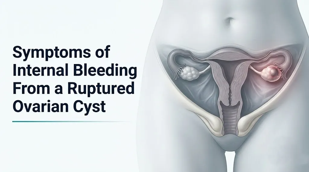
Symptoms of Internal Bleeding From a Ruptured Ovarian Cyst
The symptoms of internal bleeding from a ruptured ovarian cyst include sudden severe pelvic pain, referred pain to the shoulder tip, dizziness, nausea, rapid heart rate, pallor, and in serious cases, fainting or loss of consciousness from hemodynamic collapse. Not every ruptured ovarian cyst causes internal bleeding, but when one does, the clinical picture can progress from uncomfortable to life-threatening within hours.
The American College of Obstetricians and Gynecologists (ACOG) recognizes hemorrhagic ovarian cyst rupture as one of the most common gynecologic causes of acute abdominal pain requiring emergency evaluation in reproductive-age women. What makes this condition particularly dangerous is a deceptive early phase: the body’s compensatory mechanisms can temporarily maintain near-normal blood pressure even as substantial internal hemorrhage continues, giving a false impression of stability.
This article explains every clinically recognized symptom of internal bleeding from a ruptured ovarian cyst, the specific physiological mechanism driving each one, the critical signs that distinguish a serious hemorrhagic emergency from a self-limiting simple rupture, and exactly when to call 911.
Symptoms of Internal Bleeding From a Ruptured Ovarian Cyst
The symptoms of internal bleeding from a ruptured ovarian cyst are produced by blood accumulating in the peritoneal cavity, irritating pain-sensitive peritoneal lining, tracking to the diaphragm, and triggering the body’s emergency hemorrhagic response through the sympathoadrenal axis.
The core symptom picture includes sudden-onset pelvic pain that may be sharp, stabbing, or cramping; pain at the right or left shoulder tip (referred via the phrenic nerve); nausea and vomiting from vagal stimulation; dizziness or presyncope from blood volume loss; rapid heart rate; pallor; diaphoresis (sweating without heat exposure); and in severe cases, loss of consciousness.
What separates internal bleeding from a simple cyst rupture is the involvement of ovarian stromal blood vessels. A simple functional cyst releases follicular fluid into the peritoneal cavity, causing temporary irritation that often resolves within hours. A hemorrhagic rupture involves active arterial or venous bleeding, and that distinction determines whether a patient can wait for a scheduled appointment or needs emergency surgery tonight.
Symptom overview by severity level:
| Symptom | Mechanism | Severity Level | Action Required |
|---|---|---|---|
| Sudden pelvic pain, one side | Peritoneal nociceptor activation from cyst wall rupture | Mild to severe | Evaluate urgency by associated symptoms |
| Shoulder tip pain | Phrenic nerve (C3-C5) referred pain from sub-diaphragmatic blood | Moderate to severe | Go to ER immediately |
| Nausea and vomiting | Vagal afferent activation from peritoneal irritation | Moderate | Monitor; ER if persistent |
| Dizziness, presyncope | Blood volume depletion, reduced cerebral perfusion | Severe | Go to ER immediately |
| Rapid heart rate (tachycardia) | Sympathoadrenal compensatory response to hemorrhage | Severe | Go to ER immediately |
| Pallor and diaphoresis | Peripheral vasoconstriction from catecholamine surge | Severe | Go to ER immediately |
| Fainting or loss of consciousness | Hemodynamic decompensation, hypovolemic shock | Emergency | Call 911 now |
What Is a Hemorrhagic Ovarian Cyst Rupture?
A hemorrhagic ovarian cyst rupture occurs when a blood-filled ovarian cyst tears through the ovarian cortex and releases blood directly into the peritoneal cavity, a condition called hemoperitoneum.
The ovary contains follicles that develop each menstrual cycle under the influence of follicle-stimulating hormone (FSH) and luteinizing hormone (LH). Most follicles involute quietly without rupturing. The dominant follicle ruptures at ovulation, releasing the egg. Occasionally, a follicle develops into a larger cyst rather than releasing its egg normally, or the corpus luteum (the progesterone-secreting structure that forms after ovulation) develops into a cyst with internal bleeding, producing a hemorrhagic corpus luteum cyst.
Hemorrhagic cysts form when the thin-walled blood vessels supplying the follicle or corpus luteum bleed into the cyst interior rather than into surrounding tissue. The cyst wall becomes distended with blood. When it ruptures, it’s not just fluid that enters the peritoneal cavity. It’s blood, and sometimes active arterial bleeding continues from the torn vessel stumps in the ovarian stroma.
According to the Eunice Kennedy Shriver National Institute of Child Health and Human Development (NICHD), ovarian cysts are extremely common in reproductive-age women, with most being functional and self-resolving. However, hemorrhagic cysts that rupture with significant vessel involvement represent a distinct clinical entity requiring a fundamentally different management approach than a simple follicular cyst rupture.
Women in their late teens and twenties have the highest follicular activity and therefore face the highest incidence of hemorrhagic follicular cyst ruptures. Women in their late twenties through early forties are more likely to develop corpus luteum cyst-related hemorrhage, typically in the second half of their menstrual cycle (the luteal phase, days 14 to 28).
Ruptured Ovarian Cyst Pain: Location and Character
The pain from a ruptured ovarian cyst producing internal bleeding begins as sudden, often severe pain in the lower abdomen on the side of the affected ovary, typically the right or left iliac fossa, which is the lower right or lower left region of the abdomen.
The initial pain has a distinct character: most patients describe it as sharp and stabbing at onset, sometimes so abrupt that it stops them mid-activity. This sudden onset reflects the moment the cyst wall tears and blood contacts the parietal peritoneum, the body wall lining of the abdominal cavity, which contains dense somatic nerve endings from the lower thoracic and lumbar spinal nerves. Contact with blood activates these nociceptors immediately.
Within minutes to hours, the pain often shifts in character from a localized sharp stab to a more diffuse, constant, cramping or aching pain across the lower abdomen. This shift reflects progression from initial peritoneal contact to a broader inflammatory response as blood spreads through the pelvic cavity and prostaglandins amplify nociceptor sensitivity across a wider peritoneal area.
Pain that initially seems to be only on one side but spreads to involve the entire lower abdomen within 30 to 60 minutes is a clinical flag for significant hemoperitoneum, not a reassuring sign that symptoms are spreading evenly.
To accurately describe your pain to an emergency provider:
- Note the exact time the pain started and whether it was sudden or gradual.
- Identify the original location: lower right, lower left, or central lower abdomen.
- Note whether the pain has spread from its original location since it started.
- Rate the pain on a scale of 0 to 10 at onset and at its current level.
- Note whether any movement, deep breathing, or position change makes the pain worse (peritoneal guarding).
- State whether the pain has improved, stayed the same, or worsened since it began.
Women taking opioid pain medications chronically, or those with chronic pelvic pain from endometriosis or interstitial cystitis, may report lower pain scores despite significant hemoperitoneum because of altered central pain processing. Clinicians should interpret pain scores in the context of all associated symptoms, not pain score alone, in these populations.
Ruptured Ovarian Cyst Shoulder Pain
Shoulder tip pain from a ruptured ovarian cyst is not a musculoskeletal injury. It’s a neurological referral pattern produced by blood pooling under the diaphragm.
This symptom, known clinically as Kehr’s sign, occurs through a specific anatomical pathway. When blood accumulates in the peritoneal cavity, gravity and body position draw it toward the lowest available space (the pouch of Douglas in the pelvis initially) and then, as volume increases, superiorly through the paracolic gutters (lateral channels on either side of the colon) toward the sub-diaphragmatic space beneath the right and left hemidiaphragm.
The diaphragm is innervated by the phrenic nerve, which originates from spinal nerve roots C3, C4, and C5. Those same spinal levels also supply sensory innervation to the skin of the shoulder. When blood irritates the diaphragmatic peritoneum, the brain interprets the phrenic nerve signal as coming from the shoulder, because both the diaphragm and the shoulder are served by the same spinal segment. The result is real, reproducible shoulder pain with no injury to the shoulder itself.
The shoulder pain is typically felt at the very tip of the shoulder (the acromial region), rather than in the shoulder joint or the upper arm. Left-sided sub-diaphragmatic blood typically produces left shoulder tip pain. Right-sided accumulation produces right shoulder tip pain. In large hemoperitoneum, both sides may be affected.
Quick Tip:
- Shoulder tip pain appearing within minutes to hours of sudden pelvic pain is a red flag for significant hemoperitoneum regardless of pain severity in the pelvis.
- Do not assume shoulder tip pain means a shoulder injury. If it coincides with pelvic pain, this combination warrants immediate emergency evaluation.
- Women who have been lying flat (sleeping, resting) may notice shoulder tip pain more intensely when blood distributes more evenly sub-diaphragmatically in the supine position.
Key Takeaway: Sudden shoulder tip pain appearing alongside lower abdominal pain is not a coincidence or a muscle strain. It’s a neurological signal that blood is under your diaphragm, and it means you need emergency evaluation right now.
Ruptured Ovarian Cyst Nausea and Vomiting
Nausea and vomiting from a ruptured ovarian cyst with internal bleeding are produced by two simultaneous mechanisms: vagal afferent nerve activation from peritoneal irritation, and the systemic response to blood volume loss.
The peritoneum contains a dense network of vagal afferent fibers. When blood contacts and irritates the peritoneal lining, these fibers carry signals to the nucleus tractus solitarius in the medulla oblongata, which activates the adjacent vomiting center (the area postrema). This is the same pathway that produces nausea with any cause of peritoneal inflammation, including appendicitis or peritonitis.
A second mechanism operates in parallel. As blood volume drops from hemorrhage, the body reduces blood flow to non-essential organs including the gastrointestinal tract. This splanchnic vasoconstriction can itself produce nausea, and the systemic catecholamine surge (epinephrine from the adrenal medulla) also activates nausea pathways.
Nausea with vomiting alongside pelvic pain is therefore a symptom of the overall systemic response to hemorrhage, not just a minor gastrointestinal complaint.
According to a clinical review published in the Annals of Emergency Medicine, nausea and vomiting are present in a substantial proportion of patients with confirmed hemoperitoneum from ruptured ovarian cysts presenting to emergency departments, and their presence alongside pelvic pain should increase clinical suspicion for significant intraperitoneal hemorrhage rather than being dismissed as a nonspecific complaint.
Adolescents may present with more pronounced nausea and vomiting relative to their reported pain level, because their central pain modulation pathways process visceral and peritoneal pain differently from adults. This can cause the overall symptom picture to be attributed incorrectly to gastroenteritis or anxiety, delaying diagnosis.
Ruptured Ovarian Cyst Dizziness and Fainting
Dizziness and fainting from a ruptured ovarian cyst signal that blood volume loss is affecting cerebral perfusion, and these symptoms require immediate emergency evaluation without exception.
Orthostatic hypotension (a drop in blood pressure when moving from lying to sitting or standing) is one of the earliest objective signs of clinically meaningful blood volume depletion. The body normally compensates for positional changes through rapid sympathetic nervous system adjustments. When circulating blood volume is reduced, that compensation is impaired. The result is a sudden drop in blood pressure when upright, reducing cerebral blood flow and producing dizziness, tunnel vision, or near-fainting (presyncope).
Frank syncope (loss of consciousness) occurs when cerebral perfusion falls below the threshold needed to maintain consciousness, typically when mean arterial pressure drops sufficiently to reduce cerebral blood flow by approximately 35% below normal. This represents advanced hemorrhagic compromise and should be treated as a medical emergency equivalent to shock.
The danger in this symptom is timing. A patient may feel dizzy when she first stands up, then feel slightly better after lying down (because supine position reduces the circulatory burden). This temporary improvement does not mean the hemorrhage has stopped. Active bleeding may continue while apparent symptoms ease in the lying position.
Dizziness red flag pattern to recognize:
- Feeling lightheaded or dizzy when moving from lying to sitting: this is orthostatic change, suggesting blood volume loss
- A brief blackout or “grey-out” when standing: this is presyncope requiring emergency evaluation
- Complete loss of consciousness: Call 911 immediately
- Dizziness that returns each time you try to stand, even if it partially resolves lying down: the bleeding is likely ongoing
Women who take beta-blockers for hypertension, migraine, or anxiety may not develop the expected tachycardia response to hemorrhage because these medications blunt the sympathetic heart rate increase. Dizziness may be their primary early warning sign, without the accompanying racing heart that otherwise alerts patients and clinicians to significant blood loss.
Ruptured Ovarian Cyst Rapid Heart Rate and Low Blood Pressure
A rapid heart rate from a ruptured ovarian cyst is the body’s emergency response to blood volume loss, and it often appears before blood pressure drops.
When hemorrhage reduces circulating blood volume, pressure sensors called baroreceptors in the aortic arch and carotid sinus detect the change. They signal the brainstem’s cardiovascular control center to activate the sympathetic nervous system. The adrenal medulla releases epinephrine and norepinephrine. Heart rate accelerates (clinical threshold: above 100 beats per minute is tachycardia). Peripheral blood vessels constrict to redirect blood to the brain and heart. Blood pressure is initially maintained through this vasoconstriction.
This is the deceptive phase of hemorrhagic shock. A patient may have lost 750 to 1,000 milliliters of blood into the peritoneal cavity and still present with a blood pressure in the low-normal range, because the body’s compensatory mechanisms are working effectively. The tachycardia is the giveaway. Normal resting heart rate for adult women is 60 to 100 beats per minute. A heart rate of 110 to 120 beats per minute in a woman with pelvic pain and no fever is a clinical red flag for active hemorrhage even with a technically normal blood pressure reading.
As blood loss progresses beyond compensatory capacity, typically exceeding 1,500 milliliters in adults, blood pressure falls. The clinical picture now shows both tachycardia and hypotension. This is hypovolemic shock and is a surgical emergency.
Hemodynamic response stages in hemorrhagic hemoperitoneum:
| Stage | Blood Loss (Estimated) | Heart Rate | Blood Pressure | Clinical State |
|---|---|---|---|---|
| Early compensation | Under 750 mL | Slightly elevated (90-100 bpm) | Normal | Appears stable, may feel anxious |
| Active compensation | 750-1,500 mL | Tachycardia (100-120 bpm) | Low-normal | Dizzy when standing, pale, sweating |
| Compensation failing | 1,500-2,000 mL | Tachycardia (120+ bpm) | Hypotension developing | Confused, markedly pale, diaphoretic |
| Decompensated shock | Over 2,000 mL | Severe tachycardia or paradoxical bradycardia | Frank hypotension | Altered consciousness, requires immediate surgery |
Key Takeaway: A racing heart alongside pelvic pain is not anxiety. It’s your body signaling active blood loss through its most direct compensatory mechanism, and it means emergency evaluation is needed before blood pressure drops.
Signs a Ruptured Ovarian Cyst Is Serious
A ruptured ovarian cyst is clinically serious when it produces active, ongoing intraperitoneal hemorrhage that exceeds the body’s ability to compensate, rather than a self-contained fluid release that the peritoneum absorbs and resolves on its own.
The specific clinical signs that indicate a serious, hemorrhagic rupture rather than a self-limiting simple rupture are:
Red flag signs requiring immediate emergency evaluation:
- Pain that is worsening over time rather than improving in the hours after onset
- Shoulder tip pain appearing alongside pelvic pain (Kehr’s sign of sub-diaphragmatic blood)
- Dizziness, lightheadedness, or near-fainting when standing
- Heart rate above 100 beats per minute at rest
- Sweating without physical exertion or fever (diaphoresis)
- Pallor: skin that appears noticeably paler than normal, particularly around the face and lips
- Vomiting that began after the pelvic pain started (not before, which would suggest a gastrointestinal cause)
- Abdominal tenderness that is worse when pressure is suddenly released (rebound tenderness), indicating peritoneal irritation
- Rigid or board-like abdominal muscles when lying flat (guarding)
- Any episode of fainting or loss of consciousness since the pain began
A ruptured ovarian cyst that presents with improving pain, no hemodynamic symptoms, no shoulder pain, and no fever within 6 to 12 hours of onset may be managed conservatively in appropriate clinical settings. A ruptured ovarian cyst that presents with any of the above signs is a potential hemorrhagic emergency.
Women with endometriosis face a particular complication here: chronic pelvic pain from their underlying condition can mask the escalation of pain from a hemorrhagic rupture, because they are habituated to baseline pelvic pain levels. If pain quality changes (different location, sharper character, accompanied by new symptoms they don’t normally have) rather than just pain intensity, this distinction matters clinically and should be communicated specifically to the evaluating physician.
Hemorrhagic Cyst vs. Simple Cyst Rupture
The difference between a hemorrhagic cyst rupture and a simple cyst rupture is the difference between blood and fluid entering the peritoneal cavity, and that distinction determines whether a patient goes home with pain management instructions or to the operating room.
A simple follicular cyst is filled with serous (watery, protein-containing) follicular fluid. When it ruptures, that fluid enters the peritoneal cavity. The peritoneum absorbs it relatively efficiently, and the inflammatory response is limited. Most simple cyst ruptures produce pain that peaks within the first few hours and then progressively improves over 12 to 24 hours as fluid is reabsorbed. Hemodynamic stability is maintained. Shoulder pain does not develop. Nausea may occur but typically resolves within hours.
A hemorrhagic cyst is filled with blood, either from a bleeding corpus luteum or from a blood vessel that ruptured into a follicular cyst wall. When it ruptures, blood enters the peritoneal cavity. Blood is far more inflammatory than serous fluid. It activates the coagulation cascade, complement system, and prostaglandin release at the peritoneal surface, producing a more intense and sustained inflammatory response. If a torn stromal blood vessel continues to bleed after the initial cyst rupture, the hemoperitoneum grows progressively.
Comparing simple vs. hemorrhagic cyst rupture clinically:
| Feature | Simple Cyst Rupture | Hemorrhagic Cyst Rupture |
|---|---|---|
| Pain trajectory | Peaks then improves within hours | Worsening or persistent |
| Shoulder tip pain | Absent | Present if significant blood volume |
| Hemodynamic symptoms | Absent | Tachycardia, dizziness, pallor |
| Ultrasound appearance | Minimal or no free fluid | Echogenic free fluid (blood) in pelvis |
| Treatment typical | Pain management, observation | Possible surgical intervention |
| Risk of recurrence | Moderate | Higher, especially with anticoagulants |
On ultrasound, the distinction is visible: serous fluid appears anechoic (black on ultrasound), while blood appears echogenic (grey or complex texture). A trained ultrasonographer or emergency medicine physician interpreting a bedside ultrasound can often identify the nature of free pelvic fluid within minutes.
Hemoperitoneum From a Ruptured Ovarian Cyst
Hemoperitoneum refers specifically to the accumulation of blood in the peritoneal cavity, and it is the defining physiological event that produces the most serious symptoms of internal bleeding from a ruptured ovarian cyst.
The peritoneal cavity is the space inside the abdominal and pelvic cavity lined by the peritoneum. In a healthy state it contains only a few milliliters of lubricating fluid. When a hemorrhagic ovarian cyst ruptures, blood enters this space and behaves according to gravity and anatomy. In the upright position, it pools first in the pouch of Douglas (the rectouterine pouch), the lowest peritoneal recess in females. From there, as volume increases, it ascends through the paracolic gutters toward the sub-diaphragmatic space.
Think of it like spilling a liquid into a tilted bowl with a channel on each side. The liquid fills the lowest point first, then runs along the channels upward. Blood rising to the sub-diaphragmatic level is what triggers Kehr’s sign. Blood pooling in the pouch of Douglas is what produces deep pelvic pressure and pain with rectal or bladder pressure.
Clinical studies published in the American Journal of Obstetrics and Gynecology have found that the amount of free fluid detectable on pelvic ultrasound correlates with the likelihood of requiring surgical intervention. Moderate to large hemoperitoneum (free fluid extending beyond the pelvis into the paracolic gutters or sub-diaphragmatic space on imaging) is strongly associated with hemodynamic instability and surgical need.
The peritoneum also responds to hemoperitoneum by launching a fibrinolytic response that partially breaks down clots and keeps blood liquid within the cavity, which counterintuitively can perpetuate bleeding from torn vessel sites rather than promoting clotting. This is one reason significant hemoperitoneum does not reliably self-resolve.
Women with inherited bleeding disorders such as von Willebrand disease face disproportionately severe hemoperitoneum from what might otherwise be a minor cyst rupture, because their impaired platelet function prevents normal hemostasis at the torn vessel site. These women should disclose their bleeding disorder history at every gynecological and emergency evaluation.
Key Takeaway: Hemoperitoneum doesn’t stay in the pelvis. As blood volume increases it climbs toward the diaphragm, which is why shoulder pain appears, and its extent on ultrasound directly predicts how dangerous the situation is.
Ruptured Corpus Luteum Cyst Bleeding
The corpus luteum is the progesterone-secreting structure that forms from the dominant follicle after ovulation, and its rupture is the most common cause of clinically significant hemorrhagic cyst rupture in reproductive-age women.
After the dominant follicle releases its egg at ovulation, the follicle wall collapses and the granulosa and theca cells inside it transform into the corpus luteum, a highly vascularized structure. It produces progesterone and, in early pregnancy, human chorionic gonadotropin (hCG), to sustain the uterine lining. This rich blood supply is exactly what makes corpus luteum cysts prone to hemorrhage. When a corpus luteum cyst forms and its wall becomes fragile, the internal vasculature is vulnerable to rupture.
Corpus luteum cyst rupture characteristically occurs in the second half of the menstrual cycle, between approximately days 20 and 26 in a 28-day cycle. The timing is important: it means a woman may have a positive pregnancy test if the rupture occurs in early conception, because the corpus luteum is still producing progesterone. This creates a critical diagnostic challenge: a ruptured corpus luteum cyst and an ectopic pregnancy can look nearly identical on initial presentation, and serum hCG testing is mandatory to distinguish them.
Research published in Fertility and Sterility has documented that corpus luteum cyst ruptures account for the majority of hemorrhagic ovarian cyst cases requiring operative management, and they occur most frequently in the right ovary. The anatomical reason is that the right ovarian artery arises directly from the aorta (a higher-pressure origin) while the left ovarian artery arises from the left renal artery, giving right-sided corpus luteum cysts slightly more arterial pressure to contend with.
Women taking anticoagulant medications (warfarin, apixaban, rivaroxaban, dabigatran) for conditions such as deep vein thrombosis, atrial fibrillation, or hypercoagulable states face dramatically elevated risk for severe corpus luteum hemorrhage. Any woman on anticoagulation who develops sudden pelvic pain should be evaluated in an emergency setting that same day, without waiting to see whether symptoms improve.
Ruptured Ovarian Cyst During Pregnancy
A ruptured ovarian cyst during pregnancy produces the same internal bleeding symptoms as in non-pregnant women, but the clinical setting is more complex because the differential diagnosis expands and the stakes for both the patient and the pregnancy are higher.
Early pregnancy (first trimester) is the most common time for ovarian cyst complications during pregnancy, because the corpus luteum cyst that supports the pregnancy until the placenta takes over progesterone production is at peak size and vascularity between weeks 6 and 12 of gestation. Rupture of this cyst produces pain on the side of the affected ovary, with the full range of hemorrhagic symptoms if significant bleeding occurs.
The diagnostic urgency in pregnancy is intensified by the need to exclude ectopic pregnancy (implantation of the embryo outside the uterus, most commonly in the fallopian tube). Ectopic pregnancy rupture produces an identical symptom picture to hemorrhagic ovarian cyst rupture: sudden pelvic pain, internal bleeding, shoulder tip pain, dizziness, and hemodynamic instability. The clinical consequences of missing a ruptured ectopic pregnancy are potentially fatal. Serum hCG levels and transvaginal ultrasound confirming intrauterine pregnancy location are the essential first steps in any pregnant woman presenting with acute pelvic pain and signs of internal bleeding.
According to ACOG, any pregnant woman experiencing acute pelvic pain with hemodynamic instability requires emergency evaluation, and ectopic pregnancy must be excluded before an ovarian cyst rupture diagnosis can be made safely.
Physical examination of the pregnant abdomen requires modified technique, because the gravid uterus displaces abdominal organs and changes the expected location of peritoneal tenderness. Women in the second and third trimester may not demonstrate classic peritoneal guarding signs in the expected locations, making ultrasound assessment even more central to evaluation.
Ruptured Ovarian Cyst With PCOS or Endometriosis
Women with polycystic ovary syndrome (PCOS) or endometriosis face elevated risk for ovarian cyst rupture with internal bleeding, and their symptom presentation can differ from women without these underlying conditions in clinically meaningful ways.
In PCOS, the ovaries contain multiple small follicular cysts that fail to ovulate normally due to LH hyperstimulation and androgen excess. Occasionally, a dominant follicle does develop and can form a larger hemorrhagic cyst with higher rupture risk than typical follicular cysts. The baseline pelvic discomfort that many women with PCOS experience can also mask early warning symptoms, delaying recognition of a rupture.
Endometriosis creates a fundamentally different rupture risk. Women with endometriosis develop endometriomas (ovarian cysts filled with old menstrual blood and endometrial debris, often called “chocolate cysts” due to their appearance). When an endometrioma ruptures, the contents spilled into the peritoneal cavity are not fresh blood but highly inflammatory degraded blood products and prostaglandins that provoke an intense peritoneal reaction. The pain from an endometrioma rupture is typically more severe than an equivalent-sized simple hemorrhagic cyst rupture because of this heightened inflammatory response.
Research published in the Journal of Minimally Invasive Gynecology has documented that endometrioma ruptures produce higher rates of adhesion formation and require more extensive surgical management than other hemorrhagic cyst ruptures. Women with known endometriomas who develop sudden worsening of pelvic pain beyond their baseline endometriosis pain level should seek emergency evaluation.
Individual risk comparison:
| Condition | Cyst Type Most at Risk | Rupture Risk Modifier | Symptom Difference |
|---|---|---|---|
| PCOS | Large follicular or hemorrhagic cyst | Elevated follicular activity | May have blunted pain due to chronic pelvic discomfort |
| Endometriosis | Endometrioma (chocolate cyst) | High, especially with large cysts | More intense peritoneal inflammation, more severe pain |
| No underlying condition | Corpus luteum or follicular cyst | Standard baseline | Typical presentation |
| Anticoagulant use | Any cyst type | Markedly elevated hemorrhage severity | Any rupture can become significantly hemorrhagic |
Key Takeaway: For women with endometriosis, a sudden change in the quality or location of pelvic pain is more clinically meaningful than a change in pain intensity alone, because endometrioma rupture can occur at any phase of the cycle, not just mid-cycle.
Ruptured Ovarian Cyst vs. Appendicitis Symptoms
Distinguishing a ruptured ovarian cyst from appendicitis is one of the most common diagnostic challenges in emergency gynecology, and the two conditions cannot be reliably separated by symptom history alone.
Both conditions produce sudden lower abdominal pain. Both cause nausea and vomiting. Both produce peritoneal signs (guarding and rebound tenderness) on physical examination. The right side is the more common location for both: the appendix sits in the right iliac fossa, and the right ovary is the more common site of corpus luteum cyst rupture.
The key distinguishing features, where they exist, are:
Features more consistent with ruptured ovarian cyst:
- Onset that correlates with mid-cycle timing (day 12 to 16 for follicular rupture) or luteal phase (day 20 to 26 for corpus luteum rupture)
- Pain that began centrally or in the left pelvis and later moved right (less typical of appendicitis)
- Shoulder tip pain, which is characteristic of intraperitoneal blood and not typical of early appendicitis
- Known history of ovarian cysts
- Hemodynamic symptoms (tachycardia, dizziness) disproportionate to fever or white blood cell count elevation
Features more consistent with appendicitis:
- Fever above 38.5 degrees Celsius (101.3 degrees Fahrenheit)
- White blood cell count markedly elevated (above 12,000 to 15,000 cells per microliter) on complete blood count
- Pain that began around the navel and then migrated to the lower right quadrant (classic McBurney’s point migration)
- Maximum tenderness precisely at McBurney’s point (one-third of the distance from the right anterior superior iliac spine to the navel)
- No correlation with menstrual cycle timing
The bottom line: these two conditions require imaging to distinguish. A CT scan of the abdomen and pelvis with contrast is the standard diagnostic tool in the emergency setting, with sensitivity exceeding 90% for appendicitis. Pelvic ultrasound adds ovarian-specific detail. In reproductive-age women with right lower quadrant pain, serum hCG testing is mandatory before CT contrast administration and before any diagnosis is finalized, because ectopic pregnancy must always be in the differential.
Ruptured Ovarian Cyst Diagnosis
Diagnosing a ruptured ovarian cyst with internal bleeding requires a combination of clinical history, physical examination, laboratory tests, and imaging, because no single symptom or test is definitive on its own.
In the emergency setting, the diagnostic sequence typically unfolds as follows:
- Clinical history: Onset time, location and character of pain, associated symptoms (shoulder pain, dizziness, nausea, fever), menstrual cycle timing, last menstrual period date, contraceptive use, prior cyst history, medication list.
- Serum hCG (pregnancy test): Mandatory in all women of reproductive age to exclude ectopic pregnancy before other diagnoses are pursued. A positive hCG changes the entire management approach.
- Complete blood count (CBC): Hemoglobin and hematocrit values assess blood loss. Early in active hemorrhage, CBC may still be normal (hemodilution takes time), so a normal hemoglobin does not rule out significant bleeding. Serial CBCs over 4 to 6 hours provide more meaningful information.
- Transvaginal ultrasound: The primary imaging modality for ovarian cysts. Detects free fluid in the pelvis (blood appears echogenic or complex in texture rather than anechoic), identifies any remaining cyst tissue, assesses the contralateral ovary, and checks for intrauterine pregnancy.
- CT abdomen and pelvis with contrast: Used when ultrasound is inconclusive, when appendicitis is in the differential, or when the extent of hemoperitoneum needs to be assessed for surgical planning.
- Vital sign monitoring: Heart rate and blood pressure trends over time are as diagnostically important as any single laboratory value. A rising heart rate with stable blood pressure signals compensated hemorrhage that may be actively ongoing.
According to ACOG clinical guidelines, hemodynamically unstable patients with suspected hemorrhagic ovarian cyst rupture may proceed directly to the operating room for diagnostic laparoscopy without waiting for all imaging results, because surgical confirmation and treatment can occur simultaneously when the clinical picture is clear.
Postmenopausal women who develop a ruptured ovarian cyst require a different diagnostic approach, because any ovarian mass in this population requires malignancy exclusion through tumor marker testing (CA-125, HE4) and specialist referral to a gynecologic oncologist, even when acute hemorrhage is the presenting problem.
Ruptured Ovarian Cyst: When to Go to the ER
Knowing exactly when to go to the emergency room for a ruptured ovarian cyst can be the difference between conservative management and a lifesaving surgical intervention.
Not every ruptured ovarian cyst requires emergency care. A simple functional cyst rupture with mild pain that begins improving within a few hours, no shoulder pain, no dizziness, no vomiting, normal heart rate, and no fever in a hemodynamically stable woman can often be managed with pain relief and close monitoring at home, with clear return precautions.
The emergency threshold exists when any of the following are present or developing:
Go to the nearest emergency room immediately if you have:
- Pelvic pain that is worsening or not improving after 2 to 3 hours
- Pain at the tip of either shoulder alongside pelvic pain
- Any episode of dizziness, lightheadedness, or near-fainting
- Vomiting that you cannot control
- Pallor (notably pale skin, especially around the face and lips)
- Sweating without physical exertion or fever
- A positive pregnancy test alongside your pelvic pain (excludes ectopic pregnancy first)
- Pain so severe you cannot walk or stand normally
- You are on anticoagulant medications (warfarin, apixaban, rivaroxaban, dabigatran, or similar)
- You have a known bleeding disorder such as von Willebrand disease or hemophilia carrier status
Call 911 immediately if you experience:
- Fainting or loss of consciousness
- Inability to stand or walk due to weakness or dizziness
- A rapid heart rate you can feel in your chest combined with severe pelvic pain
- Confusion or disorientation alongside pelvic pain
The specific instruction when in doubt: go to the emergency room. If you are asking yourself whether your symptoms are serious enough to warrant emergency care, the safer clinical answer is always to have them assessed by an emergency medicine physician who can obtain a CBC, hCG, and ultrasound within 30 to 60 minutes rather than monitor at home and risk decompensating without clinical support.
Emergency Symptoms: When to Call 911 or Go to the ER
Certain symptoms associated with internal bleeding from a ruptured ovarian cyst require immediate emergency evaluation. Do not wait to see if these resolve on their own.
Call 911 or go to the nearest emergency room immediately if you experience:
- Fainting or loss of consciousness, which indicates that cerebral perfusion has dropped below the threshold necessary to maintain consciousness and hemodynamic collapse is occurring
- Severe inability to stand or walk, with weakness so pronounced you cannot safely move without assistance
- Sudden severe pelvic pain rated 8 or higher on a 10-point scale, with rapid onset and no prior similar episodes
- Shoulder tip pain developing alongside pelvic pain, which signals blood has reached the sub-diaphragmatic space through the paracolic gutters
- Visible pallor (unusually pale face, lips, or nailbeds) combined with sweating and pelvic pain
- A palpable rapid heartbeat in your chest alongside lower abdominal pain
- Positive pregnancy test with acute pelvic pain and any sign of bleeding: this is an obstetric emergency requiring ectopic pregnancy exclusion before all other diagnoses
- Pain that was initially mild but has progressively worsened over 2 to 4 hours without any improvement
- Any confusion, disorientation, or feeling of impending loss of consciousness
These presentations can indicate hemorrhagic hemoperitoneum progressing to hypovolemic shock, and they require emergency medical assessment by an emergency medicine physician, not an urgent care appointment or a phone call to an after-hours nurse line.
Frequently Asked Questions About Symptoms of Internal Bleeding From a Ruptured Ovarian Cyst
What are the signs of internal bleeding from a ruptured ovarian cyst?
The signs of internal bleeding from a ruptured ovarian cyst include sudden severe pelvic pain on one side, shoulder tip pain from blood irritating the diaphragm, dizziness, rapid heart rate, pallor, sweating, nausea, and in severe cases, fainting.
These symptoms are produced by blood accumulating in the peritoneal cavity and triggering the body’s emergency hemorrhagic response through the sympathoadrenal axis.
Any combination of pelvic pain with shoulder tip pain or hemodynamic symptoms (dizziness, rapid heart rate, pallor) requires immediate emergency evaluation.
Can a ruptured ovarian cyst cause internal bleeding without severe pain?
Yes, a ruptured ovarian cyst can cause internal bleeding without initially severe pain, because the body’s compensatory mechanisms can maintain apparent stability during active hemorrhage before pain escalates.
Women with chronic pelvic pain from endometriosis, or those taking opioid medications, may experience lower reported pain scores even with significant hemoperitoneum.
The absence of severe pain does not rule out significant internal bleeding; hemodynamic symptoms including dizziness and rapid heart rate are more reliable early indicators than pain intensity alone.
How do you know if a ruptured ovarian cyst is an emergency?
A ruptured ovarian cyst is an emergency when it produces any hemodynamic symptom: dizziness, presyncope, fainting, rapid heart rate above 100 beats per minute, pallor, or diaphoresis alongside pelvic pain.
Shoulder tip pain appearing alongside pelvic pain is a specific indicator that blood has reached the sub-diaphragmatic space, which typically represents a volume of hemoperitoneum sufficient to warrant emergency evaluation.
Women on anticoagulant medications or with bleeding disorders should treat any pelvic pain as a potential emergency regardless of initial severity.
What does shoulder pain from a ruptured ovarian cyst mean?
Shoulder tip pain from a ruptured ovarian cyst means that blood has pooled under the diaphragm and is irritating the diaphragmatic peritoneum, which shares nerve roots (C3-C4-C5, the phrenic nerve) with the shoulder tip.
This referred pain pattern, called Kehr’s sign, indicates a substantial volume of intraperitoneal blood has ascended through the paracolic gutters to the sub-diaphragmatic space.
This symptom is a red flag for significant hemoperitoneum and warrants immediate emergency room evaluation.
How long does pain from a ruptured ovarian cyst last?
Pain from a simple ruptured ovarian cyst (serous fluid release without significant hemorrhage) typically begins improving within 6 to 24 hours as the peritoneum reabsorbs the fluid.
Pain from a hemorrhagic cyst rupture with ongoing bleeding does not follow this improving trajectory and may worsen progressively over hours.
Pain that has not shown clear improvement within 6 hours of onset, or that is worsening at any point, should be evaluated by an emergency medicine physician.
Can a ruptured ovarian cyst heal on its own without surgery?
Many ruptured ovarian cysts, specifically simple functional cysts that release serous fluid rather than blood, do resolve without surgical intervention through peritoneal reabsorption and natural hemostasis.
Hemorrhagic cyst ruptures with large hemoperitoneum or hemodynamic instability require surgical management, typically diagnostic laparoscopy with ovarian cystectomy or hemostasis of the bleeding vessel.
The decision between conservative management and surgery is made based on hemodynamic stability, the volume of free fluid on imaging, and serial clinical assessment by an emergency medicine physician and obstetrician-gynecologist.
Recognizing the symptoms of internal bleeding from a ruptured ovarian cyst matters most in the first hours after onset, when the distinction between a self-limiting simple rupture and a progressive hemorrhagic emergency is still being established by your body’s response.
The single most actionable clinical rule from everything covered here is this: if pelvic pain is accompanied by shoulder tip pain, dizziness, rapid heart rate, or pallor, do not wait to see if it improves. Go to the nearest emergency room immediately. Bring your complete medication list, note the timing of your last menstrual period, and tell the triage nurse your first concern is ruling out internal bleeding.
The symptoms described in this article are your body communicating something specific and physiologically real. An emergency medicine physician with ultrasound access and a laboratory can give you an answer within an hour. That information is worth more than any amount of home monitoring when the clinical stakes are this high.