Jaw Cancer Symptoms: All Signs and Red Flags for 2026
Jaw cancer symptoms include persistent jaw pain, unexplained swelling or lumps in the jaw or face, numbness of the lower lip or chin, difficulty opening the mouth, loose teeth without dental cause, difficulty swallowing, and ear pain, each produced by a specific anatomical mechanism that distinguishes malignant jaw disease from the far more common benign conditions that cause similar complaints. The most important clinical fact about jaw cancer is that symptoms frequently appear weeks to months before diagnosis because they are attributed to dental problems, temporomandibular joint (TMJ) disorder, or facial nerve irritation.
According to the American Cancer Society (ACS), oral cavity cancers including those involving the jaws affect approximately 58,450 Americans each year, with the jaw being a site of both primary bone malignancy and invasion from adjacent oral mucosal cancers. The National Cancer Institute (NCI) notes that the five-year relative survival rate for localized oral cavity cancer exceeds 85%, compared to approximately 40% for cancers diagnosed after regional spread, making early symptom recognition the single most consequential factor in outcome.
This article explains every recognized jaw cancer symptom with its specific anatomical and physiological mechanism, distinguishes early from late-stage presentations, addresses which populations face the highest risk of delayed diagnosis, and gives you clear clinical thresholds for when to contact an oral and maxillofacial surgeon versus when a routine dental appointment is sufficient.
Jaw Cancer Symptoms: An Overview
Jaw cancer symptoms arise from the specific anatomical structures that malignant tumors invade, compress, or destroy as they grow within or adjacent to the mandible (lower jaw) and maxilla (upper jaw), producing a symptom pattern that reflects the tumor’s location, histological type, and stage.
The jaw is affected by two broad categories of malignant disease: primary jaw bone cancers (arising from bone cells, cartilage cells, or remnants of tooth-forming tissue) and secondary jaw cancers (where oral mucosal malignancies, most commonly squamous cell carcinoma of the gingiva, floor of mouth, or buccal mucosa, invade the adjacent jaw bone). Both categories share overlapping symptoms, but their relative frequency, typical patient demographics, and anatomical progression differ meaningfully.
| Jaw Cancer Type | Primary Origin | Most Common Age Group | Key Symptom Patterns |
|---|---|---|---|
| Squamous cell carcinoma (SCC) invading jaw | Oral mucosal epithelium | Adults over 50 (tobacco/alcohol) | Pain, mucosal ulcer, loose teeth, IAN numbness |
| Osteosarcoma | Osteoblast precursor cells in mandible/maxilla | Adolescents and adults 10 to 30 | Jaw swelling, periosteal pain, sunburst imaging pattern |
| Ameloblastoma | Odontogenic (tooth-forming) epithelium | Adults 30 to 60 | Jaw expansion, cystic swelling, displaced teeth |
| Chondrosarcoma | Cartilage cells (condyle or symphysis) | Adults 40 to 70 | TMJ-like pain, condylar mass, trismus |
| Metastatic disease | Secondary from breast, lung, kidney, thyroid | Adults over 50 | Jaw numbness (Vincent’s symptom), pathological fracture |
According to the American Association of Oral and Maxillofacial Surgeons (AAOMS), any jaw lesion that does not resolve within two to three weeks of treatment of an identifiable benign cause should be biopsied and evaluated histologically. This two-to-three-week rule is the clinical threshold that separates watchful waiting from mandatory evaluation.
Older adults over 60 with a tobacco and alcohol history account for the majority of jaw cancer cases from SCC. However, osteosarcoma predominantly affects adolescents and young adults, meaning the demographic profile of jaw cancer is not uniform and the assumption that “I’m too young for cancer” does not apply to primary jaw bone malignancies.
What Is Jaw Cancer and Which Types Affect the Jaw?
Jaw cancer refers to any malignant neoplasm arising primarily within the mandible or maxilla or invading these bones from adjacent oral mucosal sites, with squamous cell carcinoma of the oral cavity being the most frequent cancer type to produce jaw bone involvement and osteosarcoma being the most frequent primary jaw bone malignancy.
Understanding which cancer type is involved is clinically important because each type produces distinct symptoms reflecting its anatomical growth pattern. Squamous cell carcinoma invades the jaw from outside (from the gingiva or floor of mouth inward), producing early mucosal symptoms before bone destruction. Primary bone tumors like osteosarcoma arise within the jaw bone itself and may produce bone swelling and periosteal pain before any mucosal change is visible.
Key malignant conditions affecting the jaw and their anatomical growth patterns:
- Squamous cell carcinoma (SCC) of the oral cavity: Arises from the stratified squamous epithelium of the gingiva, buccal mucosa, floor of mouth, or retromolar trigone, invading the cortical bone of the mandible through the dental sockets or through direct bone erosion. Accounts for the large majority of jaw cancers in adults over 50 with tobacco and alcohol exposure.
- Osteosarcoma of the jaw: Arises from osteoblast precursor cells in the mandibular or maxillary bone, producing rapid periosteal new bone formation alongside osteolytic destruction. The mandibular body and symphysis are the most common primary sites. Produces the characteristic “sunburst” periosteal reaction on imaging.
- Ameloblastoma: Arises from odontogenic (tooth-forming) epithelium, typically within the mandibular ramus or molar region. Grows slowly, expanding the jaw through cyst-like remodeling rather than aggressive destruction, which explains why it often presents as a painless jaw expansion.
- Chondrosarcoma: Arises from cartilage cells, most commonly at the mandibular condyle (the articulating end of the lower jaw at the TMJ) or at the mandibular symphysis. Frequently mimics TMJ disorder.
Immunocompromised individuals (those with HIV infection or on prolonged immunosuppressive therapy) have elevated rates of oral malignancies, including Kaposi’s sarcoma of the palate and jaw, compared to the general population.
Early Stage Jaw Cancer Symptoms
Early stage jaw cancer symptoms are often subtle, localized to a small area, and easily attributed to dental problems or minor trauma, which is why the median duration from symptom onset to diagnosis in jaw cancer is frequently several months in published clinical series.
The clinical problem with early jaw cancer symptoms is that they mimic extraordinarily common, benign conditions. A toothache is far more likely to reflect a dental cavity or periapical abscess than early jaw cancer. A small jaw lump is far more likely to be a lymph node reaction to dental infection than a malignant mass. The clinical challenge is recognizing the specific features that differentiate a symptom that requires watchful waiting from one that requires biopsy.
Early stage jaw cancer symptoms to recognize:
- A non-healing ulcer on the gingiva (gum) or adjacent oral mucosa lasting more than two to three weeks without an identifiable cause (trauma, infection)
- A firm, non-tender lump in the jaw, floor of mouth, or adjacent face that is new and not decreasing in size over two weeks
- Mild persistent jaw aching or dull throbbing pain that does not correspond to a specific tooth and does not respond to over-the-counter analgesics
- Early loosening of one or two teeth in a specific jaw region without gum disease or recent dental trauma
- A subtle change in the fit of a denture in one specific area without obvious structural cause
According to the National Cancer Institute (NCI), erythroplakia (red mucosal patches) carries a higher malignant transformation rate than leukoplakia (white mucosal patches), with erythroplakia having malignant potential in over 50% of biopsied cases. Any red, velvety patch on the gingiva or adjacent oral mucosa that cannot be wiped off should be evaluated by a dentist or oral surgeon promptly rather than observed.
Adults who use smokeless tobacco (chewing tobacco, snuff) face particular risk for gingival squamous cell carcinoma at the site of tobacco placement, typically the mandibular vestibule (the groove between the lower gum and inner lip). Persistent mucosal thickening or an indurated area at a habitual tobacco placement site warrants prompt dental evaluation.
Key Takeaway: The two-to-three-week rule is the most practically important clinical threshold for jaw cancer: any jaw symptom (pain, swelling, mucosal lesion, loose tooth) that does not resolve or improve within two to three weeks of treating an identified cause should be evaluated by a dentist or oral and maxillofacial surgeon for biopsy consideration.
Jaw Pain as a Jaw Cancer Symptom
Jaw pain in jaw cancer is produced by three distinct mechanisms: periosteal stretching from tumor-induced bone expansion, perineural invasion of nociceptive fibers in the inferior alveolar nerve and its branches, and inflammatory prostaglandin release at the tumor-bone invasion front, where matrix metalloproteinases and cytokines activate periosteal nociceptors.
The periosteum (the dense fibrous connective tissue layer covering the outer surface of the mandible and maxilla) is richly innervated by nociceptive nerve fibers from the branches of the trigeminal nerve (cranial nerve V). When a tumor expands within the jaw bone, it stretches the overlying periosteum against its nerve supply, producing a deep, boring, aching pain that is characteristically worse at night and not relieved by position change. This pattern differs from the throbbing, position-independent pain of a dental abscess, which responds to analgesics more reliably.
The pain of jaw cancer has specific clinical characteristics that distinguish it from dental pain:
- Poorly localized: the patient often cannot point to a specific tooth as the source
- Persistent and progressive: not intermittent, not relieved by position change or dental treatment
- Worse at night: bone pain characteristically worsens with rest, unlike soft tissue pain
- Unresponsive to dental treatment: extractions or root canals in the area do not resolve the pain
- Sometimes preceded by spontaneous tingling (paresthesia) of the lower lip or chin before frank pain develops
People with a prior dental extraction in the jaw region who develop worsening rather than improving pain at the extraction site after the expected healing period (typically 2 to 3 weeks) should request re-evaluation rather than assuming normal healing variability. Persistent pain after dental treatment in an area without identifiable dental pathology is a recognized presentation of underlying jaw malignancy.
Jaw Swelling and Lumps as Jaw Cancer Symptoms
Jaw swelling and lumps in jaw cancer are produced by tumor mass occupying space within or immediately adjacent to the jaw bone, by periosteal new bone formation in bone-forming tumors like osteosarcoma, or by cystic expansion of the jaw cortex in odontogenic tumors like ameloblastoma.
The clinical appearance of jaw swelling differs by tumor type and helps orient the clinical differential diagnosis. Swelling from osteosarcoma typically has a firm, bony consistency because it involves periosteal new bone formation. The jaw expands rapidly over weeks to months and may be warm to touch from the hypervascular periosteal reaction. Swelling from ameloblastoma is often a gradual, painless jaw expansion with a smooth surface, because ameloblastoma remodels the jaw cortex slowly through internal pressure rather than aggressive infiltration. Swelling from SCC invading the jaw may be accompanied by adjacent mucosal ulceration or surface redness.
Any neck lump appearing alongside jaw symptoms warrants urgent evaluation, because enlarged cervical lymph nodes at levels I or II (submental or submandibular region, just below the jaw line) may represent regional metastasis from jaw cancer. According to the American Society of Clinical Oncology (ASCO), a neck mass persisting more than two to three weeks in an adult over 40 should be considered a possible metastatic lymph node from a head and neck primary until proven otherwise.
The distinction between a reactive lymph node (from dental infection) and a metastatic lymph node requires clinical examination and potentially fine needle aspiration cytology (FNAC) or imaging. A reactive lymph node is typically tender, mobile, and decreases in size when the source infection resolves. A metastatic lymph node is typically firm, non-tender, not changing in size, and may be fixed to surrounding structures in advanced cases.
Older adults with dentures may attribute jaw swelling to ill-fitting dentures, delaying evaluation. Any denture wearer who develops a new firm lump beneath the denture base that does not resolve after denture adjustment should be evaluated by a dentist or oral surgeon rather than accepting the explanation that the denture needs further refitting.
Jaw Numbness and Nerve Involvement in Jaw Cancer
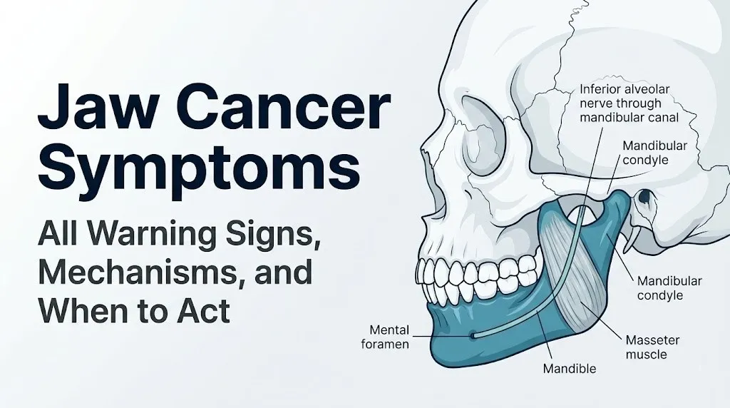
Jaw numbness in jaw cancer is produced by perineural invasion, in which tumor cells infiltrate the perineurium, the connective tissue sheath surrounding peripheral nerve fascicles, and progressively destroy the myelinated and unmyelinated nerve fibers of the inferior alveolar nerve (IAN) within the mandibular canal or the mental nerve at its exit from the mental foramen, producing paresthesia (altered sensation) followed by anesthesia (complete numbness) of the lower lip, chin, and skin overlying the mandible.
This specific symptom has a clinical name: Vincent’s symptom (or the “numb chin syndrome”), referring to the numbness of the lower lip and chin from IAN involvement. The clinical literature consistently identifies Vincent’s symptom as a red flag that warrants urgent evaluation, because it can indicate either primary jaw malignancy invading the mandibular canal from within or metastatic disease to the jaw from a distant primary tumor (most commonly breast, lung, kidney, or prostate cancer) that has seeded the mandibular bone and compressed the IAN.
According to research published in the Journal of Oral and Maxillofacial Surgery, new onset of lower lip or chin numbness in an adult without a clear dental explanation (such as recent dental injection or tooth extraction in that area) is malignancy until proven otherwise and requires imaging and biopsy evaluation by an oral and maxillofacial surgeon.
The anatomical sequence of IAN paresthesia is clinically predictable. As the tumor first impinges on the nerve, the patient notices tingling, “pins and needles,” or a burning quality in the lower lip and chin on the affected side. As invasion progresses, sensation becomes reduced and ultimately absent. Pain may accompany the paresthesia phase because the nociceptive C fibers are activated by perineural inflammation before they are destroyed.
People with known breast, lung, kidney, or prostate cancer who develop new jaw or lower lip numbness should contact their oncologist the same day, as this symptom may represent jaw metastasis and requires immediate evaluation rather than a scheduled follow-up appointment.
Key Takeaway: New unexplained numbness of the lower lip or chin (Vincent’s symptom) in an adult without a dental explanation is a clinical red flag for jaw malignancy or metastatic disease and requires evaluation by an oral and maxillofacial surgeon within one week, not a routine future appointment.
Trismus and Difficulty Opening the Mouth in Jaw Cancer
Trismus in jaw cancer is defined clinically as a maximum interincisal opening (the distance between the upper and lower front teeth when the mouth is fully open) of less than 35 millimeters, produced by tumor invasion and fibrotic replacement of the masseter muscle, medial pterygoid muscle, or pterygomandibular space, all of which are anatomically essential to the normal mechanics of jaw opening and closing.
The pterygomandibular space is an anatomically compact region bounded by the medial surface of the mandibular ramus laterally, the medial pterygoid muscle medially, the pterygoid hamulus superiorly, and the mylohyoid muscle inferiorly. When tumor from the retromolar trigone or posterior buccal mucosa extends into this space, it encounters and infiltrates the medial pterygoid muscle, producing pain with jaw movement initially and then progressive restriction of opening as fibrotic replacement of muscle tissue occurs.
Think of the jaw-opening mechanism like a pair of hinges on either side of a door. The lateral pterygoid muscles are the primary “openers,” pulling the condyles forward and down. When tumor infiltrates adjacent musculature, it’s as if someone poured concrete around those hinges. The jaw still functions until the restriction reaches the threshold of clinical trismus, but the restriction progresses steadily as tumor infiltration expands.
The clinical significance of trismus in jaw cancer is twofold: it indicates tumor extension into the masticator space (a clinically advanced finding that worsens the staging and prognosis) and it makes diagnostic biopsy, dental care, and subsequent anesthesia for surgical treatment technically challenging.
People being treated for jaw cancer should be informed that radiation therapy, when used as part of treatment, can worsen trismus through radiation-induced fibrosis of the same masticatory muscles, and that jaw stretching exercises during and after radiation therapy are a standard component of functional rehabilitation. This is a discussion to have with the radiation oncologist before treatment begins.
Loose Teeth and Dental Changes as Jaw Cancer Symptoms
Loose teeth in jaw cancer are produced by tumor-driven destruction of the periodontal ligament and alveolar bone surrounding the tooth roots, in which tumor-secreted matrix metalloproteinases (MMP-2 and MMP-9) degrade the collagen fiber bundles of the periodontal ligament and osteoclast-activating factors (RANKL, IL-6, and PTHrP released by tumor cells) recruit and activate osteoclasts to resorb the alveolar bone that anchors the tooth.
The periodontal ligament is a specialized connective tissue structure consisting of densely packed collagen fiber bundles (Sharpey’s fibers) that suspend the tooth root within the alveolar socket, absorbing occlusal forces and maintaining tooth position. When the ligament’s collagen infrastructure is degraded by tumor-secreted MMPs, the tooth loses its primary anchoring mechanism and becomes progressively mobile.
Clinically, the pattern of tooth loosening in jaw cancer has distinguishing features:
- Loosening of teeth in a specific localized area of the jaw, not generalized across all teeth (which would suggest periodontal gum disease)
- Loosening occurring in teeth with healthy-appearing surrounding gum tissue (no signs of the bleeding, recession, or pocket formation typical of periodontal disease)
- Loosening associated with jaw pain or swelling in the same region
- Loosening that persists or worsens after professional dental cleaning and scaling (which would normally improve mobility from periodontal disease)
Any tooth that becomes loose without periodontal disease, recent trauma, or a clear orthodontic explanation should prompt the dentist to obtain a periapical radiograph and panoramic radiograph of the jaw. These images can identify early osteolytic bone loss surrounding the root that may indicate tumor invasion of the alveolar bone before the lesion is clinically palpable.
Adults wearing partial or full dentures who notice that their prosthesis is suddenly fitting differently in one specific area should request a dental examination rather than proceeding directly to denture adjustment, as a change in denture fit can indicate alveolar bone resorption from an underlying jaw lesion.
Jaw Cancer Symptoms Affecting Swallowing and the Throat
Jaw cancer symptoms affecting swallowing occur when tumor extends beyond the jaw bone into the floor of mouth (the soft tissue region inferior to the tongue, supported by the mylohyoid muscle and genioglossus muscle), the tongue base, or the pharyngeal wall, compressing or infiltrating the muscular structures coordinating the swallow reflex and producing dysphagia (difficulty swallowing) and odynophagia (painful swallowing).
The act of swallowing requires coordinated contraction of the mylohyoid, genioglossus, geniohyoid, hyoglossus, and pharyngeal constrictor muscles in a precise temporal sequence. When tumor invades the floor of mouth musculature, this coordination is disrupted. The patient typically notices progressive difficulty initiating swallowing, a sensation of food “sticking” in the throat or base of the tongue, or the need to wash food down with liquids more frequently than before.
These symptoms overlap with symptoms of many benign conditions including acid reflux (gastroesophageal reflux disease), pharyngitis, and epiglottitis. The feature that distinguishes jaw cancer-related dysphagia from these benign causes is the progressive, non-fluctuating course alongside other jaw-specific symptoms (jaw pain, numbness, swelling) in a person with jaw cancer risk factors.
According to the Mayo Clinic, dysphagia that is progressive, accompanied by weight loss, and occurring in a person with a history of tobacco or alcohol use warrants evaluation by an otolaryngologist (head and neck surgeon) rather than being managed with dietary modification alone.
People experiencing dysphagia alongside any of the jaw-specific red flag symptoms (jaw numbness, trismus, palpable neck lump) should be evaluated by an oral and maxillofacial surgeon and an otolaryngologist concurrently, because the combination suggests locally advanced disease requiring multidisciplinary assessment.
Key Takeaway: Dysphagia (difficulty swallowing) alongside jaw pain, numbness, or a palpable neck lump is not a benign symptom cluster to attribute to reflux or sore throat. This combination requires evaluation by both an oral and maxillofacial surgeon and a head and neck otolaryngologist, not a primary care appointment alone.
Ear Pain and Referred Pain as Jaw Cancer Symptoms
Ear pain in jaw cancer is almost always referred pain (otalgia), in which nociceptive signals from the tumor-invaded jaw or floor of mouth travel along the auriculotemporal nerve (a branch of cranial nerve V3) or the glossopharyngeal nerve (cranial nerve IX) to the ear, producing pain perceived at the ear while the actual source of nociception is in the jaw or pharynx.
Referred otalgia from jaw malignancy occurs because the ear shares its sensory innervation with multiple head and neck structures through a convergent pattern in the trigeminal brainstem nuclear complex. The auriculotemporal nerve, which supplies the anterior ear and external auditory meatus, originates from the same trigeminal mandibular trunk (V3) that supplies the lower jaw, floor of mouth, and TMJ. When nociceptive signals from a jaw tumor travel along V3, the brain may misattribute the pain to the ear because both structures share the same nerve origin.
Clinically, referred otalgia from jaw cancer has a specific presentation pattern:
- Ear pain present without any ear examination findings (normal tympanic membrane, no discharge, no hearing loss on audiometry)
- Ear pain that is constant and progressive rather than episodic
- Ear pain occurring alongside jaw or throat symptoms
- Ear pain not responding to antibiotic treatment for a presumed ear infection
The clinical problem is that referred otalgia from jaw cancer is frequently misdiagnosed as recurrent otitis media (middle ear infection) or eustachian tube dysfunction, leading to multiple antibiotic prescriptions without evaluation of the underlying head and neck cause. According to clinical guidance from the American Academy of Otolaryngology-Head and Neck Surgery (AAO-HNS), any adult with persistent otalgia and a normal ear examination requires endoscopic nasopharyngoscopy and evaluation of the oral cavity, oropharynx, and hypopharynx to rule out a head and neck malignancy as the source of referred pain.
Jaw Cancer Symptoms at the TMJ and Condylar Region
Jaw cancer symptoms at the temporomandibular joint (TMJ) and condylar region are among the most frequently misdiagnosed presentations in jaw malignancy, because cancer of the mandibular condyle or the adjacent parotid gland and infratemporal fossa produces symptoms including jaw joint pain, restricted mouth opening, and clicking or crepitus that are clinically indistinguishable from temporomandibular disorder (TMD) without imaging evaluation.
The mandibular condyle is the rounded protrusion at the upper end of the mandibular ramus that articulates with the mandibular fossa of the temporal bone through the articular disc of the TMJ. Cancer arising from the condylar cartilage (chondrosarcoma), from the bone of the condylar head or neck (osteosarcoma or SCC invasion), or from the adjacent parotid gland compressing the condyle can produce pain felt at the preauricular (in front of the ear) region, which is the classic location of TMJ pain.
The distinguishing clinical features that point toward condylar malignancy rather than benign TMD include:
- Progressive restriction of mouth opening that does not fluctuate day to day (TMD typically varies with stress and jaw use)
- Preauricular pain that is constant and does not respond to rest, NSAIDs, or occlusal splinting
- A palpable mass at the preauricular or retromandibular region (in front of or just behind the ear)
- Facial asymmetry from condylar mass expanding into the face
- Numbness of the ear, preauricular skin, or temporalis scalp (from auriculotemporal nerve involvement)
Any patient with a working diagnosis of TMD who does not improve within four to six weeks of appropriate conservative treatment (occlusal splint, physical therapy, anti-inflammatory medications) should have a panoramic radiograph and ideally a CT scan of the jaw ordered before assuming the diagnosis is correct.
Women are disproportionately diagnosed with TMD, and condylar jaw cancer in women is therefore more likely to be attributed to TMD for longer before the correct diagnosis is reached.
Late Stage Jaw Cancer Symptoms and Disease Progression
Late stage jaw cancer symptoms reflect tumor extension beyond the primary jaw site into adjacent structures, regional lymph node involvement, and the systemic effects of advancing malignancy, producing a symptom pattern that involves multiple anatomical regions simultaneously and significantly impairs basic functions including eating, speaking, and swallowing.
As jaw cancer advances through local invasion, the following symptom categories develop:
- Skin involvement: Tumor erosion through the cortical bone, periosteum, and overlying soft tissue to the external facial skin produces a visible skin ulceration or sinus tract over the jaw. Once skin is involved, the disease has breached all local tissue barriers.
- Pathological fracture: Extensive tumor destruction of the mandibular cortical and trabecular bone can cause the mandible to fracture spontaneously or with minimal force (such as chewing firm food). This represents a surgical emergency requiring immediate maxillofacial evaluation.
- Uncontrolled bleeding: Tumor erosion into the facial artery, inferior alveolar artery, or other vascular structures of the jaw and floor of mouth can produce life-threatening hemorrhage.
- Airway compromise: Tumor extension into the floor of mouth, sublingual space, or parapharyngeal space can displace the tongue posteriorly and superiorly, narrowing the oropharyngeal airway. Progressive difficulty breathing, particularly when supine, in a person with known or suspected jaw cancer requires immediate emergency room evaluation.
According to the American Cancer Society, the overall five-year relative survival rate for oral cavity cancer drops from over 85% when localized to approximately 64% with regional lymph node involvement and approximately 38% with distant metastasis. This gradient underscores why late-stage symptoms, while clinically obvious, represent a diagnostic opportunity that has been missed.
People with a known jaw cancer diagnosis who develop any sudden worsening of symptoms (new bleeding, sudden increase in swelling, breathing difficulty) should go to the emergency room rather than waiting for a scheduled oncology appointment.
Jaw Cancer Symptoms in Women and Sex-Specific Presentation
Women with jaw cancer face a specific clinical challenge: their jaw symptoms are disproportionately attributed to temporomandibular disorder (TMD), a condition that is approximately two to three times more prevalent in women than in men due to sex-based differences in estrogen’s effects on joint laxity and trigeminal pain sensitization, leading to longer delays from symptom onset to jaw cancer diagnosis in women compared to men.
The biological reason for this sex-specific misdiagnosis pattern is that the symptoms of jaw malignancy (preauricular pain, restricted mouth opening, jaw clicking, referred ear pain) overlap almost exactly with the symptoms of TMD, and women are far more likely than men to carry a pre-existing or concurrent TMD diagnosis. When a woman presents with jaw pain and restricted opening, the clinical prior probability in her physician’s or dentist’s mental model defaults to TMD, and jaw cancer may not be considered unless a specific red flag forces reconsideration.
The clinical features that should prompt consideration of jaw malignancy over TMD in a woman with jaw symptoms are:
- Age over 50 with tobacco or alcohol use history (raises SCC risk above TMD as primary diagnosis)
- Jaw symptoms that have been progressively worsening despite four to six weeks of standard TMD management
- The development of new jaw numbness (which is not a feature of TMD)
- A palpable new mass in the jaw, face, or neck region
- Weight loss, fatigue, or other systemic symptoms alongside the jaw complaints
Women also represent a growing proportion of HPV-associated oropharyngeal cancers, which can present with jaw involvement. According to data from the NCI SEER database, the incidence of HPV-associated oropharyngeal cancer in women has been increasing in recent decades, and this population may have no traditional tobacco or alcohol risk factors, making the clinical index of suspicion lower for a malignant cause of jaw symptoms.
HPV-Associated Jaw and Oropharyngeal Cancer Symptoms
HPV-associated oropharyngeal cancer caused by human papillomavirus 16 (HPV16) presents with a distinct symptom pattern compared to traditional tobacco- and alcohol-related jaw cancers, often beginning with minimal local symptoms and instead manifesting first as a painless neck mass from a metastatic lymph node, while producing jaw-related symptoms only when the primary tumor at the tongue base or tonsillar region extends anteriorly or inferiorly to involve the posterior jaw and floor of mouth.
The mechanism of HPV-related carcinogenesis differs from tobacco-related carcinogenesis. HPV16 and HPV18 encode the oncoproteins E6 and E7, which degrade p53 (TP53) and retinoblastoma protein (Rb), the two primary tumor suppressor proteins controlling cell cycle arrest and DNA repair. By inactivating these gatekeepers, the virus allows cells with DNA damage to continue dividing unchecked, producing a rapidly growing, poorly keratinized squamous cell carcinoma with high sensitivity to radiation therapy.
The symptoms that distinguish HPV-associated oropharyngeal cancer from traditional jaw SCC include:
- A painless neck mass as the first presenting symptom (in approximately 60 to 80% of cases, per clinical series in Head and Neck journal)
- Throat fullness or a persistent sensation of something in the throat
- Odynophagia (painful swallowing), particularly with solid foods
- Referred otalgia (ear pain) without otoscopic findings
- Minimal visible intraoral mucosal changes despite a palpable neck mass
- Often occurring in adults aged 40 to 60 without tobacco or alcohol history
The jaw becomes involved when HPV-associated base of tongue or tonsillar tumors extend anteriorly into the floor of mouth and mandibular region, at which point jaw pain, numbness, and trismus can develop. The American Cancer Society reports that HPV-positive oropharyngeal cancers have substantially better outcomes than HPV-negative cancers at equivalent stages, making early identification of the HPV status of a jaw or oropharyngeal tumor important for treatment planning.
Key Takeaway: A painless neck mass in a non-smoking adult between 40 and 60, especially with persistent throat fullness or mild swallowing difficulty, is more likely to be HPV-associated oropharyngeal cancer with cervical lymph node metastasis than an infectious reactive lymph node, and requires evaluation by a head and neck otolaryngologist the same week it is discovered.
How Jaw Cancer Symptoms Are Diagnosed and Evaluated
Jaw cancer symptoms are evaluated through a systematic clinical and radiological workup that begins with a comprehensive oral examination and panoramic radiograph and progresses through cross-sectional imaging (CT and MRI) and tissue biopsy to achieve a histological diagnosis, which is the only method that definitively confirms or excludes malignancy.
No symptom pattern, imaging finding, or blood test can diagnose jaw cancer without a tissue biopsy confirming the histological type and cellular characteristics. This is the most important clinical fact about jaw cancer diagnosis: clinical evaluation and imaging stratify the risk and guide the biopsy, but only pathology confirms the diagnosis.
The standard diagnostic pathway for suspected jaw cancer:
- Clinical oral examination by a dentist or oral and maxillofacial surgeon: Assessment of mucosal lesions, jaw swelling, tooth mobility, lymph node examination, and assessment of mouth opening (measured with a ruler between upper and lower incisors). Documentation of lesion size, location, surface characteristics, and symptom duration.
- Panoramic radiograph (OPG/OPT): First-line jaw imaging showing the entire mandible and maxilla in a single view. Can identify osteolytic bone destruction, periosteal reactions, tooth root resorption, and loss of lamina dura around tooth roots. Does not provide the cross-sectional detail needed for surgical planning.
- Computed tomography (CT) scan of the jaw and neck: Provides axial and coronal cross-sections showing the degree of cortical bone destruction, extent of soft tissue invasion, and presence of cervical lymphadenopathy. Essential for staging and surgical planning.
- MRI of the jaw and neck: Superior to CT for assessing soft tissue extent, perineural invasion of the IAN, floor of mouth involvement, and tongue base involvement. Ordered when soft tissue detail is clinically needed beyond what CT provides.
- Incisional biopsy: A small piece of tissue is removed from the suspicious lesion under local anesthesia and sent for histological examination. This is the gold standard for diagnosis. Fine needle aspiration cytology (FNAC) of a neck mass may precede open biopsy to assess lymph node status.
- PET-CT: Used in confirmed malignancy cases to assess distant metastasis and identify the primary site in cases presenting with neck mass alone.
People whose primary care physician or dentist orders a panoramic radiograph that shows any osteolytic lesion, periosteal reaction, or root resorption without a dental explanation should be referred to an oral and maxillofacial surgeon for further evaluation within two weeks of that imaging finding.
Jaw Cancer Red Flags and When to See a Doctor Urgently
Jaw cancer red flags are specific symptom combinations or individual symptoms that, in an adult with any jaw cancer risk factors, require evaluation by an oral and maxillofacial surgeon or an otolaryngologist within one to two weeks rather than a routine appointment scheduled months away.
The following symptom patterns require urgent evaluation within one to two weeks by a board-certified oral and maxillofacial surgeon or head and neck otolaryngologist:
- New unexplained numbness or paresthesia of the lower lip, chin, or anterior tongue in an adult over 40 without a dental explanation (possible IAN or mental nerve involvement from jaw malignancy or metastatic disease)
- A persistent oral mucosal ulcer that has not healed after three weeks, particularly if indurated (firm to touch) or if associated with jaw pain
- A new firm, non-tender neck mass in an adult over 40 that has not decreased in size over three weeks
- Progressive trismus (restricted mouth opening) developing over weeks without a clear dental cause (abscess, post-extraction swelling)
- Tooth loosening in a localized jaw area without periodontal disease or trauma
- A preauricular or jaw mass that is progressively enlarging over weeks
Go to the emergency room immediately for the following jaw cancer-related presentations:
- Difficulty breathing or swallowing due to progressive floor of mouth or sublingual swelling in a person with known or suspected jaw tumor
- Spontaneous fracture of the jaw with minimal or no trauma in a person with a known jaw lesion
- Uncontrolled oral hemorrhage from a jaw or floor of mouth lesion
These presentations can indicate airway compromise from tumor mass displacement of the tongue, pathological jaw fracture from tumor bone destruction, or tumor vascular erosion, all of which require emergency surgical or interventional management at a hospital equipped for head and neck surgical emergencies.
Frequently Asked Questions About Jaw Cancer Symptoms
What are the first signs of jaw cancer?
The first signs of jaw cancer are typically a persistent oral mucosal ulcer not healing after two to three weeks, unexplained localized jaw pain or dull aching that does not correspond to a specific tooth, or a new firm lump in the jaw or adjacent neck region.
Early loosening of teeth in a localized jaw area without gum disease and subtle changes in jaw sensation (tingling of the lip or chin) can also represent early jaw cancer symptoms, particularly when no benign dental cause is identified.
Any of these symptoms persisting beyond two to three weeks in an adult with tobacco, alcohol, or HPV exposure warrants evaluation by a dentist or oral and maxillofacial surgeon rather than continued observation.
Can jaw cancer be mistaken for TMJ disorder?
Jaw cancer, particularly cancer of the mandibular condyle or adjacent parotid gland, can produce symptoms including preauricular jaw pain, restricted mouth opening, and jaw clicking that are clinically identical to temporomandibular disorder (TMD) without imaging evaluation.
Women are disproportionately affected by this diagnostic confusion because TMD is two to three times more prevalent in women and is frequently the initial working diagnosis when a woman presents with jaw pain and restricted opening.
Any patient with a presumed TMD diagnosis who does not improve within four to six weeks of appropriate conservative treatment should have a panoramic radiograph and CT scan of the jaw before the TMD diagnosis is confirmed.
What does jaw cancer pain feel like?
Jaw cancer pain is typically described as a deep, boring, aching pain that is poorly localized, not reliably tied to a specific tooth, worse at night, and progressive over weeks rather than intermittent.
This pattern differs from dental abscess pain (throbbing, localized, often associated with temperature sensitivity and tenderness on percussion of a specific tooth) and from TMJ pain (variable, related to jaw use, often worsening with chewing or stress).
Jaw cancer pain that is accompanied by lower lip or chin numbness represents a specific neurological red flag (IAN or mental nerve involvement) requiring urgent evaluation by an oral and maxillofacial surgeon.
Is jaw numbness a sign of cancer?
New unexplained numbness or tingling of the lower lip or chin (Vincent’s symptom or numb chin syndrome) in an adult without a recent dental injection or extraction in that area is a recognized clinical red flag for jaw malignancy or metastatic disease to the jaw.
The inferior alveolar nerve, running through the mandibular canal, can be compressed or invaded by primary jaw tumors or by metastatic deposits from breast, lung, kidney, or prostate cancer that have seeded the jawbone.
Any adult with new unexplained lower lip or chin numbness should be evaluated by an oral and maxillofacial surgeon within one week and should have a panoramic radiograph and CT scan of the jaw ordered as part of that evaluation.
How quickly do jaw cancer symptoms progress?
The rate of symptom progression in jaw cancer depends on the tumor type: squamous cell carcinoma invading the jaw and osteosarcoma typically progress over weeks to a few months, while ameloblastoma may expand the jaw over months to years due to its slower biological behavior.
HPV-associated oropharyngeal cancers extending to the jaw can appear to progress rapidly because the primary tumor at the tongue base or tonsil may have been growing undetected for months before jaw symptoms develop.
Any jaw symptom that is measurably worsening over a four-to-six-week period, regardless of the rate, warrants clinical imaging and evaluation rather than further observation.
At what stage should I see a doctor for jaw symptoms?
Any jaw symptom that has been present without resolution or improvement for two to three weeks should prompt evaluation by a dentist or primary care physician, not continued watchful waiting.
Symptoms including unexplained jaw numbness, a new neck lump, progressive difficulty opening the mouth, or a non-healing oral ulcer should prompt evaluation by an oral and maxillofacial surgeon within one to two weeks of symptom onset, not at the next available routine appointment.
Earlier evaluation directly affects survival outcomes: the National Cancer Institute reports that localized oral cavity cancers have five-year survival rates exceeding 85%, compared to approximately 38% for cancers that have spread to distant sites at the time of diagnosis.
Closing
Jaw cancer symptoms are identifiable, and early identification changes outcomes in a measurable, documented way. The symptom that requires the most consistent clinical vigilance is new unexplained numbness of the lower lip or chin. That one symptom, appearing without a dental explanation in an adult, should never be attributed to a minor nerve irritation and monitored. It requires imaging and a specialist evaluation within one week.
For any jaw symptom lasting longer than two to three weeks without resolution after treating an identifiable cause, request a panoramic radiograph from your dentist and a referral to a board-certified oral and maxillofacial surgeon if no obvious benign cause is confirmed. Bring a timeline of your symptoms, a note of which symptoms have changed over time, and a list of your tobacco, alcohol, and HPV vaccination history to that appointment.
The clinical window between an early and a late jaw cancer diagnosis is the difference between surgical cure and complex, multimodal treatment with a substantially lower probability of disease-free survival.