Benign Adrenal Gland Tumor Symptoms: Full 2026 Guide
Benign adrenal gland tumor symptoms range from completely absent to a distinct cluster of hormonal effects, depending entirely on whether the tumor secretes excess hormones. Most are found accidentally on imaging done for unrelated reasons, but those that do produce symptoms can cause high blood pressure, unexplained weight gain, muscle weakness, and metabolic changes that many people attribute to aging or stress for years before the real cause is found.
According to the National Institute of Diabetes and Digestive and Kidney Diseases (NIDDK), adrenal tumors are among the most common incidentally discovered masses in the human body, with estimates suggesting they are present in approximately 3 to 7 percent of adults when abdominal imaging is performed for any reason. The rate rises steeply with age, reaching 10 percent or higher in adults over 70 in some radiological series. That prevalence makes understanding their symptom patterns genuinely important for a large number of people.
This article covers every major symptom type by the specific hormone involved, explains the physiological mechanism behind each symptom, addresses how the clinical picture differs by sex and age, describes the full diagnostic workup, and identifies the specific warning signs that require urgent evaluation.
Benign Adrenal Gland Tumor Symptoms: What to Know First
Benign adrenal gland tumor symptoms depend almost entirely on whether the tumor actively secretes hormones, and if so, which hormone is being overproduced in excess of the body’s regulated range.
Two broad categories define the clinical picture. A hormone-secreting adrenal adenoma produces a recognizable symptom cluster tied to the specific excess hormone: cortisol, aldosterone, androgens, or catecholamines. A non-functioning adenoma produces no hormonal symptoms at all and is typically found during CT or MRI imaging ordered for an unrelated complaint.
The challenge, clinically, is that symptoms from hormone-secreting adenomas often develop gradually. Mild cortisol excess can look like ordinary weight gain, fatigue, and poor sleep for two to four years before the pattern becomes recognized as a hormonal problem. Mild aldosterone excess can look like treatment-resistant hypertension. Recognizing the symptom pattern matters because it changes treatment.
Key clinical symptoms by type of hormone secretion:
| Hormone Secreted | Characteristic Symptoms | Organ Systems Primarily Affected |
|---|---|---|
| Cortisol | Weight gain (central), thin skin, fatigue, high blood sugar, poor wound healing | Metabolic, musculoskeletal, immune, cardiovascular |
| Aldosterone | High blood pressure, muscle cramps, fatigue, excessive urination at night | Cardiovascular, renal, electrolyte balance |
| Androgens (men and women) | Acne, hirsutism in women, early puberty in children, menstrual irregularity | Reproductive endocrine axis, skin |
| Catecholamines | Episodic headache, sweating, rapid heart rate, severe hypertensive episodes | Cardiovascular, autonomic nervous system |
| None (non-functional) | No hormone-related symptoms | None |
People with diabetes, hypertension, or obesity should take note: these conditions are both risk factors for adrenal incidentalomas and common consequences of undetected cortisol or aldosterone excess, which means the existing diagnosis may be obscuring the underlying cause.
What Is an Adrenal Adenoma?
An adrenal adenoma is a benign, non-cancerous growth arising from the cells of the adrenal cortex, the outer layer of the adrenal gland seated at the superior pole of each kidney.
The term “adenoma” specifically indicates a benign growth of glandular cells. This distinguishes it from adrenocortical carcinoma, which is the malignant version, and from pheochromocytoma, which arises from the adrenal medulla rather than the cortex. Most adrenal adenomas are small, typically between one and three centimeters, though some reach four or five centimeters. Size matters clinically because the Endocrine Society Clinical Practice Guidelines (2023 update) use a four-centimeter threshold as one of the imaging criteria influencing the decision between surgical removal and active surveillance.
Adrenal adenomas are not a single uniform entity. They are better understood as a spectrum: some are hormonally silent, some produce borderline-excess hormone levels that cause subtle metabolic effects, and some produce overt hormone excess with a fully developed clinical syndrome. That spectrum is why two people with “adrenal adenomas” can have completely different symptom experiences.
Important distinction: an adrenal adenoma is not adrenal cancer. The malignancy rate for adrenal masses under four centimeters with benign imaging characteristics is estimated at less than two percent, according to data reviewed in the Journal of Clinical Endocrinology and Metabolism. The primary clinical concern with most adrenal adenomas is not cancer but undetected hormone excess.
Adrenal Gland Anatomy and Hormone Function
The adrenal gland functions like a layered hormone production facility. Each of its four distinct zones makes a different hormone class, and a benign tumor arising in any one zone will overproduce that zone’s specific hormone.
The adrenal cortex has three concentric layers. The zona glomerulosa (outermost layer) produces aldosterone, the hormone that governs sodium retention and potassium excretion in the kidney. The zona fasciculata (middle and largest layer) produces cortisol, the body’s primary glucocorticoid and stress hormone. The zona reticularis (innermost cortex layer) produces androgens, primarily dehydroepiandrosterone (DHEA) and its sulfate form, DHEA-S, which are precursors to testosterone and estrogen in peripheral tissues.
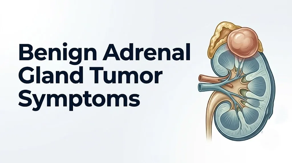
Inside the cortex sits the adrenal medulla, which contains chromaffin cells that produce epinephrine and norepinephrine, the catecholamines. Strictly speaking, a tumor of the adrenal medulla producing catecholamines is called a pheochromocytoma rather than an adenoma, but because pheochromocytomas are typically discovered during an adrenal mass workup, understanding their symptoms is essential for anyone evaluating an adrenal tumor.
Think of the adrenal gland like a four-department manufacturer. An adenoma is essentially a rogue department that keeps producing its product regardless of whether headquarters (the pituitary gland and hypothalamus) sends the order. The result is a factory floor flooded with one type of hormone, producing downstream effects in every organ that responds to that hormone.
According to the NIDDK, the adrenal glands collectively process signals from the pituitary ACTH (adrenocorticotropic hormone) to regulate cortisol output, and from renin-angiotensin signaling to regulate aldosterone, with the adenoma’s cells largely bypassing this regulatory feedback.
Functional vs. Non-Functional Adrenal Adenoma
A functional adrenal adenoma secretes hormone at levels sufficient to produce measurable physiological effects; a non-functional adrenal adenoma does not produce any hormone excess and has no direct symptom consequence.
The distinction defines the entire clinical management plan. A non-functional adenoma under four centimeters with benign imaging characteristics requires imaging surveillance only. A functional adenoma requires biochemical confirmation, specialist co-management, and often surgical or pharmacological intervention to prevent cardiovascular, metabolic, and skeletal complications.
The proportion that are functional varies by hormone type. According to a 2021 meta-analysis published in the European Journal of Endocrinology, approximately 30 to 50 percent of adrenal incidentalomas show some degree of autonomous cortisol secretion, though only a fraction of these meet the threshold for full Cushing syndrome. About 1 to 5 percent of incidentalomas are aldosterone-producing (with higher rates, up to 30 percent, among patients with primary hypertension). Catecholamine-secreting tumors account for roughly 4 to 5 percent of adrenal masses found incidentally.
Determining functionality requires biochemical testing. Imaging characteristics can suggest a lipid-rich, likely benign adenoma, but they cannot confirm whether it is hormonally active. The Endocrine Society 2023 Clinical Practice Guidelines recommend biochemical screening for hormone excess in every patient with an adrenal incidentaloma regardless of symptoms, because subclinical hormone excess can be clinically consequential even without overt symptoms.
For people with difficult-to-control blood pressure, unexplained low potassium levels, or new-onset diabetes, awareness that an adrenal adenoma might be functionally contributing to these problems can prevent years of inadequate treatment.
Key Takeaway: Whether a benign adrenal tumor causes any symptoms depends entirely on whether it secretes excess hormone. Imaging alone cannot determine this. Biochemical testing is required in every case.
Cortisol-Secreting Adrenal Adenoma Symptoms
A cortisol-secreting adrenal adenoma produces excess cortisol autonomously from the zona fasciculata, bypassing the normal pituitary ACTH regulatory signal and delivering a sustained hormonal excess that disrupts multiple organ systems simultaneously.
The full clinical picture of cortisol excess is called Cushing syndrome. When the source is an adrenal adenoma specifically, it is classified as ACTH-independent Cushing syndrome (because the pituitary is not the driver). The hallmark features emerge from cortisol’s natural physiological roles taken to pathological excess.
Central weight gain occurs because cortisol stimulates adipogenesis specifically in visceral and truncal fat depots through glucocorticoid receptor activation in adipocytes, while simultaneously driving peripheral fat breakdown. The characteristic result is truncal obesity with relatively thin limbs, sometimes called “lemon on sticks” distribution. The face may develop a rounded appearance (“moon face”) from fat redistribution to facial fat pads.
Skin changes are mechanistically distinct. Cortisol suppresses fibroblast synthesis of collagen and elastin in the dermis, thinning the skin and making subcutaneous blood vessels more visible through the skin surface. Wide purple striae (stretch marks), typically wider than one centimeter and purple rather than the white or silver seen in ordinary weight gain, appear most commonly on the abdomen, thighs, and underarms. Bruising occurs easily from minor trauma because vessel walls lack collagen structural support.
Symptoms of cortisol-secreting adrenal adenoma:
- Central (truncal) fat accumulation with relatively thin arms and legs
- Round, full facial appearance with fat accumulation in cheeks and at the base of the neck
- Purple stretch marks wider than one centimeter on abdomen, thighs, or upper arms
- Easy bruising from minor skin contact
- Proximal muscle weakness, particularly difficulty rising from a chair or climbing stairs (reflects cortisol’s catabolic effect on type II skeletal muscle fibers in the quadriceps and hip flexors)
- High blood pressure (cortisol activates mineralocorticoid receptors at high concentrations, promoting sodium retention)
- Elevated blood glucose or new-onset type 2 diabetes (cortisol drives hepatic gluconeogenesis via phosphoenolpyruvate carboxykinase upregulation and suppresses insulin-mediated glucose uptake in skeletal muscle)
- Poor wound healing (immune and fibroblast suppression)
- Frequent infections (cortisol suppresses T-lymphocyte proliferation and interleukin-2 production)
- Mood changes including irritability, anxiety, depression, and cognitive slowing
- Menstrual irregularity or cessation in women (cortisol suppresses GnRH pulse frequency at the hypothalamus)
- Insomnia with disrupted circadian cortisol pattern (loss of normal evening cortisol nadir)
Postmenopausal women are particularly vulnerable to the skeletal consequences: cortisol excess suppresses osteoblast activity via Wnt signaling pathway inhibition and increases osteoclast activity, producing accelerated bone loss on top of the estrogen-withdrawal bone loss already occurring after menopause.
Aldosterone-Secreting Adrenal Adenoma Symptoms
An aldosterone-secreting adrenal adenoma, also called an aldosteronoma or the tumor responsible for primary hyperaldosteronism (historically Conn syndrome), arises from the zona glomerulosa and drives the renin-angiotensin-aldosterone system (RAAS) into persistent overdrive independently of the normal renin signal.
The physiological mechanism is specific. Aldosterone binds to mineralocorticoid receptors (NR3C2) in the principal cells of the renal distal convoluted tubule and cortical collecting duct. Receptor activation upregulates ENaC sodium channels on the luminal membrane and increases Na/K-ATPase pump density on the basolateral membrane. Sodium reabsorption increases, plasma volume expands, and vascular resistance rises: this is the hypertension mechanism. Simultaneously, potassium exits via ROMK potassium channels and is lost in urine, producing hypokalemia.
Symptoms of aldosterone excess:
| Symptom | Mechanism | Clinical Note |
|---|---|---|
| Resistant hypertension | Plasma volume expansion via ENaC-driven sodium retention | Blood pressure often does not respond adequately to three or more medications |
| Muscle weakness and cramps | Hypokalemia impairs muscle cell membrane potential; affects proximal muscles and can cause rhabdomyolysis at severe levels | Potassium below 3.0 mEq/L is clinically significant |
| Fatigue and low energy | Chronic hypokalemia impairs mitochondrial energy production and alters cardiac conduction | May be mistaken for depression or burnout |
| Nocturia (urinating at night) | Aldosterone-mediated changes in renal water handling reduce normal nocturnal urinary concentration | Often an early, overlooked symptom |
| Polydipsia (excessive thirst) | Compensatory response to renal volume changes and mild sodium elevation | |
| Headaches | Secondary to hypertension, particularly early morning | |
| Palpitations or irregular heartbeat | Hypokalemia lowers resting cardiac membrane potential, increasing arrhythmia risk |
A critical clinical point: the Endocrine Society 2023 guidelines estimate that primary hyperaldosteronism is present in 5 to 10 percent of all hypertensive patients, making aldosterone-secreting adenomas one of the most common correctable causes of secondary hypertension. Many patients with this condition have been treated for essential (primary) hypertension with multiple medications for years before the underlying adenoma is found.
People with African ancestry have a higher baseline prevalence of hypertension, which can mask the aldosterone-driven component. A low serum potassium level alongside resistant hypertension in any demographic should prompt aldosterone screening.
Androgen-Secreting Adrenal Adenoma Symptoms
An androgen-secreting adrenal adenoma arises from the zona reticularis and produces excess DHEA, DHEA-S, and androstenedione, which are converted in peripheral tissues (particularly skin, fat, and gonads) into active androgens including testosterone.
The symptom picture depends heavily on the person’s baseline sex hormone environment. In women, even a modest increase in androgen production from an adrenal adenoma tips the androgen-estrogen ratio enough to produce virilizing effects. In men, the effect is less conspicuous because physiological testosterone levels from testicular production are already much higher.
Symptoms in women with androgen-secreting adrenal adenomas:
- Hirsutism: coarse, dark terminal hair growth in androgen-sensitive areas including the upper lip, chin, chest, and lower abdomen, driven by androgen receptor activation in hair follicles
- Acne of adult onset or worsening, particularly on the jaw, chin, and back
- Scalp hair thinning or male-pattern hair loss (androgenetic alopecia in a female pattern, affecting the crown)
- Clitoral enlargement in cases of significant androgen excess
- Menstrual irregularity or cessation (androgen excess suppresses GnRH pulsatility and reduces LH/FSH output, impairing ovulation)
- Deepening of voice in pronounced cases (irreversible once it occurs)
- Reduced breast tissue in severe or prolonged excess
In men, androgen-secreting adenomas produce fewer noticeable symptoms because the baseline androgen environment is already high. Children, however, present dramatically: androgen excess in prepubertal boys causes accelerated growth, penile enlargement, and premature pubic hair appearance (precocious pseudopuberty). In prepubertal girls, it causes clitoral enlargement, pubic hair development, and accelerated bone age. Pediatric presentations of virilization or precocious puberty always require urgent evaluation by a pediatric endocrinologist.
Estrogen-secreting adenomas are rare but produce feminizing symptoms in men, including gynecomastia (breast tissue development) and testicular atrophy.
Key Takeaway: Cortisol-secreting adenomas affect metabolism and immunity across the whole body; aldosterone-secreting adenomas primarily drive cardiovascular and electrolyte problems; androgen-secreting adenomas produce sex-hormone-related changes most visible in women and children.
Catecholamine Symptoms From an Adrenal Tumor
Catecholamine-producing adrenal tumors are technically pheochromocytomas, arising from chromaffin cells of the adrenal medulla rather than the cortex, but because they are found as part of the adrenal mass workup, their symptoms belong in this clinical picture.
Pheochromocytomas produce epinephrine and norepinephrine through the enzymatic pathway beginning with tyrosine, converting through DOPA, dopamine, and norepinephrine (via dopamine beta-hydroxylase) to epinephrine (via phenylethanolamine N-methyltransferase). When released in excess, these catecholamines produce dramatic adrenergic stimulation.
The classic presentation is episodic rather than constant, which distinguishes it from the persistent symptoms of cortisol or aldosterone excess. Episodes may last minutes to an hour and can be triggered by physical pressure on the tumor, certain medications, emotional stress, urination (for extra-adrenal pheochromocytomas), or physical exertion.
Classic episodic symptom triad of pheochromocytoma:
- Severe headache (pulsating, throbbing, produced by acute hypertensive surges)
- Profuse sweating (adrenergic stimulation of eccrine sweat glands via cholinergic sympathetic innervation)
- Palpitations and rapid heart rate (direct beta-1 adrenergic receptor stimulation in the sinoatrial node and ventricular myocardium)
Additional symptoms during episodes:
- Severe, sometimes crisis-level hypertension (systolic above 200 mmHg possible)
- Pallor rather than flushing (norepinephrine-mediated alpha-1 adrenergic peripheral vasoconstriction)
- Tremor
- Anxiety or sense of impending doom
- Chest tightness or chest pain
- Nausea
Between episodes, some patients have persistent hypertension, fatigue, and weight loss.
Catecholamine-secreting adrenal tumors carry the highest immediate safety urgency of any adrenal mass type. An undiagnosed pheochromocytoma during surgery, certain medication administration (including some anesthetics, beta-blockers given without alpha-blockade, and glucocorticoids), or an emotional stress event can trigger a hypertensive crisis or fatal cardiac arrhythmia. Biochemical screening for catecholamine excess (plasma free metanephrines or 24-hour urinary metanephrines) is mandatory in every adrenal mass workup.
Adrenal Incidentaloma: The Tumor With No Symptoms
An adrenal incidentaloma is defined as an adrenal mass found on imaging performed for a reason completely unrelated to adrenal disease. It carries no inherent symptom of its own when it is non-functional.
The person has no pain from the adenoma. No palpable mass. No hormonal symptom. They may have had a CT scan for abdominal pain, a kidney stone evaluation, or a pre-surgical abdominal survey, and the radiologist notes “adrenal nodule, right side, 1.8 cm” in the report. The clinical challenge begins at that point.
According to JAMA Internal Medicine, the prevalence of adrenal incidentalomas detected on imaging has risen substantially as CT scanning has become more common in clinical practice, with some estimates now placing incidental discovery at more than one million new cases per year in the United States. This means primary care physicians and radiologists encounter them regularly.
Non-functional does not mean no evaluation needed. The Endocrine Society 2023 Clinical Practice Guidelines recommend:
- Biochemical screening for all three major hormone excess syndromes (cortisol, aldosterone, catecholamines) regardless of symptoms
- CT imaging characterization using Hounsfield unit (HU) measurement to estimate lipid content: a mass measuring 10 HU or below on unenhanced CT is highly likely to be a benign lipid-rich adenoma
- Follow-up imaging at 6 to 12 months if the mass is over 4 cm or has indeterminate imaging characteristics, and again at 24 months
- Repeat biochemical testing at 2 years regardless of initial result, because some initially non-functional adenomas develop autonomous hormone secretion over time
A non-functional incidentaloma does not cause symptoms today. But it requires professional evaluation to confirm that status and establish an appropriate monitoring plan.
Key Takeaway: An adrenal incidentaloma found on imaging requires biochemical hormone screening and imaging characterization even when no symptoms are present, because subclinical hormone excess can exist without obvious symptoms and progress over time.
Adrenal Adenoma Symptom Progression Over Time
Adrenal adenoma symptoms do not typically appear suddenly. They develop over months to years as the hormone excess gradually accumulates above the threshold where the body’s compensatory mechanisms can mask the effect.
Subclinical hypercortisolism illustrates this progression. The 1-mg overnight dexamethasone suppression test is used to screen for autonomous cortisol secretion. A normal result means cortisol is suppressed below 1.8 mcg/dL after the dexamethasone dose. A result between 1.8 and 5.0 mcg/dL suggests possible autonomous secretion without full Cushing syndrome. A result above 5.0 mcg/dL indicates clinically meaningful cortisol excess.
A person with a morning cortisol of 2.5 mcg/dL after suppression has subclinical hypercortisolism. They may not look like they have Cushing syndrome. Their weight gain may seem modest. Their blood pressure may be mildly elevated but not alarmingly so. Their bone density may be slowly falling. Their blood sugar may be inching upward. Each individual finding is easily attributed to aging. Collectively, the trajectory represents years of undetected metabolic damage.
Research published in the Journal of Clinical Endocrinology and Metabolism in 2022 found that patients with subclinical hypercortisolism had significantly higher rates of type 2 diabetes, hypertension, and cardiovascular events compared to matched controls with non-functional adenomas, even without overt Cushing syndrome features. The damage is real even when the symptoms are subtle.
Symptom progression can also move in the other direction after treatment. Following adrenalectomy for a cortisol-secreting adenoma, the contralateral adrenal gland (which has been suppressed by chronic ACTH suppression from the autonomous cortisol excess) needs weeks to months to recover normal function. During this recovery period, patients are at risk for adrenal insufficiency and must receive glucocorticoid replacement therapy under strict specialist management. The transition from hormone excess to potential hormone deficiency is itself a clinical window requiring careful monitoring.
Adrenal Adenoma Symptoms in Women and Older Adults
Women represent the majority of patients diagnosed with adrenal adenomas, particularly cortisol-secreting types. Postmenopausal women face a compounded clinical challenge: the natural metabolic and hormonal changes of menopause, including central weight gain, bone loss, and blood pressure changes, overlap precisely with the early symptoms of cortisol-secreting adenoma.
A postmenopausal woman gaining abdominal weight, developing mild hypertension, and experiencing fatigue is managing what appears to be a common menopausal trajectory. The adrenal adenoma contributing subclinical cortisol excess to that picture may go undetected for years precisely because nothing seems out of the ordinary. Research in the European Journal of Endocrinology has highlighted that postmenopausal women with adrenal incidentalomas and subclinical hypercortisolism have accelerated bone loss beyond what menopause alone produces.
Sex-specific considerations for adrenal adenoma symptoms:
| Biological Sex | Unique Clinical Features | Why the Mechanism Differs |
|---|---|---|
| Women | Cortisol-secreting adenomas more prevalent; androgen excess produces more visible virilizing symptoms; menstrual disruption is an early indicator | Lower baseline androgen levels mean smaller excess is clinically significant |
| Men | Androgen-secreting adenomas produce fewer visible symptoms; estrogen-secreting adenomas (rare) cause gynecomastia; aldosterone-driven symptoms are similar | Higher baseline androgen masking effect; lower rate of cortisol-secreting types relative to women |
| Older adults (over 60) | Higher overall incidence of incidentalomas; subclinical hypercortisolism worsens age-related osteoporosis, sarcopenia, and insulin resistance; hypertension from aldosterone excess may compound pre-existing cardiovascular disease | Age-related declines in cortisol clearance amplify relative hormone excess |
| Children | Androgen-secreting tumors produce precocious puberty; cortisol-secreting tumors cause growth deceleration despite weight gain; adrenal carcinoma must be ruled out aggressively in pediatric adrenal masses | Pediatric adrenal masses have higher malignancy rates than adult masses |
People with multiple endocrine neoplasia type 1 (MEN1), a genetic syndrome involving mutations in the MEN1 tumor suppressor gene, have a substantially elevated risk of adrenal adenomas alongside parathyroid and pituitary tumors. Individuals with a family history of MEN1 should discuss adrenal surveillance with their physician proactively.
How Adrenal Adenoma Is Diagnosed
Adrenal adenoma diagnosis follows a two-track process: imaging characterization to assess size, density, and growth pattern, and biochemical testing to determine hormone secretion status.
Imaging characterization uses computed tomography (CT scan) as the primary modality. The key measurement is the Hounsfield unit (HU) density on unenhanced CT. A mass measuring 10 HU or below is lipid-rich and carries very high probability of being a benign adenoma. A mass measuring above 10 HU is lipid-poor and requires further characterization with contrast-enhanced CT washout protocol or MRI chemical shift imaging (which assesses lipid content using in-phase and out-of-phase signal intensity changes). Size is also relevant: the European Society of Endocrinology 2023 guidelines recommend surgical consultation for any adrenal mass exceeding four centimeters because the probability of malignancy increases above this threshold.
Biochemical hormone screening tests:
- 1-mg overnight dexamethasone suppression test: detects autonomous cortisol secretion; post-suppression cortisol above 1.8 mcg/dL is abnormal
- Late-night salivary cortisol (two measurements on separate nights): assesses loss of normal cortisol circadian rhythm
- 24-hour urinary free cortisol: measures total daily cortisol output; elevation confirms excess but is less sensitive for subclinical cases
- Aldosterone-to-renin ratio (ARR): initial screening test for primary hyperaldosteronism; a ratio above 30 (with aldosterone in ng/dL and renin in ng/mL/hr) suggests autonomous aldosterone production and warrants confirmatory testing
- Plasma free metanephrines (or 24-hour urinary metanephrines and catecholamines): screens for pheochromocytoma; highly sensitive and specific for catecholamine excess
- DHEA-S measurement: screens for androgen excess from adrenal source
When aldosterone-to-renin ratio is elevated and confirmatory testing (salt loading or fludrocortisone suppression test) confirms primary hyperaldosteronism, adrenal vein sampling (AVS) is the gold standard for determining whether the excess comes from a unilateral adenoma or bilateral adrenal hyperplasia. This distinction is critical because unilateral adenoma is surgically curable while bilateral hyperplasia is managed medically.
Adrenal Adenoma Complications and Secondary Conditions
Undetected or inadequately treated adrenal adenoma complications extend well beyond the primary symptoms. The sustained hormone excess reshapes multiple organ systems over time in ways that do not fully reverse even after the adenoma is removed.
Cardiovascular complications from aldosterone excess go beyond hypertension. The Endocrine Society has documented that aldosterone activates NF-kB-mediated inflammatory signaling in cardiac fibroblasts and vascular smooth muscle cells, promoting left ventricular hypertrophy and arterial wall stiffening that persist even after blood pressure is controlled. Patients with primary hyperaldosteronism have higher rates of myocardial infarction, stroke, and atrial fibrillation compared to patients with essential hypertension of similar blood pressure severity, according to data published in JAMA Internal Medicine.
Cortisol excess complications include:
- Type 2 diabetes or prediabetes: from gluconeogenesis upregulation and peripheral insulin resistance
- Osteoporosis: cortisol suppresses RANKL/OPG ratio in bone, shifting toward osteoclast dominance and net bone resorption; vertebral fracture risk increases even in subclinical hypercortisolism
- Sarcopenia: sustained cortisol-mediated catabolism of skeletal muscle protein, most evident in proximal muscle groups
- Cardiovascular disease: independent of blood pressure, cortisol excess promotes dyslipidemia and endothelial dysfunction
- Immune suppression: increased susceptibility to bacterial and fungal infections, reactivation of latent tuberculosis in endemic populations
- Psychiatric comorbidities: depression, anxiety, and cognitive impairment are direct glucocorticoid receptor-mediated effects in the prefrontal cortex and hippocampus, where cortisol excess reduces dendritic branching and synaptic density
The psychiatric effects of cortisol excess are particularly underrecognized. Many patients with cortisol-secreting adrenal adenomas carry a diagnosis of major depressive disorder or generalized anxiety disorder for years before the endocrine source is identified. Adrenal adenoma should be considered in patients with treatment-resistant depression accompanied by metabolic features like central weight gain or unexplained hypertension.
Key Takeaway: The complications of undetected hormone-secreting adrenal adenomas, including cardiovascular remodeling, bone loss, and metabolic disease, can persist after the adenoma is removed and are reason enough to screen thoroughly even when symptoms seem mild.
Emergency Symptoms: Adrenal Crisis Signs
Certain symptoms associated with benign adrenal gland tumors and their treatment require immediate emergency evaluation. Do not wait to see if these resolve on their own.
Call 911 or go to the nearest emergency room immediately if you experience:
- Severe, sudden-onset abdominal pain accompanied by vomiting and profound weakness following adrenal surgery or in a person known to have adrenal adenoma with cortisol excess: this can represent acute adrenal insufficiency (adrenal crisis), a life-threatening emergency caused by sudden withdrawal of cortisol
- Extreme low blood pressure (systolic below 90 mmHg), dizziness, and fainting especially after adrenalectomy or during acute illness in a person on glucocorticoid replacement: this pattern indicates acute cortisol deficiency requiring immediate intravenous hydrocortisone
- Sudden severe hypertension (blood pressure above 180/120 mmHg) with pounding headache, sweating, and chest pain or palpitations: this can represent a pheochromocytoma crisis, a catecholamine storm that can cause cardiac arrhythmia, myocardial infarction, or hemorrhagic stroke
- Loss of consciousness or unresponsiveness in a person with known adrenal disease or recent adrenal surgery: presume adrenal crisis until proven otherwise
- Severe chest pain or sudden difficulty breathing in a person with known aldosterone-producing adenoma with hypokalemia: extreme potassium depletion can cause dangerous cardiac arrhythmias including ventricular tachycardia
- High fever with confusion and hypotension in a person on glucocorticoid replacement after adrenalectomy: any physiological stress (infection, trauma, surgery) can precipitate adrenal crisis in these patients
These presentations can indicate acute adrenal insufficiency, pheochromocytoma hypertensive crisis, or life-threatening cardiac arrhythmia from severe electrolyte disturbance and require emergency medical assessment, not an urgent care appointment or a phone call to a clinic.
When to See an Endocrinologist for Adrenal Adenoma
A board-certified endocrinologist should evaluate any person who has been told they have an adrenal mass on imaging, regardless of symptoms, and any person with unexplained symptoms strongly suggesting hormone excess from the adrenal gland.
The referral threshold is specific. Any adrenal mass found incidentally on imaging warrants endocrinology evaluation because the initial workup, including biochemical hormone screening and imaging characterization, requires specialized interpretation. Primary care physicians should not attempt to manage adrenal incidentalomas without endocrinology co-management.
Circumstances that specifically warrant urgent or prioritized endocrinology referral:
- Any adrenal mass over four centimeters in size (higher malignancy risk requiring surgical consultation alongside endocrinology)
- Biochemical evidence of any hormone excess on initial screening tests
- Blood pressure unresponsive to three or more medications alongside low serum potassium (primary hyperaldosteronism workup)
- New-onset diabetes, osteoporosis, or proximal muscle weakness in a person with known adrenal mass
- Episodic hypertension with headache, sweating, and palpitations (pheochromocytoma screening priority)
- Any virilizing symptoms in a woman or feminizing symptoms in a man with an adrenal mass (sex hormone-secreting adenoma or carcinoma workup)
- Pediatric patients with any adrenal mass (higher malignancy rate, requires pediatric endocrinology evaluation)
Before the endocrinology appointment, bring:
- All imaging reports and the actual CT or MRI images on disc or electronic format
- A complete medication list including over-the-counter medications, herbal supplements, and inhaled or topical steroids (which can affect cortisol testing results)
- Blood pressure logs from home monitoring if hypertension is present
- A written description of all symptoms with dates when they started
- Any prior laboratory results including potassium levels, blood glucose, and lipid panels
Follow-up imaging schedule (for non-functional adenomas under four centimeters with benign CT characteristics) follows the European Society of Endocrinology 2023 guidelines: repeat CT at 6 to 12 months, then at 24 months. If no growth and no biochemical change, further imaging follow-up may not be needed. Repeat biochemical hormone screening at 2 years is standard.
Key Takeaway: Every person with an adrenal mass on imaging needs endocrinology evaluation and biochemical hormone screening. This is not optional monitoring. It is the standard of care because subclinical hormone excess can be silently damaging even without obvious symptoms.
Frequently Asked Questions About Benign Adrenal Gland Tumor Symptoms
Can a benign adrenal gland tumor cause weight gain?
A benign adrenal tumor that secretes excess cortisol can cause weight gain, specifically central (truncal) fat accumulation driven by cortisol’s activation of glucocorticoid receptors in visceral adipocytes.
This type of weight gain concentrates in the abdomen, face, and upper back, while the arms and legs may remain thin, producing a distinctive body distribution.
Weight gain from a non-functional adrenal adenoma that does not secrete hormones is not caused by the tumor itself, and other causes should be investigated.
What does an adrenal adenoma feel like physically?
Most adrenal adenomas produce no pain, pressure, or physical sensation because they are small (typically one to three centimeters) and located in the retroperitoneal space where there is no somatic sensory nerve coverage for early-stage masses.
Symptoms that do arise come from the hormones the tumor secretes, not from the physical mass itself: fatigue, weakness, palpitations, or high blood pressure rather than localized pain.
Very large adenomas (above five centimeters) may occasionally cause a dull flank or upper abdominal discomfort due to local compression, but this is uncommon with benign adrenal adenomas.
How do you know if an adrenal tumor is benign or cancerous?
CT imaging using Hounsfield unit measurement is the first step: a mass measuring 10 HU or below on unenhanced CT is almost certainly benign, while higher density or irregular borders raises suspicion for malignancy.
Size is also relevant: the probability of malignancy increases substantially in masses over four centimeters, and any mass over six centimeters is considered surgically suspicious regardless of imaging characteristics.
Definitive confirmation of benign status requires pathological examination of removed tissue after adrenalectomy; imaging can assess probability but not certainty.
Can an adrenal adenoma go away on its own?
Adrenal adenomas do not typically resolve spontaneously; they are benign neoplasms of adrenocortical cells that do not regress without treatment.
Non-functional adenomas often remain stable in size for years and may never require intervention beyond monitoring, but this stability is not the same as resolution.
Functional adenomas that secrete hormones will continue producing excess hormone and their associated complications until treated, whether surgically through adrenalectomy or medically through medications targeting the hormone pathway.
What blood tests detect a benign adrenal gland tumor?
No single blood test detects an adrenal adenoma, but biochemical screening identifies whether one is secreting excess hormones.
The 1-mg overnight dexamethasone suppression test screens for cortisol excess, the aldosterone-to-renin ratio screens for aldosterone excess, and plasma free metanephrines screen for catecholamine-secreting tumors.
These tests are recommended for every person with an adrenal mass found on imaging, regardless of whether symptoms are present, according to the Endocrine Society 2023 Clinical Practice Guidelines.
Is it safe to leave an adrenal adenoma untreated?
A confirmed non-functional adrenal adenoma under four centimeters with benign imaging characteristics can be safely managed with scheduled surveillance imaging and repeat biochemical testing rather than immediate surgery.
A functional adrenal adenoma secreting excess cortisol, aldosterone, or catecholamines should not be left untreated because the persistent hormone excess causes progressive cardiovascular, metabolic, and skeletal damage that worsens over time.
The decision between active treatment and surveillance should be made by a board-certified endocrinologist reviewing both imaging characteristics and biochemical results, not based on symptoms alone.
Closing
Most people who find out they have a benign adrenal gland tumor through an incidental CT scan feel more alarmed about the word “tumor” than about any symptom they are experiencing. The clinical reality is more nuanced: the tumor itself is almost never the immediate danger. What matters is whether it is secreting hormones, at what level, and for how long.
If you have been told you have an adrenal nodule or mass, the most practical next step is to request a referral to a board-certified endocrinologist and ask specifically about biochemical hormone screening. Bring your imaging report, your medication list, your blood pressure readings, and any symptoms you have noticed over the past two years including weight changes, fatigue, and muscle weakness. These details form the clinical picture that guides every subsequent decision.
The science on this is clear: catching hormone excess early, before it has spent years reshaping your blood pressure, metabolism, and bones, produces better outcomes than discovering it only after complications have developed.