Pulsatile Tinnitus Symptoms: Full Clinical Guide 2026
Pulsatile tinnitus symptoms are a specific category of auditory perception: a rhythmic whooshing, thumping, or heartbeat-like sound in one or both ears that synchronizes with your own pulse, produced by turbulent or abnormal blood flow near the cochlea rather than by any external sound. Unlike regular tinnitus, which is a constant or variable ringing, buzzing, or hissing, pulsatile tinnitus has a vascular or structural cause that can almost always be identified with the right diagnostic workup.
According to the National Institute on Deafness and Other Communication Disorders (NIDCD), pulsatile tinnitus accounts for approximately 3 to 5 percent of all tinnitus cases, but it carries a disproportionately higher rate of underlying medical conditions requiring treatment compared to non-pulsatile tinnitus. The American Academy of Otolaryngology-Head and Neck Surgery (AAO-HNS) clinical practice guideline on tinnitus specifically identifies pulsatile tinnitus as a presentation that warrants expedited medical evaluation rather than watchful waiting.
This guide covers every recognized pulsatile tinnitus symptom with its specific physiological mechanism, explains the full range of causes from benign venous hum to serious intracranial pathology, maps the symptom patterns that distinguish low-urgency from high-urgency presentations, and tells you precisely when you need same-day emergency evaluation versus a scheduled specialist appointment.
Pulsatile Tinnitus Symptoms: What Your Ears Are Actually Detecting
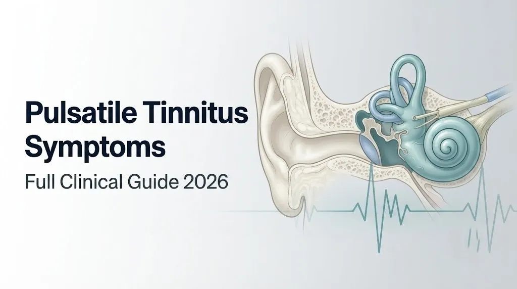
Pulsatile tinnitus symptoms occur when abnormal or turbulent blood flow in vessels near the temporal bone generates mechanical vibrations that are transmitted to the cochlea and perceived by the brain as a rhythmic sound synchronized with the cardiac cycle.
The core mechanism is bone conduction. Blood flowing through vessels adjacent to the inner ear, specifically the sigmoid sinus, transverse sinus, internal carotid artery, and jugular bulb, normally produces laminar (smooth) flow that generates no audible vibration. When that flow becomes turbulent, whether from vessel wall abnormalities, elevated flow velocity, or elevated pressure, it creates mechanical oscillations. Those oscillations travel through the petrous temporal bone into the perilymph and endolymph of the cochlea. The fluid movement then deflects the stereocilia of the outer hair cells in the organ of Corti, generating electrical impulses that travel along cranial nerve VIII (the vestibulocochlear nerve) to the auditory cortex. The cortex interprets those impulses as sound.
The key clinical feature that distinguishes this from regular tinnitus is synchrony with the heartbeat. Every time the heart contracts and drives a pulse wave through the arterial and venous system, the abnormal flow generates a new wave of vibration, and the brain registers a new sound. The result is a rhythmic, repeating sound rather than a constant tone.
The primary recognized pulsatile tinnitus symptoms include:
- A rhythmic whooshing, thumping, swooshing, or rushing sound in one or both ears
- Synchrony with the pulse: the sound beats at exactly the same rate as the heart
- A sound described variably as a “heartbeat in the ear,” “ocean waves,” or “wind gusts”
- Fluctuation in volume with changes in body position (especially lying down)
- Possible accompanying symptoms: headache, vision changes, hearing loss, ear fullness
What Is Pulsatile Tinnitus and How Is It Different From Regular Tinnitus?
Pulsatile tinnitus is a form of tinnitus defined by its rhythmic, pulse-synchronous character, produced by an identifiable mechanical source in the cardiovascular or cerebrospinal fluid system, which distinguishes it fundamentally from the more prevalent non-pulsatile tinnitus that has no identifiable acoustic generator.
Non-pulsatile tinnitus, which affects an estimated 15 percent of adults according to the NIDCD, is typically generated within the auditory system itself: changes in cochlear hair cell function after noise exposure, age-related sensorineural hearing loss, or central auditory pathway changes. It produces a constant or fluctuating tone, hiss, or buzz. It does not beat with the pulse.
Pulsatile tinnitus, by contrast, has an external acoustic generator located in the vascular system or, in the case of elevated intracranial pressure, in the cerebrospinal fluid dynamics adjacent to the dural venous sinuses. This is a critical distinction because it means pulsatile tinnitus is almost always a symptom of an underlying medical condition rather than a primary auditory system disorder.
| Feature | Pulsatile Tinnitus | Non-Pulsatile Tinnitus |
|---|---|---|
| Sound character | Rhythmic, beats with pulse | Constant tone, hiss, or buzz |
| Acoustic generator | Vascular or structural source | Auditory system dysfunction |
| Synchrony with heartbeat | Yes, by definition | No |
| Identifiable medical cause | Present in the majority of cases | Often no identifiable cause |
| Diagnostic urgency | Higher: requires vascular workup | Lower: often monitored |
| Audible to examiner | Sometimes (objective type) | Never |
People taking aspirin at high doses, quinine-containing medications, or certain loop diuretics may develop non-pulsatile tinnitus as a medication side effect. That presentation does not produce the rhythmic quality of pulsatile tinnitus and represents a different clinical pathway entirely.
Objective vs Subjective Pulsatile Tinnitus
Objective pulsatile tinnitus is a sound audible to both the patient and a clinician using a stethoscope or tuning fork placed near the ear, while subjective pulsatile tinnitus is heard only by the patient, making the objective/subjective distinction the most clinically important categorization in the entire diagnostic workup.
Objective pulsatile tinnitus accounts for a smaller proportion of pulsatile tinnitus cases but carries a higher likelihood of finding a treatable structural cause. When a clinician can actually hear a bruit (a vascular sound) over the ear, temple, neck, or mastoid bone, the sound is being generated by a true mechanical source with enough energy to radiate through tissue. This strongly points to an arteriovenous malformation (AVM), a dural arteriovenous fistula (DAVF), a glomus tumor (paraganglioma), or a highly vascular neoplasm near the skull base.
Subjective pulsatile tinnitus, heard only by the patient, is more often associated with elevated intracranial pressure, venous sinus stenosis, or sigmoid sinus wall abnormalities. The Cleveland Clinic notes that the majority of pulsatile tinnitus cases presenting to neurotology clinics are of the subjective type, which still requires thorough vascular imaging to exclude serious structural pathology.
To document which type you are experiencing before your appointment:
- Sit quietly in a silent room and confirm whether the sound is present.
- Locate your pulse at your wrist and compare its rate to the sound in your ear.
- Ask a family member to press a stethoscope or their ear against your mastoid bone (the bony prominence behind your ear) while you are experiencing the sound.
- Note whether the sound changes when you press gently on the internal jugular vein in your neck (on the same side as the tinnitus). A decrease in the sound with gentle neck pressure suggests a venous source.
- Report both findings to your physician at your first appointment.
Patients with known carotid artery disease, prior neck surgery, or radiation to the head and neck region are at higher baseline risk for objective pulsatile tinnitus from vascular structural abnormalities and should specifically request vascular imaging as part of their workup.
Key Takeaway: Pulsatile tinnitus is not a random ringing. It has an acoustic generator somewhere in your vascular system or CSF dynamics, and the distinction between objective (audible to a clinician) and subjective (heard only by you) is the first step in narrowing down what that generator is.
Symptoms of Pulsatile Tinnitus: The Complete Clinical Picture
Symptoms of pulsatile tinnitus extend beyond the rhythmic ear sound itself and include a constellation of associated findings that vary by underlying cause, severity of the underlying vascular or pressure abnormality, and the duration of the condition.
The core symptom is the rhythmic auditory perception, but the complete clinical picture includes both auditory and non-auditory components. The non-auditory symptoms are, in many cases, the most diagnostically informative. A patient who reports only a whooshing sound has a different clinical picture than one who reports that sound alongside daily headache and episodes of blurred vision.
| Symptom | Mechanism | Clinical Significance |
|---|---|---|
| Rhythmic whooshing or thumping in ear | Turbulent vascular flow transmitted to cochlea | Core defining symptom; present in all cases |
| Synchrony with heartbeat | Cardiac cycle drives each pulse of turbulent flow | Confirms pulsatile rather than non-pulsatile type |
| Worsening with position change (lying down) | Increased venous pressure and jugular vein flow rate when supine | Suggests venous source or elevated ICP |
| Unilateral (one-sided) presentation | Localizing anatomical abnormality on that side | Higher suspicion for structural lesion |
| Headache (pressure-type) | Elevated intracranial pressure transmitted to pain-sensitive dura | Suggests IIH or dural venous sinus pathology |
| Visual disturbances or transient vision loss | Papilledema from elevated ICP compressing optic nerve | Requires urgent evaluation |
| Ear fullness or pressure sensation | Altered perilymph or endolymph pressure | May indicate Eustachian tube dysfunction or IIH |
| Hearing loss (low-frequency) | Cochlear hair cell damage or endolymphatic hydrops | Suggests Meniere-like component or structural lesion |
| Neck bruit or mass | Vascular lesion large enough to radiate externally | Strongly suggests AVM, DAVF, or glomus tumor |
| Pulsatile sensation without sound | Very early vascular transmission to cochlea | Early presentation; does not exclude serious cause |
According to a 2021 systematic review published in JAMA Otolaryngology-Head and Neck Surgery, an identifiable structural or vascular cause was found in approximately 70 percent of patients with pulsatile tinnitus who underwent complete radiological evaluation, underscoring the importance of thorough diagnostic workup rather than attributing the symptom to stress or normal aging.
The Whooshing or Heartbeat Sound in the Ear
The whooshing or heartbeat sound in the ear characteristic of pulsatile tinnitus is generated by turbulent blood flow in vessels adjacent to the cochlea and is transmitted to the inner ear via bone conduction through the petrous portion of the temporal bone.
Think of it this way. A garden hose with normal pressure running through a smooth, open pipe makes no sound you can hear from across the room. But crimp that hose, or run twice the water volume through it, and the turbulence generates a vibration you can feel in the pipe itself. The sigmoid sinus and internal carotid artery work the same way. Smooth laminar flow through a normal vessel is silent from the cochlea’s perspective. Turbulent flow, whether from vessel wall abnormality, stenosis, elevated flow velocity from anemia or thyroid disease, or elevated venous pressure from raised intracranial pressure, generates mechanical oscillations that the cochlear hair cells can detect.
Patients describe the sound using several specific phrases that carry diagnostic relevance:
- “Like the ocean” or “waves”: typically associated with venous sources, particularly sigmoid sinus or jugular vein turbulence
- “Like my heartbeat in my ear”: arterial source, particularly internal carotid artery turbulence or a highly vascular tumor
- “Whooshing with each pulse”: consistent with any pulsatile source; particularly noted in dural arteriovenous fistulas
- “A clicking or ticking”: may indicate stapedial artery persistence or myoclonic middle ear muscle activity (not true pulsatile tinnitus)
- “Rushing air”: commonly described in superior semicircular canal dehiscence (SSCD)
The National Institute on Deafness and Other Communication Disorders distinguishes the venous hum, a soft continuous sound that may become rhythmic, from true arterial pulsatile tinnitus, which is sharper and more percussive. Asking patients to gently compress the ipsilateral (same-side) jugular vein in the neck is a useful bedside test: if this reduces the sound, a venous source is likely.
Women in their 30s and 40s who describe a bilateral whooshing that worsens when lying down at night are at elevated risk for idiopathic intracranial hypertension as the underlying cause, particularly if they are overweight or have recently gained weight.
Pulsatile Tinnitus in One Ear vs Both Ears
Pulsatile tinnitus in one ear (unilateral) and pulsatile tinnitus in both ears (bilateral) carry different diagnostic implications, with unilateral presentation more strongly associated with a localized structural lesion and bilateral presentation more commonly linked to elevated intracranial pressure or systemic causes.
Unilateral pulsatile tinnitus directs clinical attention to the anatomy on that specific side. When the sound is confined to the right ear, the diagnostic priority is examining the right sigmoid sinus, right transverse sinus, right internal carotid artery, and right jugular bulb for structural abnormalities. A right-sided glomus tympanicum tumor (a paraganglioma arising from the middle ear floor), for example, would produce right-sided pulsatile tinnitus with no contralateral component because the acoustic generator is anatomically localized.
Bilateral pulsatile tinnitus, particularly when both ears produce sounds of similar intensity and quality, is more characteristic of a systemic or central cause. Elevated intracranial pressure affects both dural venous sinuses simultaneously, which is why idiopathic intracranial hypertension typically causes bilateral pulsatile tinnitus rather than one-sided symptoms.
Quick Tip:
- Unilateral pulsatile tinnitus: request imaging that includes the ipsilateral sigmoid sinus, transverse sinus, internal carotid artery, and temporal bone
- Bilateral pulsatile tinnitus: request evaluation that includes intracranial pressure assessment (fundoscopic exam for papilledema and, if indicated, lumbar puncture for CSF opening pressure)
- Track whether the sound is truly equal in both ears or louder on one side: asymmetry in a “bilateral” case still warrants localized structural workup on the louder side
- Patients with a prior history of stroke or carotid artery disease should report any new unilateral pulsatile tinnitus to their cardiologist or neurologist within days
Older adults over 60 with cardiovascular risk factors (hypertension, hypercholesterolemia, diabetes) presenting with new unilateral pulsatile tinnitus should have carotid artery duplex ultrasound included in their initial workup, given the elevated prevalence of carotid atherosclerotic plaque in that demographic.
Key Takeaway: Unilateral pulsatile tinnitus is more suspicious for a localized structural lesion like a glomus tumor or sigmoid sinus abnormality, while bilateral pulsatile tinnitus points more toward elevated intracranial pressure or a systemic cardiovascular cause.
Pulsatile Tinnitus With Headache and Vision Changes
Pulsatile tinnitus accompanied by headache and vision changes represents a specific and clinically urgent symptom triad that strongly suggests elevated intracranial pressure, and this combination requires prompt evaluation rather than watchful waiting.
The anatomical explanation is direct. When intracranial pressure rises above normal (above 25 cmH2O measured via lumbar puncture), that pressure propagates through the entire cerebrospinal fluid system. The dural venous sinuses are surrounded by CSF, and elevated pressure compresses their walls, narrows their lumens, and increases venous resistance. Blood forced through a narrower lumen at the same flow volume generates turbulence, and that turbulence is transmitted via the sigmoid sinus to the petrous temporal bone and then to the cochlea.
Simultaneously, elevated intracranial pressure is transmitted along the optic nerve sheaths (which are continuous extensions of the dural meningeal layers). This produces papilledema, a swelling of the optic disc that can cause transient visual obscurations (brief episodes of grayed-out or lost vision, typically lasting seconds), double vision from cranial nerve VI (abducens nerve) palsy, or in chronic unmanaged cases, permanent visual field loss.
The headache of elevated intracranial pressure is characteristically described as a daily or near-daily pressure or throbbing sensation, typically worse in the morning (when supine CSF pressure is naturally higher) and behind the eyes.
Red flags that require same-day urgent evaluation when they accompany pulsatile tinnitus:
- Headache that is new, progressively worsening over days to weeks, or described as the “worst headache of my life”
- Transient visual obscurations (brief episodes where vision grays out or goes completely dark)
- Double vision (diplopia), particularly horizontal double vision from abducens nerve palsy
- Visual field loss noticed when covering one eye
- Nausea or vomiting not explained by another cause
A 2019 study published in Neurology found that pulsatile tinnitus is present in approximately 52 to 60 percent of patients diagnosed with idiopathic intracranial hypertension, making it one of the most prevalent symptoms of that condition, not a coincidental finding.
Pulsatile Tinnitus Worse When Lying Down
Pulsatile tinnitus that worsens when lying down is produced by the positional increase in venous pressure and cerebral blood volume that occurs when the body moves from upright to supine, and this postural variation is one of the most diagnostically informative features of the symptom.
When you stand or sit upright, gravity assists venous return from the head and neck. The jugular veins drain efficiently, venous pressure in the sigmoid sinus stays relatively low, and blood flows smoothly. When you lie flat, that gravitational assistance disappears. Venous pressure in the cerebral venous system rises, blood volume in the intracranial compartment increases, and intracranial pressure increases slightly even in healthy individuals.
In a person with underlying sigmoid sinus wall abnormality, venous sinus stenosis, or elevated baseline intracranial pressure, this positional change pushes venous pressure and flow turbulence above the threshold at which the cochlea can detect it. The result is a pulsatile sound that appears at night when lying down and may quiet or disappear when sitting up in the morning.
Patients frequently describe lying awake at night unable to ignore the sound, which then diminishes or disappears once they sit on the edge of the bed. This is a clinically useful observation that supports a venous-origin or elevated ICP etiology rather than an arterial source, since arterial pulsatile tinnitus is typically present regardless of body position.
To document positional variation accurately for your physician:
- Note the time of onset when lying down (immediately upon lying flat, or after several minutes).
- Sit up and note whether the sound diminishes within 30 to 60 seconds.
- Try lying on the side with the tinnitus versus the opposite side: note any difference.
- Check whether the sound is louder during elevated heart rate (after exercise, anxiety, or caffeine) regardless of position.
- Write down all of these observations before your appointment, as positional and circumstantial details significantly narrow the differential diagnosis.
Pregnant women in the third trimester frequently report new-onset pulsatile tinnitus worse when lying on their back. This is partly explained by inferior vena cava compression from the gravid uterus increasing venous backpressure into the jugular system, and partly by the physiological increase in blood volume (approximately 40 to 50 percent above baseline) that occurs during normal pregnancy.
Pulsatile Tinnitus Causes and Vascular Mechanisms
Pulsatile tinnitus is caused by any condition that creates turbulent, abnormally fast, or abnormally pressured blood flow near enough to the cochlea that the mechanical vibration is transmitted through the temporal bone to the inner ear.
The cause is not always a dangerous one. A venous hum from a normal but high-flow jugular vein in an anemic or thyrotoxic patient is benign once the underlying condition is treated. But the full spectrum of causes includes serious structural lesions that require vascular surgery or endovascular intervention.
| Cause | Mechanism | Who It Affects Most | Diagnostic Approach |
|---|---|---|---|
| Idiopathic intracranial hypertension (IIH) | Elevated CSF pressure narrows dural venous sinuses, generating turbulent venous flow | Women 20 to 45, BMI over 30 | MRI brain, MRV, lumbar puncture |
| Sigmoid sinus wall anomaly/diverticulum | Outpouching of sigmoid sinus wall vibrates with each heartbeat | Women, any age | CT temporal bone (high-resolution) |
| Dural arteriovenous fistula (DAVF) | Abnormal arteriovenous shunting creates high-velocity, high-pressure flow audible near cochlea | Adults 40 to 60, any sex | CT angiography, digital subtraction angiography |
| Arteriovenous malformation (AVM) | Direct arteriovenous connection generating high-flow turbulence | Younger adults, congenital | MRI/MRA, DSA |
| Glomus jugulare or glomus tympanicum tumor | Highly vascular paraganglioma generates pulsatile blood flow adjacent to middle ear | Women 40 to 60 | CT temporal bone, MRI with gadolinium |
| Superior semicircular canal dehiscence (SSCD) | Absent or thinned bone over superior semicircular canal allows sound transmission | Any age, more common in adults | CT temporal bone |
| Carotid artery atherosclerosis or stenosis | Turbulent arterial flow through narrowed carotid lumen near temporal bone | Adults over 55, cardiovascular risk factors | Carotid duplex ultrasound, CTA |
| High-output cardiovascular states (anemia, thyrotoxicosis, pregnancy) | Elevated cardiac output increases flow velocity in all cranial vessels | Varies by underlying condition | CBC, thyroid function tests |
| Hypertension (systemic arterial) | Elevated arterial pressure increases flow velocity and vessel wall pulsation | Adults over 40 | Blood pressure monitoring, clinical exam |
Mayo Clinic notes that the most treatable causes of pulsatile tinnitus include hypertension, anemia, and thyroid dysfunction, making a basic blood panel and blood pressure measurement appropriate first steps before proceeding to specialized imaging.
Idiopathic Intracranial Hypertension and Pulsatile Tinnitus
Idiopathic intracranial hypertension (IIH), also called pseudotumor cerebri, is the single most common identifiable cause of pulsatile tinnitus in women of childbearing age, occurring when elevated cerebrospinal fluid pressure narrows the dural venous sinuses and generates turbulent venous flow detectable by the cochlea.
The mechanism works like a partially blocked drain. Normally, CSF produced by the choroid plexus of the cerebral ventricles flows around the brain and spinal cord and is reabsorbed at arachnoid granulations near the superior sagittal sinus at a rate that keeps intracranial pressure steady. In IIH, this reabsorption is impaired, or production is increased, and pressure builds throughout the entire intracranial compartment. The pressure is transmitted to the walls of the transverse sinus and sigmoid sinus, narrowing their lumens. Blood forced through a narrower lumen at the same cardiac output generates turbulence, and that turbulence travels through the petrous temporal bone into the cochlea.
According to research published in Neurology, IIH affects approximately 1 to 2 per 100,000 people overall, but in women aged 20 to 44 with a body mass index over 30, the incidence rises to approximately 19 per 100,000, representing a 15 to 20-fold increase over the general population rate. The association with obesity is strong and consistent across multiple epidemiological studies.
Key symptoms of IIH that accompany the pulsatile tinnitus include:
- Daily headache, typically pressure-type, worse in the morning
- Transient visual obscurations lasting seconds, often triggered by bending over or straining
- Horizontal diplopia from abducens nerve palsy
- Neck and shoulder pain
- Papilledema visible on fundoscopic examination (optic disc swelling from elevated CSF pressure)
- Cognitive slowing or “brain fog” as a patient-reported but not yet formally classified symptom per current IIH clinical criteria
IIH in men exists but is uncommon. When it does occur in men, it tends to present without the strong obesity association, and clinicians should have a lower threshold for alternative diagnoses including intracranial mass lesion or dural venous sinus thrombosis in male patients with the IIH symptom pattern.
Key Takeaway: If you are a woman between 20 and 45 with a body mass index over 30 who has pulsatile tinnitus, daily headache, and any visual symptoms, idiopathic intracranial hypertension is the most likely cause and requires urgent evaluation by a neurologist, not a routine otolaryngology referral.
Pulsatile Tinnitus in Women and During Pregnancy
Pulsatile tinnitus in women occurs at higher rates than in men for multiple physiological reasons, with the sex disparity most pronounced in the 20 to 45 age group due to the strong association between female sex hormones, body weight dynamics, and idiopathic intracranial hypertension.
Estrogen and progesterone influence the rate of CSF production by the choroid plexus epithelium and the sensitivity of arachnoid granulations to CSF reabsorption pressure. These hormonal effects partly explain why IIH is predominantly a condition of women in their reproductive years and why symptom onset or worsening is sometimes tied to menstrual cycle phase, oral contraceptive use, or hormonal changes in early pregnancy.
During pregnancy specifically, three physiological changes converge to increase the risk of pulsatile tinnitus. Blood volume increases by approximately 40 to 50 percent above pre-pregnancy baseline by the third trimester, according to data from the American College of Obstetricians and Gynecologists. Cardiac output rises in parallel. Both changes increase the velocity of blood flow through all cranial vessels, including the sigmoid sinus and internal carotid artery, increasing the likelihood that flow becomes turbulent enough to reach the cochlea. Gestational weight gain further elevates ICP in women with underlying IIH.
Pregnant women who develop new pulsatile tinnitus should notify their obstetrician rather than waiting until after delivery. Pulsatile tinnitus in pregnancy accompanied by headache and visual symptoms warrants evaluation for IIH and for cerebral venous sinus thrombosis (CVST), a serious clotting condition that occurs at elevated rates during pregnancy and the postpartum period.
Pulsatile tinnitus that appears after certain medications should be reported to the prescribing physician. Tetracycline-class antibiotics (including doxycycline), vitamin A derivatives (including isotretinoin, used for acne), and withdrawal from prolonged corticosteroid use are all associated with secondary intracranial hypertension that can produce pulsatile tinnitus.
Pulsatile Tinnitus and Anxiety
Anxiety does not cause pulsatile tinnitus directly, but the physiological effects of anxiety on the cardiovascular system can amplify a pre-existing pulsatile tinnitus symptom and create a reinforcing cycle that makes the sound feel more severe than its underlying cause warrants.
The mechanism is specific and well-documented. Acute anxiety activates the sympathetic nervous system, releasing epinephrine and norepinephrine from the adrenal medulla. These catecholamines increase heart rate (tachycardia) and cardiac output. Higher cardiac output means higher blood flow velocity through the sigmoid sinus, jugular vein, and internal carotid artery. If a person already has a mild sigmoid sinus wall abnormality or borderline elevated intracranial pressure that was not generating enough turbulence to be audible at rest, the added velocity from sympathetic activation may push flow past the turbulence threshold. The previously unnoticeable sound becomes audible.
This mechanism explains why many patients first notice pulsatile tinnitus during periods of stress or after a panic attack. The anxiety did not create the underlying vascular abnormality. It revealed it by raising cardiac output.
The clinical implication is important. A physician who attributes pulsatile tinnitus solely to anxiety without completing a vascular workup is making a potentially dangerous diagnostic error. According to the AAO-HNS clinical practice guideline on tinnitus, anxiety is a comorbidity that influences tinnitus severity and patient distress, but it does not constitute a diagnosis that explains the pulsatile quality. A full vascular and structural evaluation remains necessary even in patients with known anxiety disorders.
Quick Tip:
- Tell your physician both facts: you have anxiety AND the tinnitus synchronizes with your pulse
- Do not accept anxiety as the complete explanation for pulsatile tinnitus without vascular imaging
- Pulsatile tinnitus that began or worsened during a period of high stress still requires the same structural evaluation as tinnitus that appeared without stress
- Patients with panic disorder who are taking stimulant medications (or high-dose caffeine) may experience amplified pulsatile tinnitus due to chronically elevated sympathetic tone increasing cardiac output
Pulsatile Tinnitus and Hearing Loss
Pulsatile tinnitus accompanied by hearing loss represents a more complex clinical picture than pulsatile tinnitus alone, as the hearing loss indicates that the underlying process has begun affecting the mechanical or neural function of the cochlea itself, not merely transmitting vibration to it.
The distinction matters because two different mechanisms can produce this combination, and they have very different causes. The first is direct cochlear damage. A glomus tympanicum tumor arising from the floor of the middle ear can erode into the cochlear structures, producing both the pulsatile sound (from its own highly vascular blood supply) and a conductive hearing loss (from middle ear space occupation). The second is cochlear ischemia. Severe carotid artery stenosis that significantly reduces blood flow through the cochlear artery (a branch of the anterior inferior cerebellar artery, AICA) can produce both sensorineural hearing loss and pulsatile tinnitus.
Superior semicircular canal dehiscence (SSCD) is a particularly instructive example. In SSCD, a thinned or absent bone cap over the superior semicircular canal creates an abnormal “third window” in the inner ear. This allows sound energy and intracranial pressure pulses to dissipate through the dehiscence rather than being fully directed to the oval window and round window. The result can include autophony (hearing one’s own voice or internal sounds at abnormal volume), low-frequency conductive hearing loss on audiometry, and pulsatile tinnitus from transmission of vascular pulsations through the dehiscent bone.
Patients with the combination of pulsatile tinnitus and hearing loss should bring these specific details to their appointments:
- Which ear (or both ears) has the hearing loss
- Whether the hearing loss is for high-pitched sounds, low-pitched sounds, or all frequencies
- Whether the ear feels full or plugged
- Whether the hearing loss preceded the tinnitus or appeared simultaneously
- Any history of noise exposure, ear infections, or ear surgery
Cleveland Clinic recommends that all patients with pulsatile tinnitus and concurrent hearing loss receive formal audiometry (pure-tone audiogram with bone conduction testing) as part of their initial evaluation, not simply a clinical hearing screen.
Key Takeaway: Pulsatile tinnitus combined with hearing loss is more clinically urgent than pulsatile tinnitus alone, because it suggests the underlying process has begun affecting cochlear function, which points toward structural lesions like glomus tumors, carotid stenosis, or superior semicircular canal dehiscence.
How Pulsatile Tinnitus Is Diagnosed
Pulsatile tinnitus is diagnosed through a staged clinical and imaging workup that begins with a detailed history and physical examination and progresses to targeted vascular and structural imaging based on the specific characteristics of the symptom.
No single test diagnoses pulsatile tinnitus in isolation. The goal of the workup is to identify the specific anatomical source of the sound, because different causes require completely different treatments.
The typical diagnostic pathway unfolds in this order:
- Primary care physician evaluation: Complete medical history including symptom onset, laterality, positional variation, associated headache and vision symptoms, medication list, and cardiovascular risk factors. Blood pressure measurement. Basic blood panel including complete blood count (CBC) to check for anemia and thyroid function tests (TSH) to exclude thyrotoxicosis.
- Otoscopic examination: Direct visualization of the ear canal and tympanic membrane. A reddish pulsatile mass visible behind the tympanic membrane is characteristic of a glomus tympanicum tumor.
- Audiometry (pure-tone audiogram): Assesses for concurrent sensorineural or conductive hearing loss, which narrows the differential.
- Fundoscopic examination: Inspection of the optic disc for papilledema. Papilledema confirms elevated intracranial pressure and directs immediate attention to the IIH or CVST pathway.
- CT temporal bone (high-resolution, without contrast): The single highest-yield initial imaging study for pulsatile tinnitus. Identifies sigmoid sinus diverticulum, superior semicircular canal dehiscence, glomus tumors, and bony abnormalities.
- MRI brain with gadolinium and MR venography (MRV): Assesses for dural venous sinus stenosis, intracranial mass, and DAVF. The MRV specifically evaluates the transverse and sigmoid sinus caliber.
- MR angiography (MRA) of head and neck: Evaluates for AVM, DAVF, carotid stenosis, and other arterial lesions.
- Digital subtraction angiography (DSA): The definitive gold-standard study for vascular lesions. Reserved for cases where MRI/MRA findings are indeterminate or where an AVM or DAVF is suspected based on prior imaging.
- Lumbar puncture (spinal tap): Measures CSF opening pressure directly. Indicated when IIH is suspected clinically. Opening pressure above 25 cmH2O (in the lateral decubitus position with legs extended) confirms elevated intracranial pressure.
The AAO-HNS clinical practice guideline on tinnitus states that pulsatile tinnitus warrants a clinical evaluation by an otolaryngologist or neurotologist rather than primary care management alone when initial workup does not identify a clear systemic cause.
When to See a Doctor for Pulsatile Tinnitus
Any new-onset pulsatile tinnitus warrants evaluation by a primary care physician, and this applies regardless of symptom severity, because the rhythmic, pulse-synchronous character of the sound is itself a clinical indicator of an underlying condition that needs to be identified.
This is where pulsatile tinnitus differs from regular tinnitus management. Non-pulsatile tinnitus of mild severity can reasonably be monitored without immediate evaluation in many cases. Pulsatile tinnitus cannot, because approximately 70 percent of cases have an identifiable structural or vascular cause, according to the 2021 systematic review in JAMA Otolaryngology-Head and Neck Surgery.
The specific urgency level depends on the full symptom picture:
| Symptom Pattern | Urgency Level | Appropriate Provider |
|---|---|---|
| Pulsatile tinnitus alone, new onset | Within 1 to 2 weeks | Primary care physician first, then otolaryngologist |
| Pulsatile tinnitus plus daily headache | Within days | Primary care physician, request urgent neurology referral |
| Pulsatile tinnitus plus vision changes | Same day | Neurologist or ophthalmologist urgently |
| Pulsatile tinnitus plus hearing loss | Within 1 week | Otolaryngologist with audiometry |
| Pulsatile tinnitus in pregnancy | Within days | Obstetrician; request neurology consultation |
| Pulsatile tinnitus plus neck mass or bruit | Within days | Otolaryngologist or vascular surgeon |
| Sudden-onset pulsatile tinnitus plus neurological symptoms | Immediately | Emergency department, call 911 |
Before your appointment, prepare this specific information:
- The exact character of the sound (whooshing, thumping, clicking)
- Whether it is unilateral or bilateral
- Whether it varies with body position
- Whether it synchronizes with your heartbeat (time it against your pulse)
- Any associated headache, vision changes, ear fullness, or hearing loss
- Your complete current medication list
- Any recent weight gain, especially if you are a woman aged 20 to 45
Emergency Symptoms: When Pulsatile Tinnitus Requires Urgent Care
Certain presentations associated with pulsatile tinnitus indicate a potential neurological or vascular emergency. Do not wait to see if these resolve on their own.
Call 911 or go to the nearest emergency room immediately if pulsatile tinnitus is accompanied by any of the following:
- A sudden, severe headache that you would describe as the worst headache of your life (this is the thunderclap headache of subarachnoid hemorrhage until proven otherwise)
- Sudden loss of vision in one or both eyes, or sudden appearance of a persistent dark spot in your visual field
- Sudden double vision that was not present before this episode
- Weakness, numbness, or tingling in the face, arm, or leg on one side of the body
- Sudden difficulty speaking, understanding speech, or finding words
- Sudden loss of balance or inability to walk normally
- Sudden onset pulsatile tinnitus following a head injury, fall, or trauma
- Loss of consciousness or near-fainting associated with the tinnitus onset
- A pulsatile sound accompanied by blood draining from the ear canal
These presentations can indicate subarachnoid hemorrhage, intracranial arteriovenous malformation rupture, dural venous sinus thrombosis, or posterior fossa stroke, all of which require emergency neurosurgical or neurological assessment within minutes, not an urgent care appointment or a telephone advice line.
Key Takeaway: Pulsatile tinnitus with sudden severe headache, any new neurological deficit, or sudden vision loss is a neurological emergency. These symptoms combined require 911 activation, not a same-day doctor call.
Frequently Asked Questions About Pulsatile Tinnitus Symptoms
What does pulsatile tinnitus feel like?
Pulsatile tinnitus feels like a rhythmic whooshing, thumping, or heartbeat-like sound in one or both ears that beats in time with your own pulse.
Most people describe it as hearing their own heartbeat in their ear, or as a rushing or ocean-wave sound that accelerates when their heart rate increases.
The sound is typically more noticeable in quiet environments and when lying down, and it may diminish or change character when gentle pressure is applied to the jugular vein on the same side.
Is pulsatile tinnitus dangerous?
Pulsatile tinnitus is not always dangerous, but approximately 70 percent of cases have an identifiable underlying structural or vascular cause that requires medical evaluation, according to a 2021 systematic review in JAMA Otolaryngology-Head and Neck Surgery.
Causes range from benign (high-output states like anemia or thyroid overactivity that resolve with treatment) to serious (dural arteriovenous fistula, arteriovenous malformation, or elevated intracranial pressure threatening vision).
Any pulsatile tinnitus accompanied by headache, vision changes, or neurological symptoms requires prompt evaluation, not watchful waiting.
Can pulsatile tinnitus go away on its own?
Pulsatile tinnitus caused by a systemic, reversible condition such as anemia, hyperthyroidism, or pregnancy-related increased blood volume may resolve once the underlying condition is treated.
Pulsatile tinnitus caused by structural abnormalities like sigmoid sinus diverticulum, dural arteriovenous fistula, or glomus tumor does not resolve without specific treatment of that structural lesion.
Because the cause cannot be determined without medical evaluation, pulsatile tinnitus should never be assumed to be self-resolving until a physician has completed a workup.
What type of doctor should I see for pulsatile tinnitus?
Begin with a primary care physician, who can order initial blood work, check blood pressure, examine the ear, and perform or refer for fundoscopic examination.
If no systemic cause is identified, a referral to an otolaryngologist (ENT specialist) or, ideally, a neurotologist (an otolaryngologist with subspecialty training in ear and skull base conditions) is the appropriate next step.
If IIH, DAVF, or AVM is suspected based on imaging, a neurologist or interventional neuroradiologist will become part of the evaluation team.
Can anxiety or stress cause pulsatile tinnitus?
Anxiety does not directly cause pulsatile tinnitus, but it can amplify the volume of a pre-existing pulsatile sound by raising heart rate and cardiac output through sympathetic nervous system activation, which increases blood flow velocity in cranial vessels.
A person may first notice pulsatile tinnitus during a stressful period because elevated cardiac output pushes borderline turbulent flow past the threshold of cochlear detection.
The AAO-HNS clinical practice guideline states that anxiety as a comorbidity does not replace the need for vascular imaging in pulsatile tinnitus; structural evaluation is required even in patients with confirmed anxiety disorders.
What does it mean if pulsatile tinnitus is only in one ear?
Unilateral pulsatile tinnitus more strongly suggests a localized structural lesion on that side of the head, such as a sigmoid sinus diverticulum, glomus tumor, internal carotid artery stenosis, or ipsilateral dural arteriovenous fistula.
The Mayo Clinic notes that asymmetric or unilateral pulsatile tinnitus warrants more thorough imaging than bilateral presentations, because the localization narrows the differential to structures on that specific side.
A high-resolution CT temporal bone and MRI with MR venography focused on the ipsilateral sigmoid sinus, transverse sinus, and temporal bone are the priority imaging studies for new-onset unilateral pulsatile tinnitus.
Closing
Pulsatile tinnitus is not a variant of normal aging and it is not an anxiety symptom. It is a specific auditory signal produced by a specific mechanical source in your vascular or cerebrospinal fluid system, and in the majority of cases, that source can be identified. The diagnostic workup starts with a primary care visit and a blood pressure check, but for most patients it will proceed to targeted imaging of the temporal bone and cranial vessels.
The next step is straightforward: book an appointment with your primary care physician, bring the symptom details you have documented (which ear, whether it synchronizes with your pulse, whether it worsens lying down, any associated headache or vision changes), and ask specifically for a referral to an otolaryngologist or neurotologist if no systemic cause is found on initial evaluation.
If you have pulsatile tinnitus with new headache, any visual disturbance, or any neurological symptom appearing at the same time, that appointment should be today.