Spinal Infection Symptoms: Signs You Must Not Ignore
Spinal infection symptoms include persistent, worsening back pain that does not improve with rest, fever above 38.3 degrees Celsius (101 degrees Fahrenheit), unexplained weight loss, and, in advanced cases, neurological changes including leg weakness, numbness, or sudden loss of bladder or bowel control. These symptoms differ from ordinary back pain in a way that matters urgently: spinal infections can compress or destroy the spinal cord if not caught early.
According to the Infectious Diseases Society of America, the annual incidence of vertebral osteomyelitis has risen over recent decades, driven by aging populations, increasing rates of intravenous drug use, and more frequent spinal procedures. Research published in The Lancet Infectious Diseases has documented that delayed diagnosis, often by 6 to 12 weeks after symptom onset, is the single most common factor driving permanent neurological damage from spinal infections.
This article covers every clinically recognized symptom category of spinal infection: the early presentations that often get misread as muscle strain, the late-stage red flags that indicate an evolving emergency, the specific differences across spinal regions, and the precise clinical threshold at which you need emergency care rather than a scheduled appointment.
Spinal Infection Symptoms: What They Are and Why They Happen
Spinal infection symptoms are produced when bacteria, fungi, or mycobacteria colonize the vertebral body, intervertebral disc, or epidural space, triggering a cascade of inflammation, bone destruction, and, in the worst cases, direct compression of the spinal cord or cauda equina.
The key anatomical reason the spine is vulnerable to infection is the nature of its blood supply. The vertebral endplate, the thin cartilaginous layer capping each vertebral body, is supplied by end-capillary loops that create sluggish, low-flow zones where blood-borne bacteria can lodge and multiply without being efficiently cleared. Think of these terminal capillary zones like a slow backwater in a river: debris settles there precisely because the current is weakest.
Once bacteria lodge in the vertebral endplate, the body’s immune response floods the area with neutrophils. Interleukin-1 beta (IL-1B), IL-6, and tumor necrosis factor alpha (TNF-alpha) are released from activated macrophages at the infection site. These cytokines travel via the bloodstream to the hypothalamus, where prostaglandin E2 resets the body’s thermal set-point upward, producing fever. Locally, the same inflammatory cascade drives bone resorption and disc destruction.
Key symptoms produced by this mechanism include:
- Deep, aching back pain that worsens with movement and does not improve with standard pain relief
- Fever and chills from systemic cytokine release
- Night sweats as the fever breaks and resets repeatedly
- Point tenderness directly over the infected vertebral segment when pressed
- Progressive neurological symptoms if epidural abscess develops
Older adults over age 65 may present with a blunted fever response because of age-related immune senescence, making back pain the only presenting symptom for weeks before the infection is recognized.
Types of Spinal Infection: Vertebral Osteomyelitis, Discitis, and Epidural Abscess
Spinal infections occur in three anatomically distinct patterns, each with a distinct mechanism, a somewhat different symptom profile, and a different urgency timeline.
Vertebral osteomyelitis (ICD-10 M46.2), also called pyogenic spondylitis, is infection of the vertebral body itself. It is the most common form in adults and typically originates from hematogenous seeding through Batson’s venous plexus, the valveless paravertebral venous network connecting pelvic, prostatic, urinary, and abdominal venous drainage to the vertebral bodies. This is why urinary tract infections, dental abscesses, and skin infections can each seed the spine: the venous pathway is open and valveless, allowing retrograde bacterial flow.
Spondylodiscitis or discitis (ICD-10 M46.4) refers to infection of the intervertebral disc and adjacent vertebral endplates together. In adults, the disc itself is avascular (it lost its direct blood supply by approximately age 30, leaving it dependent on diffusion from the vertebral endplate). Adult discitis therefore almost always results from spread from an adjacent infected endplate. In children under age 5, the disc retains its own vascular channels, making primary hematogenous discitis possible.
Spinal epidural abscess (ICD-10 G06.1) is the most neurologically dangerous form. Pus collects in the epidural space between the posterior vertebral ligament and the dura mater, directly adjacent to the spinal cord or cauda equina. Compression can be mechanical (from the abscess volume itself) or ischemic (from thrombosis of radicular or spinal arteries).
| Type | Primary Structure Infected | Common Age Group | Neurological Risk |
|---|---|---|---|
| Vertebral osteomyelitis | Vertebral body and endplate | Adults over 50 | Moderate (if untreated) |
| Spondylodiscitis | Disc space and adjacent endplates | Adults; children under 5 | Moderate to high |
| Spinal epidural abscess | Epidural space | Any age, highest in older adults | High; can be hours to days |
People taking TNF-alpha inhibitors (biologic medications for rheumatoid arthritis, Crohn’s disease, or psoriasis) face a higher risk of all three types, particularly infections caused by atypical organisms including mycobacteria and fungi, where fever may be absent or minimal at presentation.
Back Pain from Spinal Infection: What It Feels Like and Why
Back pain is present in over 90 percent of adults with vertebral osteomyelitis, according to retrospective case series published in Spine, making it the most consistent and earliest symptom of spinal infection.
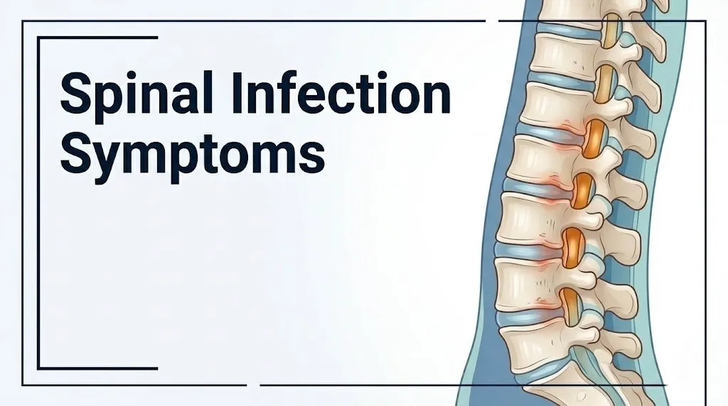
The character of the pain distinguishes it from mechanical back pain in several clinically meaningful ways. Mechanical back pain (from a herniated disc, strained paraspinal muscle, or facet joint degeneration) typically improves with rest and worsens with movement. Spinal infection pain is constant. It worsens with position changes, is often present at night when lying flat, and does not improve with rest or standard over-the-counter analgesics. The pain is deep and gnawing rather than sharp and shooting.
Direct percussion tenderness over the spinous process of the infected vertebral segment is a classically described clinical sign. When a clinician taps the bony prominence of the spine at the infected level, the patient reports a localized, sharp, reproducible pain that is disproportionate to the gentle force applied. This is caused by the periosteal inflammatory response within the infected vertebral body, where the periosteum (the pain-sensitive outer membrane covering bone) is under inflammatory pressure.
Pain location tracks the infected spinal level:
- Infection at the lumbar spine (L1 to L5) produces low back and buttock pain, sometimes with groin or anterior thigh referral from the psoas muscle if a psoas abscess develops
- Infection at the thoracic spine (T1 to T12) produces mid-back pain, sometimes with chest wall referral that mimics cardiac or pleural pain
- Infection at the cervical spine (C1 to C7) produces neck pain, shoulder pain, or occipital headache depending on the level involved
In people with type 2 diabetes mellitus, peripheral neuropathy may blunt the intensity of spinal pain, causing the infection to be more advanced before the pain becomes alarming.
Fever, Chills, and Night Sweats as Spinal Infection Symptoms
Fever, chills, and night sweats in combination with back pain represent the classic triad that should immediately raise clinical concern for a spinal infection over a simple mechanical back problem.
Fever in spinal infection is driven by systemic cytokine release from the infected vertebral tissue. IL-6 and TNF-alpha produced at the infection site enter the systemic circulation and act on the hypothalamic thermoregulatory center, triggering prostaglandin E2 synthesis via the cyclooxygenase-2 (COX-2) pathway. The set-point rises, and the body produces fever by increasing heat generation (shivering, peripheral vasoconstriction) until core temperature reaches the new set-point. Night sweats occur when the set-point temporarily drops, triggering heat dissipation through sweating.
According to a retrospective analysis published in Annals of Internal Medicine, fever is present in only approximately 50 to 60 percent of adult vertebral osteomyelitis cases at the time of initial medical evaluation. This means a significant minority of patients have no fever at all at presentation, which is a major reason spinal infection is underdiagnosed for weeks to months.
Groups where fever is most commonly absent or blunted include:
- Older adults over 70 (reduced hypothalamic sensitivity and immune senescence)
- People on chronic systemic corticosteroids (suppress IL-6 and TNF-alpha signaling)
- People on biologic immunosuppressants including TNF-alpha inhibitors
- People with advanced diabetes mellitus (impaired cytokine signaling)
Chills (rigors) that accompany fever spikes suggest bacteremia, meaning bacteria are actively circulating in the bloodstream. This distinction matters because bacteremia drives both the severity of spinal seeding and the risk of seeding other organs including the heart valves (infective endocarditis) and kidneys.
Key Takeaway: Fever is absent in nearly half of confirmed spinal infection cases at first presentation, which means back pain alone, particularly constant pain worse at night and unresponsive to rest, warrants medical evaluation even if temperature is normal.
Neurological Symptoms of Spinal Cord Infection
Neurological symptoms of spinal cord infection develop when a spinal epidural abscess or severe vertebral collapse compresses or ischemia compromises the spinal cord (in the cervical and thoracic spine) or the cauda equina (in the lumbar spine below the L1 vertebral level).
The neurological presentation depends on the anatomical level of compression. Cervical and thoracic epidural abscesses compress the spinal cord directly, producing an upper motor neuron syndrome: increasing leg stiffness (spasticity), weakness, hyperreflexia, and in severe cases a positive Babinski sign (upward great toe extension when the lateral sole is stroked). Lumbar abscesses compress the cauda equina nerve roots rather than the cord itself, producing a lower motor neuron syndrome: flaccid weakness, reduced reflexes, and the particularly alarming symptom of cauda equina syndrome (bilateral leg weakness, saddle anesthesia of the perineum, and loss of voluntary bladder and bowel control).
The National Institute of Neurological Disorders and Stroke (NINDS) describes neurological deficits from spinal epidural abscess as progressing through four stages: back pain only, radicular pain radiating along the nerve root distribution, motor weakness or sensory changes, and finally paralysis. The critical clinical window is stages 2 to 3. Patients who receive surgical decompression at stage 2 or 3 have substantially better neurological outcomes than those who deteriorate to paralysis before intervention.
Neurological symptoms that require immediate evaluation include:
- New weakness in one or both legs
- Numbness or tingling from the waist down
- Sudden difficulty walking
- Loss of voluntary control of bladder (urinary retention or incontinence)
- Loss of voluntary control of bowel
- Saddle anesthesia (numbness in the groin, perineum, and inner thighs)
In people with pre-existing conditions such as multiple sclerosis or peripheral neuropathy from diabetes, new neurological symptoms may be attributed to the known condition rather than a new spinal infection, causing dangerous delays in diagnosis.
Early-Stage Spinal Infection Symptoms
Early spinal infection symptoms are almost universally nonspecific, which is the primary reason the average time from symptom onset to confirmed diagnosis ranges from 6 to 12 weeks in published case series.
In the first one to four weeks of vertebral osteomyelitis or spondylodiscitis, the most common presentation is back pain that feels similar to a muscle strain or disc flare. The pain is aching rather than sharp, worsens with movement, and may be attributed to lifting, prolonged sitting, or a recent minor injury. At this stage, fever is often low-grade or absent. Inflammatory markers (ESR and CRP) are elevated but not dramatically so.
Think of early spinal infection like a smoldering fire inside a wall. The outside surface shows no flame. The wall may feel faintly warm. The destruction is happening internally, silently, and the structural damage accumulates long before the building shows any visible sign of collapse.
Early features that distinguish a spinal infection from ordinary back pain at this stage, even before fever becomes prominent, include:
- Pain that is present and constant through the night rather than relieved by lying flat
- Pain that does not improve after two to three weeks of rest and standard analgesics
- Unexplained fatigue and low-grade malaise accompanying the back pain
- Unexplained loss of appetite or modest unintentional weight loss
- A recent history within the past four to eight weeks of a skin infection, urinary tract infection, dental procedure, intravenous line placement, or any other potential bacteremia source
According to Mayo Clinic, one of the most common settings for delayed spinal infection diagnosis is a patient with known degenerative disc disease or prior back problems whose new infection symptoms are assumed to be a recurrence of their known condition rather than a new and different process.
Late-Stage and Advanced Spinal Infection Symptoms
Late-stage spinal infection symptoms are those that appear after weeks of uncontrolled infection have produced substantial vertebral destruction, paravertebral abscess formation, or epidural abscess with neural compression.
By this stage, the pain is typically severe and constant, rated 7 to 10 on a Visual Analog Scale by most patients in clinical series. Movement in any direction provokes sharp pain. The spine may be held rigidly in a protective posture as the paraspinal muscles splint the infected segment. High fever is more consistently present. Weight loss may be 5 to 10 percent of body weight from the combined effects of systemic infection and reduced appetite.
Vertebral body collapse from bone destruction can produce visible spinal deformity, most notably a gibbus deformity (an angular kyphotic hump) in thoracic Pott’s disease (tuberculous spondylitis, ICD-10 A18.01). In pyogenic infection, acute vertebral fracture from bone erosion can cause sudden severe worsening of pain and immediate onset of neurological deficits.
A psoas abscess is a late complication specific to lumbar spinal infections. Pus tracks anteriorly from an infected lumbar vertebra along the fascial plane of the psoas major muscle, producing swelling and tenderness in the groin or anterior thigh. Patients with a psoas abscess typically walk with a flexed hip posture (hip flexion deformity) because extending the hip stretches the inflamed psoas and provokes pain.
| Stage | Timeframe | Key Symptoms | Urgency |
|---|---|---|---|
| Early | Weeks 1 to 4 | Vague back pain, low or no fever, fatigue | Same-day primary care |
| Intermediate | Weeks 4 to 8 | Severe constant pain, higher fever, night sweats | Urgent evaluation within 24 hours |
| Advanced | Weeks 8 plus | Neurological deficits, vertebral collapse, psoas abscess | Emergency department immediately |
Older adults with advanced spinal infection may present with acute confusion or delirium as the primary presentation of systemic sepsis, with the back pain as a secondary complaint that is not volunteered until specifically asked about.
Key Takeaway: Advanced spinal infection that has produced vertebral collapse, psoas abscess, or any neurological deficit requires emergency department evaluation, not a scheduled appointment.
Lower Back Infection Symptoms Specifically
Lower back infection symptoms are the most common presentation of spinal infection overall because the lumbar spine (L1 to L5) is the most frequently affected region, accounting for approximately 50 percent of vertebral osteomyelitis cases according to published clinical series in the Journal of Bone and Joint Surgery.
The lumbar region’s vulnerability relates to its blood supply. The lumbar vertebral bodies receive a high volume of blood flow from the lumbar arteries, and the adjacent paravertebral venous plexus communicates directly with the pelvic venous system. Urinary tract infections, prostate infections, gynecological infections, and pelvic inflammatory disease all drain into this same venous network, creating a direct seeding route to the lower lumbar vertebral endplates.
Lower back infection pain is characteristically located in the midline or paravertebral region, without the radicular shooting character (down the leg in an L4, L5, or S1 dermatomal distribution) that typically accompanies a herniated lumbar disc. When radicular symptoms do appear in lumbar spinal infection, they often indicate either nerve root involvement from inflammatory edema or, more urgently, early cauda equina compression from an epidural abscess.
Signs specifically associated with lower lumbar spinal infection include:
- Midline lower back pain with point tenderness over the L3-L4, L4-L5, or L5-S1 spinous processes on palpation
- Groin or anterior thigh pain from psoas muscle irritation adjacent to an infected L2-L4 vertebral body
- Difficulty sitting upright or walking without marked pain
- Rectal or perineal pressure if a paraspinal abscess has tracked into the pelvis
Females with recurrent pelvic inflammatory disease or recent gynecological procedures carry an elevated lower lumbar infection risk, as pelvic venous drainage communicates directly with the lower lumbar Batson’s plexus.
Spinal Infection Symptoms After Surgery or Medical Procedures
Spinal infection symptoms that develop after a surgical procedure or medical intervention constitute a distinct clinical category known as post-procedural or iatrogenic spinal infection, and the timeline and responsible organisms differ from hematogenous infections.
After spinal surgery (laminectomy, discectomy, spinal fusion, instrumented fixation), postoperative infection can develop within days to weeks of the procedure. Early postoperative infections (within 30 days) are typically caused by skin organisms introduced at the time of surgery, most commonly Staphylococcus aureus or Staphylococcus epidermidis. Late postoperative infections (30 days to 1 year post-surgery) may involve lower-virulence organisms seeded during the procedure but manifesting slowly.
After epidural steroid injections or spinal cord stimulator implantation, the incidence of epidural abscess is low but not negligible. A 2019 retrospective analysis published in Spine found that the incidence of epidural abscess following interventional spinal procedures ranged from 0.1 to 0.5 percent, with most cases involving S. aureus. Symptoms in this context typically begin 3 to 14 days after the procedure.
Warning symptoms after any spinal procedure that should not be attributed to normal postoperative discomfort include:
- Fever above 38.3 degrees Celsius (101 degrees Fahrenheit) appearing after an initial afebrile recovery period
- Sudden worsening of back pain that had been improving since surgery
- Redness, swelling, or discharge at the surgical incision site
- Any new neurological symptom including weakness, tingling, or bladder change
- Return of severe pain weeks after initial post-surgical improvement
People who are immunocompromised at the time of spinal surgery, including those on systemic corticosteroids or biologic immunosuppressants, carry a substantially higher risk of post-procedural spinal infection and may develop it with minimal fever and fewer inflammatory signs.
Spinal Infection Symptoms in Children
Spinal infection symptoms in children differ meaningfully from the adult presentation, primarily because the most common form in children is discitis rather than vertebral osteomyelitis, and the age-related vascular anatomy of the intervertebral disc creates a mechanistically distinct infection pathway.
In children under age 5, the intervertebral disc retains vascular channels that allow hematogenous seeding directly into the disc tissue. This makes primary pediatric discitis a distinct clinical entity from the secondary disc involvement seen in adult spondylodiscitis. The lumbar disc is the most frequently affected in children, particularly L3-L4 and L4-L5 levels.
The clinical presentation in young children is often dramatically different from back pain in adults. Toddlers and preschool-age children may present with:
- Refusal to walk or stand (the most consistent early sign in children under 3)
- Crying or irritability with position changes, particularly when picked up under the arms
- Refusal to bend forward to pick up objects from the floor
- Low-grade fever or no fever at all in many cases
- Irritability and reduced appetite rather than any direct complaint of back pain
Research published in Pediatrics has documented that the average age at diagnosis for pediatric discitis is 2 to 7 years, and the condition is frequently missed for weeks because the child cannot localize or describe pain, and the presentation of gait refusal is misattributed to developmental or orthopedic causes.
Older children aged 8 to 16 may present more like adults with back pain and fever. However, even in adolescents, spinal infection symptoms can be subtle and may initially mimic athletic injuries or growing pains, particularly if fever is low-grade.
A pediatrician or pediatric emergency medicine physician should evaluate any child under age 10 who refuses to walk, develops an unusual gait, or presents with midline spine tenderness, particularly if they have had a recent febrile illness in the preceding four to eight weeks.
Key Takeaway: In children under age 5, refusal to walk or bend forward is a more reliable early sign of discitis than fever or back pain, and should prompt evaluation by a pediatrician or pediatric emergency physician within 24 hours.
Spinal Infection Risk Factors: Who Is Most Vulnerable
Spinal infection risk factors cluster around three broad mechanisms: conditions that create bacteremia (blood-borne bacteria available to seed the spine), conditions that impair the immune response’s ability to clear those bacteria, and conditions that create direct access routes bypassing normal skin and mucosal barriers.
Bacteremia risk factors include:
- Intravenous drug use (direct injection of non-sterile material, skin flora into the bloodstream)
- Hemodialysis with repeated vascular access
- Indwelling central venous catheters
- Urinary tract infections, particularly in people with recurrent UTIs or indwelling urinary catheters
- Infective endocarditis (bacteria from heart valve vegetation seed the spine continuously)
- Recent dental procedures in people with known bacteremia risk
Immune impairment risk factors include:
- Type 1 or type 2 diabetes mellitus (impaired neutrophil chemotaxis and oxidative burst)
- HIV infection or other causes of T-cell deficiency
- Organ transplant recipients on immunosuppressive therapy
- Long-term systemic corticosteroid use
- Biologic therapy including TNF-alpha inhibitors, IL-6 inhibitors, and JAK inhibitors
- Active malignancy or chemotherapy-induced immunosuppression
Direct inoculation risk factors include:
- Spinal surgery within the past year
- Epidural steroid injections within the past 30 days
- Lumbar punctures or myelography procedures
According to the Infectious Diseases Society of America, males are affected approximately twice as often as females across published vertebral osteomyelitis case series, a disparity attributed in part to higher rates of IV drug use and hemodialysis access in men and in part to the contribution of prostatic venous drainage to Batson’s plexus seeding.
How Spinal Infection Is Identified and Diagnosed
Spinal infection is diagnosed through a combination of clinical assessment, laboratory inflammatory markers, imaging studies, and microbiological confirmation, with MRI with gadolinium contrast representing the gold standard imaging modality.
The diagnostic process typically unfolds in this sequence:
- Clinical assessment: A physician evaluates the specific character of the back pain, its duration, any associated fever or neurological symptoms, and the presence of known risk factors. Point tenderness over the spinous process and any neurological deficits are documented.
- Laboratory tests: Erythrocyte sedimentation rate (ESR) and C-reactive protein (CRP) are ordered immediately. ESR is elevated in over 90 percent of vertebral osteomyelitis cases. CRP is more sensitive for acute-phase infection. A complete blood count (CBC) looks for leukocytosis. Blood cultures (drawn from two separate sites before any antibiotic is given) are positive in approximately 50 to 70 percent of cases and can identify the causative organism, avoiding the need for invasive biopsy in confirmed cases.
- MRI with gadolinium contrast: MRI is the imaging study that identifies spinal infection with the highest sensitivity and specificity. It shows vertebral endplate edema and erosion, disc space signal change, paravertebral soft tissue edema, and epidural fluid collections (abscess). A CT scan is used when MRI is contraindicated (certain pacemakers, severe claustrophobia).
- CT-guided bone biopsy: When blood cultures are negative and the diagnosis is not confirmed by imaging alone, a CT-guided percutaneous bone biopsy of the infected vertebral segment provides tissue for microbiological culture and histopathology. This should be performed before antibiotics are started whenever possible.
- Procalcitonin: A serum procalcitonin level helps distinguish bacterial from fungal or mycobacterial infection and tracks treatment response over time.
People who have already received antibiotics before arriving at medical care will have falsely negative blood cultures in a higher proportion of cases, making the bone biopsy more likely to be necessary.
Spinal Infection vs Herniated Disc: How to Tell the Difference
Spinal infection and a herniated intervertebral disc can produce overlapping back and leg pain symptoms, but several clinical features reliably distinguish one from the other when assessed together.
A herniated nucleus pulposus (disc herniation) occurs when the gel-like nucleus pulposus of the intervertebral disc extrudes through a tear in the outer annulus fibrosus and presses against a nerve root. The pain is typically sharp and radicular: it follows a specific dermatomal distribution down one leg (sciatica for L4-L5 or L5-S1 herniations). It is often provoked by specific movements (coughing, sneezing, bending forward) and may have had a clear mechanical onset (lifting, twisting).
Spinal infection pain is different in character and context. It is constant, deep, and aching. It is present at rest and at night. It does not follow a neat dermatomal distribution in early or intermediate cases. And critically, it is accompanied by systemic features (fever, malaise, night sweats, weight loss) that herniated discs simply do not produce.
| Feature | Spinal Infection | Herniated Disc |
|---|---|---|
| Pain character | Constant, deep, aching, worse at night | Sharp, shooting, dermatomal |
| Response to rest | Does not improve | Often improves |
| Fever | Present in 50 to 60 percent of cases | Absent |
| Night sweats | May be present | Absent |
| Weight loss | Possible with prolonged infection | Absent |
| Onset | Gradual over days to weeks | Often acute, movement-related |
| Neurological symptoms | Diffuse, bilateral in advanced cases | Typically unilateral, dermatomal |
| Point tenderness | Midline over spinous process | Variable, often paraspinal |
The most dangerous clinical error is attributing infection back pain in a patient with known degenerative spine disease to a disc flare. If a patient has known disc disease but develops constant night pain, fever, or new bilateral leg symptoms, a spinal infection must be excluded before the symptoms are attributed to the disc.
Spinal Infection and Sepsis: When Infection Spreads Beyond the Spine
Spinal infection can progress to sepsis when the infectious process seeds the systemic bloodstream beyond the controlled zone of local vertebral infection, producing a whole-body inflammatory response that threatens multiple organ systems.
Sepsis occurs when the body’s response to an infection becomes dysregulated, causing organ dysfunction at sites distant from the primary infection. According to the National Institute of Neurological Disorders and Stroke, patients with spinal epidural abscess who present with altered mental status, rapid heart rate, low blood pressure, or respiratory distress may already be in septic shock, which carries a mortality risk exceeding 20 percent without immediate intensive care.
The pathway from spinal infection to sepsis typically involves:
- Uncontrolled bacteremia from the primary vertebral infection site
- Seeding of secondary sites including the heart valves (infective endocarditis), kidneys (renal abscess), or lung (septic emboli)
- Systemic release of cytokines (particularly IL-6 and TNF-alpha) that cause vasodilation, increased capillary permeability, and relative hypovolemia, dropping blood pressure
Signs that a spinal infection has progressed to sepsis include:
- Sudden change in mental status or confusion
- Heart rate above 100 beats per minute (tachycardia)
- Rapid breathing (respiratory rate above 22 breaths per minute)
- Low blood pressure (systolic below 90 mmHg)
- Skin that is pale, mottled, or cool and clammy despite fever
- Reduced or absent urine output
People over age 70 with spinal infection are at the highest risk of rapid deterioration to sepsis, partly because their physiological reserves are reduced and partly because their baseline vital signs differ from younger adults, making early sepsis harder to identify without close monitoring.
Key Takeaway: Any patient with known or suspected spinal infection who develops confusion, rapid heart rate, low blood pressure, or reduced urine output is in a potential septic emergency and needs 911 called immediately, not transport to an urgent care clinic.
How to Describe Spinal Infection Symptoms to a Doctor
Accurately describing your symptoms to a primary care physician or emergency medicine physician is the single most effective way to prompt the diagnostic workup that catches a spinal infection before neurological damage occurs.
To give a clinically useful symptom description:
- State the duration precisely: “This back pain started approximately three weeks ago,” not “my back has been hurting.” A duration of more than two weeks with no improvement is a red flag for a non-mechanical cause.
- Describe the pain character specifically: Say “the pain is constant and present even when I’m lying still” rather than “my back hurts.” The constant, non-positional nature of infection pain is what separates it from mechanical pain.
- State any fever or temperature readings you have taken: Provide the number. “I measured 38.6 Celsius at 2 a.m. twice this week” is far more actionable than “I felt hot.”
- List any recent potential bacteremia events: Tell the doctor about any recent skin infections, urinary tract infections, dental procedures, IV lines, injections, or surgical procedures within the past two to three months.
- Describe neurological symptoms explicitly: Do not say “my leg feels weird.” Say “my left leg feels weaker than before when I walk up stairs” or “I have tingling in both feet from the knee down.”
- List your relevant medical history and medications: Specifically name any immunosuppressant drugs, diabetes diagnosis, dialysis status, or prior spinal surgery. These details immediately change the pre-test probability of spinal infection in any clinician’s mind.
Things to bring to your appointment or emergency evaluation:
- A written list of all current medications including biologics, steroids, and immunosuppressants
- Any prior imaging reports (MRI, CT) of your spine from the past two years
- A temperature log if you have been monitoring at home
- The name of any recent procedures or hospitalizations
Emergency Symptoms: When Spinal Infection Requires Immediate Care
Certain spinal infection symptoms require immediate emergency evaluation. Do not wait to see if these resolve on their own.
Call 911 or go to the nearest emergency room immediately if you experience:
- New weakness in one or both legs combined with back pain and fever (possible spinal cord or cauda equina compression from epidural abscess, a surgical emergency where hours matter)
- Loss of bladder control (urinary retention or incontinence) or bowel control combined with back pain (classic presentation of cauda equina syndrome, which requires neurosurgical decompression within hours to prevent permanent paralysis)
- Saddle anesthesia: numbness in the groin, inner thighs, or perineum (a specific sign of cauda equina compression)
- Sudden severe worsening of back pain in a person already diagnosed with a spinal infection (may indicate vertebral fracture from bone destruction)
- Confusion, rapid heart rate, low blood pressure, or pale/mottled skin in a person with known or suspected spinal infection (signs of septic shock)
- Inability to walk that develops over hours to days in a person with back pain and fever
- High fever above 39.4 degrees Celsius (103 degrees Fahrenheit) with severe back pain in any person with diabetes, immunosuppression, IV drug use, or recent spinal procedure
- Neck stiffness combined with fever and back pain (possible concurrent meningitis from spread of infection to the subarachnoid space)
These presentations can indicate spinal epidural abscess with active cord compression, cauda equina syndrome, septic shock, or vertebral osteomyelitis with acute instability, and they require emergency medical and surgical assessment within minutes to hours, not an urgent care appointment or a phone call.
Frequently Asked Questions About Spinal Infection Symptoms
What are the first signs of a spinal infection?
The first signs of a spinal infection are typically constant back pain that does not improve with rest, low-grade fever, and unexplained fatigue.
These early symptoms often appear weeks before the infection is recognized, as they closely resemble common mechanical back pain or a disc flare.
Any back pain lasting more than two weeks without improvement, particularly with fever or night sweats, warrants evaluation by a primary care physician to exclude a spinal infection.
Can you have a spinal infection without a fever?
Yes. Fever is absent in approximately 40 to 50 percent of confirmed vertebral osteomyelitis cases at initial evaluation, according to retrospective case series published in Annals of Internal Medicine.
People who are older, immunosuppressed, or taking chronic corticosteroids or biologic medications are especially likely to have no fever despite active spinal infection.
The absence of fever does not rule out spinal infection when back pain is constant, severe, and accompanied by elevated inflammatory markers on blood testing.
How serious is a spinal infection?
Spinal infection is a serious, potentially life-threatening condition that can cause permanent paralysis if the infection forms an epidural abscess that compresses the spinal cord before surgical decompression.
Without treatment, vertebral osteomyelitis can produce vertebral bone destruction, vertebral fracture, and progressive neurological deficit over weeks to months.
With early diagnosis and appropriate antibiotic therapy, most patients recover without permanent neurological damage, which is why prompt evaluation is the most important factor in outcomes.
How do doctors diagnose a spinal infection?
Doctors diagnose spinal infection using MRI with gadolinium contrast, which is the most sensitive and specific imaging test for identifying vertebral endplate infection, disc space involvement, and epidural abscess.
Blood cultures drawn before antibiotics are started identify the responsible organism in approximately 50 to 70 percent of cases.
When blood cultures are negative, a CT-guided bone biopsy of the infected vertebral segment provides tissue for culture and confirms the diagnosis.
What does spinal infection pain feel like compared to normal back pain?
Spinal infection pain is constant, deep, and aching, present even at rest and typically worse at night, rather than the movement-provoked, positional pain of a muscle strain or herniated disc.
It does not improve with anti-inflammatory medications or lying flat, and it is often accompanied by systemic symptoms including fever, night sweats, and unexplained weight loss that ordinary back pain does not cause.
Point tenderness directly over the infected vertebral spinous process when pressed is a clinical sign that distinguishes infection from mechanical back pain.
Can a spinal infection cause permanent paralysis?
Yes. A spinal epidural abscess that is not surgically decompressed in time can cause permanent paralysis by compressing or causing ischemia to the spinal cord or cauda equina.
According to the National Institute of Neurological Disorders and Stroke, patients who already have complete paralysis at the time of surgery have significantly lower rates of neurological recovery than those treated during the weakness or sensory-loss stage.
This is why any new leg weakness, numbness, or bladder or bowel dysfunction combined with back pain requires immediate emergency department evaluation, not a scheduled appointment.
Back pain and fever together are not a combination to wait out. A spinal infection that is caught in the early weeks, before the vertebral body has significantly eroded or the epidural space has filled with pus, is treated with antibiotics and close monitoring. That same infection, left unaddressed until neurological symptoms appear, becomes a race against the clock in a neurosurgical suite.
If your back pain is constant, present through the night, unresponsive to over-the-counter medication, and accompanied by fever, weight loss, or night sweats, contact a primary care physician for same-day evaluation and ask specifically about blood tests for ESR, CRP, and blood cultures. If any neurological symptom appears, that changes the timeline entirely.
Go to the nearest emergency room immediately if you have back pain with leg weakness, bladder or bowel changes, or confusion. The window for preserving neurological function is measurable in hours, and you are the first one who has to decide to act.