Fractured Nose Symptoms: Signs, Pain, and When to Act
Fractured nose symptoms typically include sharp pain at the site of impact, rapid swelling across the nasal bridge, bruising that spreads under one or both eyes, and a sensation of nasal blockage that can develop within minutes of the injury. These symptoms occur because the nasal bones are the most frequently fractured bones in the human face, and the structures involved in breaking produce an intense, fast-moving inflammatory response that affects both the nose and the surrounding facial tissue.
According to the National Institutes of Health, nasal fractures account for approximately 39 to 45 percent of all facial bone fractures, making them the single most common facial fracture seen in emergency departments across the United States. That prevalence matters because it means a large number of people experience this injury every year without knowing exactly what to expect from it or when it crosses into emergency territory.
This article covers every clinically recognized symptom of a nasal fracture in plain, mechanistically accurate language. It explains why each symptom happens at the anatomical level, how symptoms change over time, which populations experience different presentations, and which specific signs require immediate emergency evaluation. By the end, you will know what your body is signaling, whether your situation can wait for a clinic appointment, and when it cannot.
Fractured Nose Symptoms: The Complete Clinical Picture
Fractured nose symptoms span a predictable range from immediate pain and swelling to delayed bruising, breathing obstruction, and in serious cases, complications requiring emergency intervention. The specific symptoms you experience depend on the force of impact, the direction of the blow, which nasal structures were involved, and your individual anatomy.
The core symptoms recognized in clinical practice include:
- Sudden, sharp pain at the nose, nasal bridge, or nasal dorsum, present immediately after impact
- Tenderness to touch along the nasal bones, worsening with any pressure on the nasal bridge
- Rapid swelling across the nose, cheeks, and upper lip, beginning within minutes of injury
- Bruising below the eyes and along the nasal sidewalls, often appearing 12 to 48 hours after the injury
- Nasal obstruction producing difficulty breathing through one or both nostrils
- Epistaxis (nosebleed) from disruption of the nasal mucosa and its underlying vascular supply
- Visible deformity, including a crooked nasal bridge, a depressed nasal dorsum, or asymmetry of the nasal tip
- Crepitus, a grinding or crackling sensation when the nose is touched or gently moved, produced by bone fragment movement
- Nasal discharge, sometimes blood-tinged, from mucosal irritation and bleeding
Not every person with a nasal fracture experiences all of these symptoms. According to a clinical review published in Otolaryngology-Head and Neck Surgery, some patients present with a single dominant symptom (pain only, or obstruction only) without visible external change, particularly in low-energy impacts to the nasal tip or alar region where cartilage rather than bone is primarily involved.
People who use blood-thinning medications (warfarin, apixaban, rivaroxaban, clopidogrel) typically present with epistaxis that is heavier, lasts longer, and may require medical management rather than simple direct pressure.
What Is a Nasal Fracture?
A nasal fracture is a break in one or more of the bony or cartilaginous structures that form the nasal skeleton, occurring when blunt force applied to the nose exceeds the structural tolerance of these tissues. The medical term is nasal bone fracture, classified under ICD-10 code S02.2.
“Fractured nose” refers to the same condition whether the break involves the bone, the cartilage, or both. The distinction between bony and cartilaginous fractures matters clinically because they have different healing properties, different complication risks, and slightly different physical examination findings.
Nasal fractures are classified by pattern:
| Fracture Type | Description | Common Cause |
|---|---|---|
| Lateral fracture | One nasal bone displaced sideways | Side-impact blow, sports collision |
| Frontal fracture | Both nasal bones driven posteriorly (inward) | Direct frontal blow, motor vehicle impact |
| Comminuted fracture | Multiple bone fragments, often with septal involvement | High-energy impact |
| Isolated septal fracture | Septum dislocated or fractured without nasal bone involvement | Moderate frontal impact |
| Greenstick fracture | Incomplete fracture, bone bends rather than breaks | Low-energy impact, common in children |
The American Academy of Otolaryngology-Head and Neck Surgery notes that the severity of the fracture correlates with both the energy of impact and the direction of force. Frontal impacts tend to produce more severe and bilateral nasal bone disruption. Lateral impacts are more likely to produce a visibly crooked nose with unilateral bone displacement.
Older adults are at higher risk for comminuted fractures from lower-energy impacts because cortical bone density declines progressively after age 50, reducing the force threshold required to shatter rather than simply crack the nasal bones.
Nasal Bone Anatomy and How Fractures Happen
The nasal bones (os nasale) are a paired set of small, flat bones that form the upper bony bridge of the nose. They are among the thinnest bones in the entire human skeleton, measuring approximately 2 to 3 millimeters at the rhinion (the bony nasal tip junction), which is precisely why they fracture more often than any other facial bone.
These bones articulate superiorly with the frontal bone at the nasofrontal suture, laterally with the frontal process of the maxilla on each side, and posteriorly with the perpendicular plate of the ethmoid bone. Below the nasal bones, the nose transitions to a purely cartilaginous skeleton: the upper lateral cartilages (ULC) pair with the nasal bones at the rhinion, and the lower lateral cartilages (also called alar cartilages) form the mobile nasal tip and nostrils.
Think of the nose as a building with a short concrete roof (the nasal bones) sitting on top of a flexible fiberglass framework (the cartilages). A hard blow to the roof fractures the concrete. A blow aimed at the lower nose deforms or dislocates the fiberglass instead.
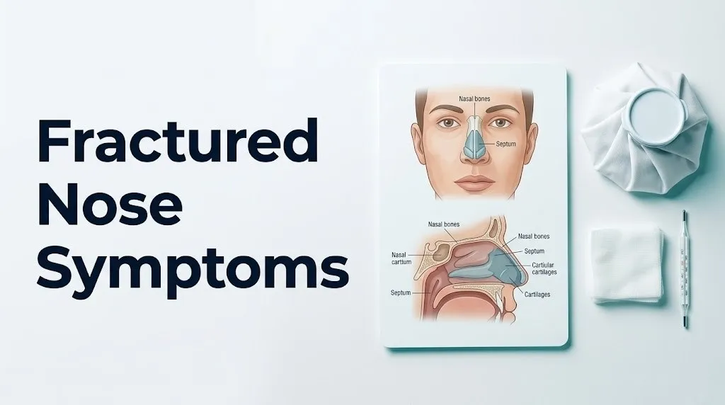
The nasal septum runs centrally through the nose, dividing it into two air passages. It is composed of the quadrangular cartilage anteriorly, the perpendicular plate of the ethmoid superoposteriorly, and the vomer inferoposteriorly. Fractures frequently involve septal displacement or fracture in addition to the nasal bones, since the septum is directly attached to the base of the nasal bones and absorbs transmitted force.
The blood supply to the nasal mucosa comes primarily from the Kiesselbach plexus (also called Little’s area) on the anterior nasal septum, a convergence of branches from the anterior ethmoid artery, superior labial artery, and sphenopalatine artery. This dense vascular network is why virtually any nasal fracture produces at least some degree of bleeding.
Broken Nose Pain: What It Feels Like and Why
Broken nose pain is typically immediate, sharp, and localized to the nasal bridge, then spreads within minutes to the cheeks, upper lip, and the area beneath both eyes. The pain arises from disruption of the periosteum (the dense connective tissue sheath covering the nasal bones) and stimulation of the trigeminal nerve branches that supply nasal sensation.
Specifically, the anterior ethmoid nerve and the external nasal branch of the anterior ethmoid nerve carry pain signals from the nasal dorsum and nasal tip. The infratrochlear nerve supplies the lateral nasal wall and inner canthus region. When the periosteum tears during fracture, these nerve endings are exposed to bradykinin from the kallikrein-kinin cascade and substance P from local trigeminal nociceptors, producing an intense burning and throbbing quality to the pain.
Pain severity does not reliably predict fracture severity. A small, minimally displaced fracture can produce intense pain because periosteal nerve density is high. A more severe fracture in someone with prior nasal trauma may produce less dramatic pain because scar tissue from previous injury has partially desensitized local nerve endings.
Pain patterns by fracture type:
| Pain Pattern | Likely Involvement | Clinical Significance |
|---|---|---|
| Sharp, localized pain at nasal bridge | Nasal bone fracture | Most common presentation |
| Pain radiating to upper teeth | Maxillary involvement or anterior ethmoid nerve | Suggests lateral wall extension |
| Pain with eye movement | Orbital floor or medial orbital wall involvement | Requires CT and ophthalmology evaluation |
| Pain deep to nasal bridge (between eyes) | Ethmoid sinus or cribriform plate involvement | Requires urgent CT imaging |
| Persistent deep aching after 72 hours | Septal hematoma, developing infection | Warrants same-day ENT evaluation |
Over-the-counter acetaminophen is the preferred pain management option in the first 48 hours after a nasal fracture. Non-steroidal anti-inflammatory drugs (NSAIDs) such as ibuprofen and naproxen inhibit thromboxane A2-mediated platelet aggregation, which can prolong or worsen nosebleeds during the acute phase of injury.
Broken Nose Swelling and Bruising Symptoms
Swelling and bruising after a nasal fracture are produced by two separate but related physiological processes happening in the same injured tissue. Understanding them helps explain why swelling peaks earlier than bruising and why each one resolves on its own timeline.
Swelling begins within minutes of the fracture. When the periosteum and nasal mucosa are injured, mast cells in these tissues degranulate, releasing histamine. This triggers vasodilation of perinasal capillaries and venules. Simultaneously, tissue macrophages activated by cellular damage release prostaglandin E2 and interleukin-1 beta, which increase vascular wall permeability. Protein-rich plasma fluid moves from blood vessels into the nasal soft tissue, producing the characteristic puffy, firm swelling across the nasal bridge and cheeks. This process is at its peak within 2 to 6 hours of injury and typically reaches maximum volume by 24 hours.
Bruising (ecchymosis) follows a different pathway. Red blood cells extravasate through the highly permeable capillary walls and track along fascial planes in the face. Gravity pulls the blood inferiorly and laterally from the fractured nasal bones, collecting in the periorbital loose areolar connective tissue beneath the eyes. This produces the classic bilateral “raccoon eyes” appearance. Because fascial plane tracking takes time, bruising typically appears 12 to 48 hours after the injury, often alarming people who did not see it immediately.
According to clinical data cited by the Mayo Clinic, periorbital ecchymosis after nasal fracture is bilateral (under both eyes) in most cases when the nasal bones are the primary site of fracture, because blood tracks symmetrically along both medial canthal areas. Unilateral periorbital bruising more often suggests an orbital floor fracture or isolated maxillary impact.
Quick Tip:
- Apply a cold compress wrapped in cloth (never ice directly on skin) for 15 to 20 minutes every hour during the first 24 hours to reduce histamine-driven swelling.
- Keep your head elevated above the level of your heart, including during sleep, to slow gravity-dependent fluid accumulation.
- For children under age 10, periorbital bruising can appear more rapidly due to thinner periorbital skin and less subcutaneous tissue between the skin surface and the fascial planes.
Key Takeaway: Swelling from a broken nose peaks within 24 hours due to the acute inflammatory cascade, while bruising under the eyes typically appears 12 to 48 hours later as blood tracks along fascial planes from the fractured nasal bones into the loose periorbital connective tissue.
Broken Nose Cartilage Symptoms
Broken nose cartilage symptoms differ from nasal bone fracture symptoms in important ways, because cartilage and bone are mechanically different tissues that fail differently under load and produce different physical examination findings. The term “broken nose cartilage” typically refers to fracture or dislocation of the quadrangular cartilage of the nasal septum, or tearing and displacement of the upper lateral cartilages or lower lateral cartilages.
Cartilage does not show up reliably on standard plain film X-ray. This is a key clinical point: a negative nasal X-ray does not rule out a significant cartilaginous injury. CT imaging with fine-cut facial bone protocol is required to fully assess septal cartilage involvement, and in many cases, clinical examination by an experienced otolaryngologist is more diagnostically informative than imaging alone.
Symptoms specific to cartilage involvement include:
- A soft, compressible deformity at the nasal tip or columella, rather than the firm deformity associated with displaced bone
- Nasal tip mobility greater than expected when gentle lateral pressure is applied (the tip moves unusually far to one side)
- A visible or palpable “step-off” at the junction between the nasal bones and the upper lateral cartilages, where these two structures have separated
- Deviation of the columella (the tissue strip between the nostrils) to one side, indicating septal base dislocation
- Pain that is localized to the lower two-thirds of the nose rather than the bony nasal bridge
- Whistling on nasal breathing, caused by air moving through a disrupted cartilaginous valve area (the region where the upper lateral cartilages meet the nasal septum at the internal nasal valve)
The internal nasal valve, formed by the angle between the upper lateral cartilage and the nasal septum (normally 10 to 15 degrees), is the narrowest point of the nasal airway. Cartilaginous injury that reduces this angle even slightly can produce disproportionate breathing difficulty, because airflow resistance through the nasal valve is extremely sensitive to small changes in its geometry.
For athletes in contact sports, isolated cartilaginous trauma without nasal bone fracture is common and often underdiagnosed because the external appearance of the nose changes minimally after a soft tissue or cartilage injury. A board-certified otolaryngologist should evaluate any nasal injury producing persistent breathing changes or visible tip asymmetry, even without bruising or X-ray findings.
Nasal Obstruction and Breathing Problems After a Fracture
Difficulty breathing through the nose after a nasal fracture is one of the most functionally disruptive symptoms, and it has two distinct causes that operate simultaneously in most patients. Distinguishing between them matters because one resolves on its own while the other may require surgical treatment.
The first cause is mucosal edema. The nasal mucosa, a richly vascularized tissue lining the interior of the nasal cavity, swells dramatically during the acute inflammatory response. Histamine-driven vasodilation and fluid extravasation cause the inferior turbinates (finger-shaped bony projections covered in mucosa on the lateral nasal wall) to swell inward toward the nasal septum, reducing or eliminating the air passage. This type of obstruction is temporary, typically resolving as swelling subsides over 5 to 10 days.
The second cause is structural displacement. When the nasal septum is fractured, dislocated from its groove in the maxillary crest, or pushed laterally, it physically narrows or blocks one nasal passage. Unlike mucosal edema, structural septal displacement does not resolve without intervention. A deviated septum after nasal fracture may require septoplasty (surgical correction of the septum) to restore adequate airflow if it persists after the acute swelling phase resolves.
Telling these two causes apart in the acute phase (first 7 to 10 days) is difficult because the swelling obscures the structural anatomy. This is one reason the American Academy of Otolaryngology-Head and Neck Surgery recommends reassessing nasal airway patency at 5 to 7 days after injury, when acute swelling has partially resolved but the fracture is not yet healed in place.
People with preexisting nasal conditions (prior deviated septum, chronic rhinitis, nasal polyps, allergic rhinitis) are more likely to have severe or prolonged nasal obstruction after a fracture. Their already-compromised nasal airway has less reserve capacity, so any additional swelling or structural shift produces a disproportionate loss of breathing function. A board-certified otolaryngologist should evaluate any patient with known preexisting nasal pathology who sustains a nasal fracture, rather than waiting to see if the obstruction resolves.
Broken Nose Symptoms Over Time and the Healing Timeline
The symptom experience of a nasal fracture changes substantially over the first two to three weeks after injury, following a predictable but sometimes alarming progression that patients frequently misinterpret as worsening or improvement when neither is happening in a medically meaningful way.
Hour 0 to 2 (Immediate Phase): Pain is maximal. Swelling begins. Epistaxis is most likely during this window due to active mucosal bleeding. The nose may or may not appear visibly deformed depending on swelling volume.
Hours 2 to 24 (Peak Swelling Phase): Swelling reaches its maximum volume, often masking deformity entirely. Breathing obstruction peaks. Pain shifts from sharp to a dull throb as the acute inflammatory mediators begin converting to the subacute inflammatory phase.
Days 2 to 4 (Bruising Appearance Phase): Periorbital ecchymosis appears and spreads. This is frequently the most visually dramatic period. Swelling begins a gradual decline from its peak. Pain typically decreases but tenderness to touch remains.
Days 5 to 7 (Assessment Window): Swelling has decreased enough for a clinician to assess the underlying deformity. This is the optimal window for clinical evaluation and potential closed reduction, because the bone remains mobile enough to reposition manually before callus formation begins.
Days 7 to 10 (Reduction Deadline): In adults, the nasal bones begin incorporating calcium into the fracture callus, progressively stiffening the fracture site. Closed reduction (manual repositioning) becomes increasingly difficult after 10 days and essentially impossible after 14 days in most adults. Beyond this point, correction requires formal surgical open reduction.
Weeks 2 to 6 (Healing Phase): Bone healing proceeds through callus formation and remodeling. Cartilaginous tissues heal by fibrocartilage scar formation (not true hyaline cartilage regeneration). Breathing may improve as structural edema resolves.
According to the Otolaryngology-Head and Neck Surgery journal, children have a shorter reduction window than adults, approximately 5 to 7 days, because pediatric bone healing is faster due to greater periosteal vascularity and higher cellular bone turnover rates.
Key Takeaway: The 5-to-10-day window after a nasal fracture is the critical period for clinical evaluation and potential closed reduction. After 14 days, correction in most adults requires surgical intervention, not a manual procedure in a clinic.
Broken Nose Without Visible Bruising
A broken nose can occur without any visible bruising, and this presentation confuses many people into believing their injury is less serious than it is. Bruising absence does not indicate cartilage-only injury, a minor fracture, or a fracture that requires no medical attention.
Several factors explain why bruising may be absent or minimal after a genuine nasal fracture:
- Fracture without significant periosteal tearing: If the fracture line is hairline (incomplete or greenstick), the periosteum may remain intact, limiting blood extravasation into soft tissue.
- Fair skin with high subcutaneous fat: Counter-intuitively, people with thicker subcutaneous facial tissue may show less surface bruising because blood disperses through a larger soft tissue volume before reaching the dermis.
- Low-energy impact: A lower-velocity blow (a child bumping into a door, for example) may fracture the nasal bone with minimal associated hemorrhage.
- Early cold compress application: Immediate vasoconstriction from cold application in the first 30 minutes can reduce erythrocyte extravasation enough to noticeably limit visible bruising.
- Dark or deeply pigmented skin: Periorbital bruising is present but clinically less visible in individuals with deeper skin tones, which can lead to delayed recognition.
The diagnostic key in the absence of bruising is tenderness to palpation along the nasal dorsum, any deviation of the nasal bridge from midline, and crepitus on gentle manipulation. A 2022 clinical review in the Journal of Craniofacial Surgery found that clinical examination by an experienced clinician was more sensitive for nasal fracture diagnosis than plain film radiography regardless of bruising status.
Any person with nasal pain, tenderness, or deformity after trauma should have a clinical evaluation by a primary care physician or emergency medicine physician, regardless of whether bruising is present.
Broken Nose Symptoms in Children
Broken nose symptoms in children follow the same general pattern as in adults, but pediatric nasal anatomy and fracture mechanics differ enough that children warrant specific clinical attention rather than a standard adult assessment approach.
Children’s noses are predominantly cartilaginous until puberty. The nasal bones are proportionally shorter and less prominent than adult nasal bones, and they sit above a larger volume of flexible cartilage. This anatomy actually provides some protection against low-energy lateral impacts but makes pediatric noses more vulnerable to frontal (anterior-posterior) compression forces, which can drive the cartilaginous nose posteriorly and dislocate the septum from the maxillary crest without a dramatic bony fracture.
Symptoms that are specific or amplified in children:
- A visible nosebleed that seems disproportionate to the injury, because children’s nasal mucosa has a proportionally higher vascular density
- Difficulty breathing through the nose that presents as mouth-breathing during sleep (parents may notice snoring or unusual sleep patterns after the injury)
- Crying and touching the nose repeatedly, which in pre-verbal or young children is a key behavioral indicator of nasal pain
- Subtle deformity that is easier to see in the frontal view after swelling subsides, because children’s nasal bridges are lower and changes in contour are more visible relative to surrounding facial structures
- Greenstick fractures, in which the bone bends rather than breaking completely, may produce no crepitus and minimal deformity, but persistent tenderness along the nasal bridge in a child who sustained nasal trauma warrants evaluation
According to the American Academy of Pediatrics, any child with nasal trauma producing deformity, persistent epistaxis, breathing difficulty, or significant pain should be evaluated by a pediatrician or pediatric emergency medicine physician promptly. Growth plate considerations near the nasofrontal suture mean that poorly managed nasal fractures in children can affect nasal growth patterns, potentially producing long-term aesthetic and functional asymmetry.
Parents should not attempt nasal manipulation in a child after trauma, even if the nose appears crooked, as this can displace fracture fragments and increase the risk of septal hematoma.
Broken Nose in Athletes and People With Prior Nasal Trauma
Athletes in contact sports, combat sports, and collision sports have the highest incidence of nasal fractures of any population group. According to data from the American College of Emergency Physicians, sports and recreational activities account for approximately 30 percent of all nasal fracture presentations in emergency departments, with boxing, martial arts, basketball, soccer, and ice hockey producing the highest rates.
The clinical picture in athletes and individuals with prior nasal trauma is complicated by altered baseline anatomy. A person who has broken their nose once or twice before may already have a deviated nasal septum, a previous callus on the nasal dorsum producing a bump, or scar tissue in the nasal soft tissue that changes how a new fracture presents.
Clinical challenges specific to athletes:
- Baseline nasal deformity from prior fractures makes it difficult to determine whether a new injury has produced additional displacement or whether the existing deviation was already present
- Scar tissue in the nasal soft tissue dampens the acute swelling response, potentially making the injury appear less severe externally than it is structurally
- Athletes may underreport or minimize symptoms due to competitive pressure, which delays evaluation beyond the closed reduction window
- Repeated trauma to the nasal cartilages progressively weakens the perichondrial blood supply, increasing the risk of septal hematoma with each subsequent injury
Athletes who sustain a nasal injury during competition should be evaluated by a sports medicine physician or otolaryngologist within 24 to 48 hours of the injury, before peak swelling obscures the clinical assessment. A pre-injury photograph of the face (standard practice in professional boxing and MMA) is invaluable for comparison.
Quick Tip:
- Athletes who wear facial protection (helmets with face shields) can still sustain nasal fractures when the face shield transmits impact energy to the nasal bridge. The absence of direct skin contact does not rule out bony injury.
- A custom nasal guard (fabricated by a sports medicine orthotist) is appropriate for return-to-sport after fracture healing is confirmed by clinical examination.
- An otolaryngologist should clear any athlete for return to contact sport after nasal fracture, not a general practitioner alone, to ensure the septal hematoma risk from return-to-contact is appropriately assessed.
Key Takeaway: Athletes with prior nasal trauma need comparison to their individual baseline anatomy when assessing a new nasal injury, because their existing deformity can mask new fracture displacement and delay appropriate evaluation.
Septal Hematoma Symptoms
Septal hematoma is the single most important complication of a nasal fracture, and recognizing it is a time-critical clinical skill. A septal hematoma forms when blood collects between the quadrangular cartilage of the nasal septum and its surrounding perichondrium (the thin fibrous membrane that covers the cartilage surface).
Here is why this is dangerous: nasal cartilage is avascular tissue. It contains no blood vessels of its own. The entire nutrient and oxygen supply of the quadrangular cartilage reaches it by diffusion from the perichondrial vessels. When blood collects between the cartilage and the perichondrium, it physically separates the cartilage from this blood supply. If the hematoma is not drained within 24 to 72 hours, the cartilage begins to undergo avascular necrosis, the progressive death of tissue from oxygen and nutrient deprivation.
The result of untreated septal hematoma is saddle nose deformity: the collapse of the nasal dorsum into a sunken, concave shape as the dead cartilage loses its structural support function. This is permanent without surgical reconstruction.
Symptoms of septal hematoma you can recognize:
- Bilateral nasal obstruction that appears or worsens after the first few hours of injury (both sides blocked, not just one)
- A smooth, rounded, bluish-red swelling on one or both sides of the nasal septum visible inside the nostril when looking in with a flashlight
- Nasal obstruction that does not improve at all with decongestant nasal spray (because structural blockage from the hematoma does not respond to vasoconstriction)
- Worsening nasal pain, particularly a deep, constant aching inside the nose rather than surface tenderness
- The sensation that the septum is “ballooning” or feels spongy rather than firm when pressing gently on the sides of the nose
If a septal hematoma is left untreated and becomes infected, it can develop into a septal abscess, which carries risk of spreading infection to the cavernous sinus through the emissary veins of the face (the so-called “danger triangle”), a potentially life-threatening complication.
Any person who suspects a septal hematoma after nasal trauma should go to an emergency department or an otolaryngology clinic that day. Do not wait for a scheduled appointment. An untreated septal hematoma is one of the few nasal conditions where hours genuinely matter.
Emergency Symptoms: When to Call 911 or Go to the ER
Certain symptoms associated with a nasal fracture require immediate emergency evaluation. Do not wait to see if these resolve on their own.
Call 911 or go to the nearest emergency room immediately if you experience:
- Nosebleed that does not stop after 20 minutes of continuous direct pressure on the soft part of the nose while sitting upright and leaning forward. This may indicate posterior epistaxis from the sphenopalatine artery, which requires nasal packing or interventional radiology embolization.
- Clear or straw-colored fluid draining from the nose after head trauma, particularly if it has a salty or slightly sweet taste and continues steadily regardless of head position. This may be cerebrospinal fluid rhinorrhea from a cribriform plate fracture, indicating an opening between the cranial vault and the nasal cavity with risk of ascending bacterial meningitis.
- Loss of consciousness, even briefly, after the facial impact. Any loss of consciousness indicates a need for full neurological evaluation to rule out traumatic brain injury.
- Neck pain or stiffness after the traumatic impact that produced the nasal injury. This requires immediate evaluation to rule out cervical spine injury, which is a contraindication to any nasal manipulation.
- Vision changes after nasal trauma, including double vision, blurred vision, or pain with eye movement. These suggest orbital floor fracture or injury to structures near the optic canal and require emergent ophthalmological and imaging evaluation.
- Facial numbness, particularly in the upper teeth, lip, or cheek on one side. This indicates involvement of the infraorbital nerve and may suggest an orbital floor or maxillary fracture beyond the nasal bones.
- A smooth, expanding swelling inside both nostrils that causes complete bilateral obstruction within hours of the injury. This is the presentation pattern of septal hematoma and must be drained the same day.
- Signs of facial fracture beyond the nose: significant cheekbone asymmetry, inability to open or close the jaw normally, or severe midface pain suggesting a Le Fort or zygomaticomaxillary complex fracture accompanying the nasal injury.
These presentations can indicate complications including posterior epistaxis, basilar skull fracture, cerebrospinal fluid leak, septal hematoma with impending cartilage necrosis, traumatic brain injury, or complex midface fracture patterns. These require emergency medical assessment, not an urgent care appointment or a phone call to your primary care physician.
How to Tell If Your Nose Is Broken or Just Bruised
Distinguishing a nasal fracture from a nasal contusion (bruise without fracture) is one of the most common diagnostic challenges after facial trauma, and it cannot be reliably done through self-assessment alone. That said, several symptom patterns are more consistent with fracture than with contusion.
Features that suggest fracture rather than contusion:
- Audible or palpable crepitus when the nose is touched or gently pressed. A grinding or crackling sensation indicates bone fragment movement and is highly specific for fracture when present.
- Visible nasal deformity: a crooked nasal bridge, a depression of the nasal dorsum, asymmetry that was not present before the injury
- Immediate epistaxis: a nosebleed occurring within seconds of the impact suggests mucosal laceration from bone edge displacement
- Step-off palpable along the nasal dorsum: a small ridge or gap felt along the bony nasal bridge, produced by fractured bone edge displacement
- Bilateral periorbital bruising appearing within 24 hours, which is more commonly associated with nasal bone fracture than with isolated soft tissue contusion
Features more consistent with contusion without fracture:
- Pain and tenderness limited to the nasal soft tissue without bony tenderness along the nasal dorsum
- Swelling without any visible or palpable deformity
- Absence of crepitus
- No epistaxis or only minimal surface bleeding from mucosa without active flow
- Pain that resolves substantially within 24 hours
The clinical reality is that swelling makes this distinction difficult even for experienced clinicians without imaging. A clinical review in JAMA Otolaryngology-Head and Neck Surgery found that initial clinical examination has a sensitivity of approximately 90 percent for detecting nasal fractures when performed by an experienced otolaryngologist, but sensitivity drops when examination is performed in the acute swelling phase before the anatomy can be properly assessed.
The responsible answer: any nasal trauma producing deformity, crepitus, significant pain, or persistent obstruction requires evaluation by a primary care physician or emergency medicine physician. Self-assessment is insufficient when fracture is possible.
Broken Nose Diagnosis and When to See a Doctor
Broken nose diagnosis is primarily clinical, meaning a physician’s physical examination is the most important diagnostic tool, not an X-ray. Plain film radiographs of the nasal bones have limited sensitivity for nasal fractures, frequently missing hairline fractures, cartilaginous injuries, and septal pathology. Their main value is medicolegal documentation rather than acute clinical decision-making.
How broken nose diagnosis typically unfolds:
- History taking: The clinician asks about the mechanism of injury (direction and force of impact), whether you heard or felt a crack, whether there was immediate epistaxis, and whether deformity appeared right away or developed gradually with swelling.
- External inspection: The clinician views the nose from the front, side, and above (bird’s eye view) with the patient seated. They are looking for asymmetry, dorsal deviation, tip asymmetry, and columellar deviation.
- Palpation: Gentle palpation along the nasal bones assesses for tenderness, step-off deformity, crepitus, and the firmness of the dorsum (soft tissue swelling vs. bony deformity vs. actual bone displacement).
- Internal examination with nasal speculum: The clinician evaluates the nasal septum for hematoma, laceration, perforation, or dislocation, and assesses the nasal mucosa and inferior turbinates.
- Imaging (when indicated): CT scanning with fine-cut axial and coronal images is the gold standard when complex fracture, orbital involvement, cribriform plate injury, or septal pathology needs characterization. Plain X-rays are generally not sufficient for treatment planning.
- Specialist referral: The American Academy of Otolaryngology-Head and Neck Surgery recommends referral to a board-certified otolaryngologist for any nasal fracture with deformity, septal involvement, septal hematoma, significant nasal obstruction, or if closed reduction may be indicated.
When to seek medical evaluation:
| Clinical Situation | Provider | Urgency |
|---|---|---|
| Deformity, crepitus, or significant pain after trauma | Primary care physician or emergency medicine physician | Within 24 hours |
| Septal swelling visible inside nostril | Emergency department or otolaryngologist | Same day, do not wait |
| Nosebleed not stopped in 20 minutes | Emergency department | Immediately |
| Clear fluid draining from nose | Emergency department (call 911) | Immediately |
| No deformity, manageable pain, no obstruction | Primary care physician | Within 3 to 5 days |
| Persistent breathing difficulty after 2 weeks | Board-certified otolaryngologist | Non-emergency, scheduled visit |
| Child with nasal trauma and any concerning symptom | Pediatrician or pediatric emergency medicine | Within 24 hours |
The appointment or ER visit should ideally occur within the 5-to-10-day window from injury to allow the closed reduction option to remain available. Tell your doctor the exact date of the injury, describe any pre-existing nasal deformity or prior nasal fractures, and bring any relevant medical history including anticoagulant medications or bleeding disorders.
Key Takeaway: Clinical examination by an experienced physician is more diagnostically useful than plain X-rays for most nasal fractures. Seek evaluation within 24 hours for any deformity, within 5 to 10 days from injury to preserve the option for closed reduction, and immediately for septal hematoma or uncontrolled nosebleed.
Frequently Asked Questions About Fractured Nose Symptoms
What does a fractured nose feel like right after the injury?
Immediately after a nasal fracture, most people feel a sharp, intense pain at the nasal bridge followed by rapid swelling beginning within minutes.
A cracking or crunching sensation (crepitus) may be noticed at the moment of impact, and a nosebleed often starts within seconds as the nasal mucosa is disrupted.
Pain typically throbs and radiates toward the cheeks and upper teeth due to stimulation of the trigeminal nerve branches (anterior ethmoid nerve and infratrochlear nerve) that supply nasal sensation.
Can you have a broken nose without bruising?
Yes, a genuine nasal fracture can occur without visible bruising, particularly with greenstick fractures, low-energy impacts, or when a cold compress is applied within the first 30 minutes after injury.
Absence of bruising does not indicate that the fracture is minor or that medical evaluation is unnecessary.
Crepitus, tenderness along the nasal dorsum, deformity, or difficulty breathing through the nose after trauma all warrant clinical evaluation regardless of bruising presence.
How do you know if your nose is broken or just bruised?
Reliable self-distinction between fracture and contusion is not possible, because swelling obscures the underlying anatomy in both cases and only clinical examination can detect crepitus, step-off deformity, or septal involvement.
Features that suggest fracture rather than contusion include visible or palpable nasal deformity, audible or tactile crepitus, immediate nosebleed, and bilateral periorbital bruising appearing within 24 hours.
Any nasal trauma producing deformity, significant pain, or breathing difficulty requires evaluation by a primary care physician or emergency medicine physician, not self-assessment alone.
What are the symptoms of broken nose cartilage?
Broken nose cartilage symptoms include pain localized to the lower two-thirds of the nose rather than the bony bridge, a soft or compressible deformity at the nasal tip, visible deviation of the columella, and whistling on nasal breathing produced by disruption of the internal nasal valve.
Unlike nasal bone fractures, cartilage injuries do not appear on standard plain film X-rays, so a negative nasal X-ray does not rule out significant cartilaginous damage.
A board-certified otolaryngologist using clinical examination and CT imaging when needed can assess cartilaginous injury and determine whether it requires intervention.
What happens if a broken nose goes untreated?
An untreated nasal fracture that heals in a displaced position can produce permanent nasal deformity, chronic nasal obstruction from a deviated septum locked in place, and chronic sinusitis from impaired nasal drainage.
The most serious untreated complication is a septal hematoma that progresses to septal abscess or avascular necrosis of the quadrangular cartilage, producing saddle nose deformity that requires surgical reconstruction.
After 14 days in adults, correction of a displaced nasal fracture requires open surgical reduction rather than the simpler closed reduction procedure available in the first 5 to 10 days.
When is a broken nose a medical emergency?
A broken nose is a medical emergency when it is accompanied by a nosebleed that does not stop after 20 minutes of direct pressure, clear fluid draining from the nose after the trauma, loss of consciousness, neck pain, vision changes, or rapidly expanding bilateral nasal obstruction from a suspected septal hematoma.
These symptoms indicate possible posterior epistaxis, cerebrospinal fluid rhinorrhea from a cribriform plate fracture, traumatic brain injury, cervical spine injury, orbital involvement, or septal hematoma with imminent cartilage necrosis.
Call 911 or go to the nearest emergency room immediately for any of these presentations. Do not drive yourself if you have lost consciousness or have vision changes.
Closing
A nasal fracture announces itself with a specific, recognizable set of physical signals. Pain at the nasal bridge, swelling that peaks within 24 hours, bruising that appears a day or two later, and breathing difficulty are the hallmark pattern. The mechanism behind each of these symptoms is real and anatomically precise, not vague or variable.
The most important clinical fact from this article is the timeline. You have approximately 5 to 10 days from the moment of injury before your options narrow from a simple clinic procedure to surgery. Getting a clinical evaluation within that window is not an optional step for anyone with deformity, significant pain, or breathing changes. Contact a primary care physician or emergency medicine physician within 24 hours of injury. If you notice bilateral nasal swelling that blocks both nostrils completely, a nosebleed that will not stop, clear fluid from the nose, or any loss of consciousness, go to the emergency room now.
You now know what your nose is doing and why. Use that to make a clear, timely decision about care