Pilonidal Cyst Symptoms: Signs, Pain, and When to Act
Pilonidal cyst symptoms range from a dull ache near the tailbone to severe pain, swelling, and purulent drainage that signals an acute infection requiring same-day medical evaluation. Recognizing the full clinical picture, not just a simple definition, is what determines whether you can wait a few days or need to be seen today.
According to the American Society of Colon and Rectal Surgeons, pilonidal disease affects an estimated 26 out of every 100,000 people annually in the United States, with the highest burden falling on young adult males between the ages of 15 and 35. It is not a minor nuisance condition for everyone who develops it: without appropriate treatment, an acute pilonidal abscess can progress to spreading cellulitis or, in rare cases, systemic infection.
This article covers every clinically recognized symptom of pilonidal cyst disease, explains the anatomical and physiological reason behind each symptom, distinguishes early from advanced presentations, and gives you a specific, evidence-grounded answer to when you need professional evaluation and what kind.
Pilonidal Cyst Symptoms: The Clinical Picture
Pilonidal cyst symptoms include pain and tenderness near the tailbone, localized swelling, redness, and in infected cases, foul-smelling drainage of pus or blood-tinged fluid from one or more openings in the skin above the natal cleft.
The symptom picture varies considerably depending on whether the cyst is currently infected or sitting in a quiet, non-inflamed state. Some people go weeks or months between symptomatic episodes. Others develop a rapid-onset abscess that becomes incapacitating within 48 to 72 hours.
The key clinical features that appear across the full spectrum of presentations include:
- Pain or pressure in the sacrococcygeal region (the area just above the cleft of the buttocks), ranging from dull and intermittent to sharp and constant
- Visible or palpable swelling, sometimes accompanied by a small pit or pore opening in the skin
- Skin redness and warmth (erythema) over and around the affected area
- Drainage of pus, blood, or serous fluid from the sinus opening
- An unpleasant or foul odor from draining sinus tracts
- Fever and chills when systemic infection is developing
- Restricted mobility, particularly difficulty sitting or bending
Adolescents and young adults often notice the first sign as pain that develops gradually after prolonged sitting. People who are immunocompromised (due to corticosteroid use, diabetes, or immunosuppressive therapy) may develop a more aggressive abscess with less localized warning before systemic symptoms appear, which is why any new pilonidal cyst presentation in these individuals warrants prompt primary care evaluation rather than watchful waiting.
What Is a Pilonidal Cyst and How Does It Form
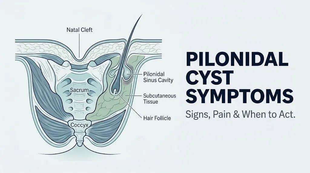
A pilonidal cyst is a subcutaneous cavity lined with keratinized squamous epithelium that forms in the natal cleft (the intergluteal fold between the buttocks) as a result of hair follicle penetration and a foreign body inflammatory response in the deep dermis.
The mechanism works like this. Loose terminal hairs, shed from the gluteal skin or back, are driven into the skin of the natal cleft by the friction and suction forces generated every time a person sits, stands, or walks. Think of the natal cleft as a natural funnel that concentrates this mechanical pressure: gluteal movement essentially creates a bellows-like vacuum that pulls loose hairs inward through hair follicle openings or small skin abrasions. The hair shaft, once embedded, acts as a foreign body.
The body’s immune system responds with a granulomatous foreign body reaction: macrophages fuse into foreign body giant cells and attempt to wall off the intruding hair. Fibroblasts lay down a fibrous capsule around the growing hair bundle. Over time, this capsule develops its own keratinized epithelial lining, essentially becoming a small sinus cavity. Research published in the Diseases of the Colon and Rectum confirms this as the primary pathological mechanism, distinct from the older theory that pilonidal cysts are congenital dermoid cysts (a theory now largely discredited in the surgical literature).
Keratin debris, sebaceous secretions, and additional hairs accumulate inside the cavity. When anaerobic bacteria — primarily Bacteroides fragilis, Staphylococcus aureus, and Peptostreptococcus species — colonize this material in sufficient numbers, an acute abscess forms.
For people with BMI above 30, a deeper natal cleft increases both the vacuum suction force and the warmth and moisture in the region, which accelerates bacterial growth. This is why obesity is a consistent, independent risk factor for both initial pilonidal disease and surgical recurrence.
Pilonidal Cyst Pain: What Does It Feel Like
Pilonidal cyst pain presents as a deep, aching tenderness in the skin and subcutaneous tissue just above the tailbone, which worsens with direct pressure, sitting, or bending forward, and can intensify rapidly to a throbbing, pulsating quality when an abscess is forming beneath the skin.
Before infection sets in, many people describe the pain as a persistent soreness, similar to a bruise that doesn’t resolve. Pressing on the area produces immediate tenderness. The pain is often worse at the end of a workday, after long drives, or after any sustained period of sitting.
Once an abscess begins to form, the pain character shifts. The throbbing quality is produced by the pressure of accumulating pus within the closed fibrous capsule. Each heartbeat increases intracavity pressure slightly, which is exactly why people with an acute pilonidal abscess often describe a rhythmic pulsating sensation synchronized with their pulse. This is a clinically useful sign: if your tailbone pain is pulsating, infection is almost certainly already present.
| Pain Type | Clinical Description | Stage |
|---|---|---|
| Dull aching soreness | Pressure-sensitive, worse with sitting, no fever | Pre-infection or early inflammation |
| Throbbing or pulsating | Rhythmic, pressure-related, worsening | Abscess forming |
| Sharp, constant, severe | Continuous regardless of position, fever present | Established abscess |
| Burning and spreading | Extends beyond the cyst site, systemic symptoms | Cellulitis or progressing infection |
People with a higher pain tolerance, particularly young men in physically demanding occupations, sometimes underreport symptom severity until the abscess is well-advanced. Women often describe the sensation as similar to a deep pelvic pressure, which can lead to a brief delay in correct identification of the source.
Pilonidal Cyst Swelling and Skin Changes
Swelling from a pilonidal cyst appears as a firm, tender, raised area in the skin directly over and just above the gluteal cleft, which may develop visible redness and warmth as the underlying inflammatory process intensifies.
Early swelling is often subtle. The overlying skin may look only slightly raised, and the firmness beneath it is due to the fibrous capsule and accumulating inflammatory tissue rather than free pus. As infection develops, the swelling becomes more pronounced, the skin tightens, and the area may develop a reddish-to-dusky discoloration.
The clinical term for the specific texture change associated with an established abscess is fluctuance: a compressible, fluid-filled quality detected by pressing on the swollen area with two fingers simultaneously on either side. When you can feel a wave-like movement within the swelling under finger pressure, that is fluctuance, and it indicates a pocket of pus is ready to be drained.
The American Academy of Family Physicians describes fluctuance as the most reliable physical examination finding for confirming an abscess and distinguishing it from pre-suppurative cellulitis. This distinction matters because cellulitis without an abscess is managed with antibiotics, while an abscess requires incision and drainage.
Key skin changes to observe and report to your provider:
- Redness (erythema) confined to the immediate cyst area, or spreading outward
- A visible pit, pore, or small dimple in the skin at the top of the natal cleft
- Skin that feels hot to the touch relative to the surrounding tissue
- Taut, shiny skin over the swollen area
- Visible hair protruding from a sinus opening
People with darker skin tones may notice warmth and firmness before visible redness becomes apparent, since erythema can be less visible on deeper skin tones. Any progressive swelling with increasing tenderness warrants evaluation regardless of whether color change is detectable.
Key Takeaway: Pain that pulses in rhythm with your heartbeat and swelling that feels fluid-filled under finger pressure are the two most reliable signs that a pilonidal abscess has formed and requires medical drainage, not home treatment.
Pilonidal Cyst Drainage and Discharge
Drainage from a pilonidal cyst occurs when the abscess or sinus tract ruptures or opens spontaneously, releasing purulent material (pus), serous fluid (clear or straw-colored), or blood-tinged discharge from one or more small openings in the skin above the natal cleft.
The drainage is one of the most diagnostically clear signs of pilonidal sinus disease. It confirms that a sinus tract has formed and is now communicating with the skin surface. The discharge may start as a small amount of clear fluid and progressively become thicker, yellow or green, and foul-smelling as bacterial load increases.
The odor associated with pilonidal cyst drainage is produced by the metabolic byproducts of anaerobic bacteria, particularly Bacteroides and Peptostreptococcus species, which release short-chain fatty acids and volatile sulfur compounds during their metabolic activity. It is clinically described as a foul, sulfurous odor that is distinct from ordinary sweat.
Spontaneous drainage can temporarily relieve the painful pressure of an abscess, which sometimes misleads people into thinking the condition has resolved on its own. It has not. The sinus tract remains, the bacteria remain, and re-accumulation of purulent material is predictable without treatment.
What to observe and report about discharge:
- Volume: Is it a few drops or steady flow?
- Color: Clear, yellow, green, blood-tinged, or dark brown
- Consistency: Watery, thick, or viscous
- Odor: Present or absent
- Duration: Hours, days, or intermittent over weeks
- Number of openings: Single pit or multiple sinus tract openings
Multiple sinus openings draining simultaneously is a sign of chronic pilonidal sinus disease with branching tract architecture, which represents a more complex surgical challenge than a single-tract acute abscess. A research review published in the British Journal of Surgery found that patients with multiple sinus tracts have approximately twice the recurrence rate after primary surgical closure compared to those with single-tract disease.
Pilonidal Cyst Infected Symptoms
Infected pilonidal cyst symptoms include rapidly intensifying pain, expanding redness and warmth in the skin around the cyst, swelling that is firm and then fluctuant, purulent drainage, and in many cases a fever above 38.3°C (101°F), all of which indicate that a bacterial abscess has developed within the pilonidal sinus cavity.
The transition from a quiet, non-infected pilonidal cyst to an acutely infected abscess can happen within days or even hours. The inflammatory cascade, driven by IL-1, IL-6, and TNF-alpha released by macrophages responding to bacterial lipopolysaccharide (LPS) from gram-negative anaerobes like Bacteroides fragilis, produces the cardinal signs of infection: calor (heat), rubor (redness), tumor (swelling), dolor (pain), and in systemic infection, the loss of normal function.
Clinically recognizable infected pilonidal cyst symptoms, distinguished from the non-infected presentation:
| Feature | Non-Infected Cyst | Infected Abscess |
|---|---|---|
| Pain | Dull aching, intermittent | Severe, constant, throbbing |
| Skin temperature | Normal to slightly warm | Distinctly hot |
| Swelling | Mild, firm | Pronounced, fluctuant |
| Skin color | Normal or mildly red | Deep red to dusky |
| Drainage | Absent | Present, purulent |
| Fever | Absent | Often present |
| Systemic symptoms | None | Possible chills, fatigue |
People with diabetes mellitus are at particular risk for accelerated infection progression. Elevated blood glucose impairs neutrophil chemotaxis and phagocytic function, allowing bacterial proliferation to outpace the immune response. A pilonidal abscess in a person with poorly controlled diabetes warrants same-day evaluation, not a scheduled appointment in several days.
Pilonidal Cyst Fever and Systemic Signs
Fever associated with a pilonidal cyst indicates that the local infection has triggered a systemic inflammatory response, and when accompanied by chills, rapid heart rate, or confusion, it signals the possibility of early sepsis requiring emergency evaluation.
The body generates fever through a prostaglandin E2-mediated pathway: bacterial toxins trigger macrophages to release IL-6 and IL-1, which act on the hypothalamic thermoregulatory center to raise the set point. A temperature at or above 38.3°C (101°F) in the setting of a painful, swollen pilonidal cyst indicates that the infection is actively driving a systemic response and that the bacteria are either spreading locally into surrounding tissue or releasing enough toxin to trigger systemic inflammation.
A fever in this context is not a minor symptom to monitor at home. The National Institutes of Health describes systemic inflammatory response syndrome (SIRS) criteria — temperature above 38°C or below 36°C, heart rate above 90 beats per minute, respiratory rate above 20 breaths per minute, and abnormal white blood cell count — as the diagnostic threshold requiring urgent evaluation. A pilonidal abscess with associated fever meeting two or more of these criteria meets the clinical definition of infection with systemic involvement.
Systemic signs that accompany infected pilonidal cysts and require urgent or emergency evaluation:
- Fever above 38.3°C (101°F) measured orally
- Chills or rigors (uncontrollable shaking)
- Heart rate above 90 beats per minute at rest
- Fatigue or general malaise disproportionate to the local wound
- Nausea or loss of appetite
- Confusion or altered mental state (a late and serious sign)
Older adults and immunocompromised individuals may not mount a classic fever response even during significant systemic infection. In these populations, an absence of fever does not rule out serious infection, and any worsening of a pilonidal cyst with increasing pain or functional decline warrants the same urgency as a fever presentation.
Key Takeaway: A fever above 38.3°C (101°F) combined with a painful, swollen pilonidal cyst is not a wait-and-see situation. It indicates active systemic infection and requires medical evaluation the same day, either through a primary care physician, urgent care, or an emergency room.
Early Symptoms of a Pilonidal Cyst
The earliest symptoms of a pilonidal cyst are subtle and often mistaken for a muscle strain, a bruise from an awkward sitting position, or general lower back fatigue: mild aching or pressure in the skin just above the tailbone that worsens with direct pressure or prolonged sitting.
At this stage, often called the pre-suppurative phase, the pilonidal sinus tract has formed but has not yet accumulated enough bacterial load to trigger a full abscess. The inflammatory process is active at a low level, but pus has not yet developed. The person may notice:
- A small, firm nodule or lump under the skin near the top of the natal cleft
- A visible pit, dimple, or pore opening in the skin (this is the sinus ostium)
- Slight redness or skin darkening around the pit
- Hair visible protruding from the pit opening
- Discomfort when clothing waistbands press against the area
This early presentation is actually the ideal window for treatment, because at this stage, some patients respond to hair removal, improved hygiene, and reduced sitting time, reducing the bacterial load and allowing the sinus to remain quiescent. A 2019 cohort study published in the World Journal of Surgery found that endoscopic pilonidal sinus treatment (EPSiT) performed during the non-infected phase produced significantly lower recurrence rates compared to emergency incision and drainage of an established abscess.
The challenge is that many people dismiss early pilonidal symptoms as insignificant. By the time they seek care, the condition has progressed to an acute abscess. Postpubertal adolescents and young adults in particular often attribute early tailbone discomfort to gym activity or sitting in class, delaying recognition of the actual condition by weeks to months.
Chronic Pilonidal Sinus Disease Symptoms
Chronic pilonidal sinus disease presents as a pattern of intermittent or persistent drainage, recurrent episodes of infection and partial resolution, multiple sinus pit openings in the natal cleft, and chronic low-grade pain that worsens during flare-ups, representing a fundamentally different clinical picture from a single acute abscess.
The distinction matters because chronic disease involves a more complex sinus architecture. Instead of one capsule with one drainage tract, chronic pilonidal sinus disease typically produces multiple branching tracts that extend laterally beneath the skin, occasionally reaching the perineum or perianal region in severe cases. Each tract is lined with keratinized squamous epithelium and harbors its own bacterial microenvironment. This is why antibiotics alone cannot cure the condition: the epithelial lining protects the bacterial colony from antibiotic penetration, similar to the way a biofilm-covered implant resists antibiotic clearance.
Symptoms that distinguish chronic pilonidal sinus disease from an isolated acute episode:
- Multiple visible sinus pits or openings in a line along the natal cleft
- Intermittent drainage over weeks or months, with periods of apparent improvement followed by recurrence
- Chronic low-grade aching that rarely fully resolves between flare-ups
- Skin changes around the sinus openings, including scarring, hyperpigmentation, or thickening of the surrounding dermis
- Episodes of acute flare where pain and discharge intensify dramatically before partially resolving on their own
- Quality of life impairment from the ongoing drainage and odor
According to the American Society of Colon and Rectal Surgeons clinical practice guidelines, recurrence after simple incision and drainage of an acute pilonidal abscess occurs in approximately 40 to 60 percent of patients without definitive surgical treatment. This high recurrence rate reflects the persistence of the epithelial-lined sinus architecture, not treatment failure in a conventional sense.
Pilonidal Cyst Symptoms in Women
Women develop pilonidal cysts less frequently than men, but when they do, the symptoms are clinically identical, and the condition is often diagnosed later because providers and patients alike are less likely to consider it as a first possibility in a woman with tailbone pain.
The lower incidence in women reflects differences in androgenic terminal hair growth: testosterone drives the proliferation of coarse terminal hairs in the natal cleft region, which is why postpubertal males have both higher hair density and higher pilonidal disease incidence. A 2021 epidemiological study published in Diseases of the Colon and Rectum found the male-to-female incidence ratio to be approximately 3:1 to 4:1, with women accounting for roughly 20 to 25 percent of all pilonidal disease cases.
That said, women with hirsutism (excess terminal hair growth due to polycystic ovary syndrome, congenital adrenal hyperplasia, or idiopathic causes) have elevated pilonidal cyst risk. This is because the underlying mechanism is the same regardless of sex: terminal hair in the natal cleft plus friction and suction forces creates the pathway for sinus formation.
Specific considerations for women with pilonidal cyst symptoms:
- Pilonidal pain in women is frequently misattributed to coccydynia, perianal fistula, Bartholin gland cyst, or pelvic floor dysfunction. This diagnostic delay means women sometimes present with more advanced disease.
- Pregnant women may experience worsening pilonidal symptoms due to increased pressure on the sacrococcygeal region and reduced local immunity. Any worsening pilonidal disease in pregnancy should be evaluated promptly by an obstetrician and a general or colorectal surgeon jointly.
- Women with pilonidal sinus near the perineum may have difficulty distinguishing symptoms from gynecological conditions. Clinical examination, and sometimes pelvic ultrasound or MRI, is needed to confirm the source.
- Pain from a pilonidal cyst typically worsens with direct pressure on the natal cleft, not with gynecological examinations or menstruation, which can help distinguish it from reproductive system sources.
Key Takeaway: Women account for approximately 20 to 25 percent of pilonidal disease cases, and diagnostic delays are common because the condition is still widely considered a male-predominant disease. Any woman with persistent tailbone pain and a visible pit or sinus opening in the natal cleft should request clinical examination regardless of whether the diagnosis is initially considered.
Pilonidal Cyst Risk Factors and Who Gets Them
The people at highest risk for developing a pilonidal cyst are young adult males aged 15 to 35 with coarse body hair, a deep natal cleft, BMI above 30, and a sedentary or seated occupation, though no single factor determines risk in isolation.
Each risk factor contributes to the underlying mechanism through a distinct pathway:
- Male sex and postpubertal age: Testosterone drives terminal hair follicle growth in the sacrococcygeal and gluteal region after puberty. The density of coarse hair in the natal cleft is the primary raw material for sinus formation.
- BMI above 30: A higher BMI deepens the natal cleft, increasing suction forces during movement and creating a warmer, more moist microenvironment that supports anaerobic bacterial growth.
- Prolonged sitting and sedentary occupations: Truck drivers, office workers, students, and military personnel who sit for extended periods generate sustained friction and pressure in the natal cleft, repeatedly driving loose hairs into the skin.
- Deep natal cleft anatomy: This is an inherited anatomical feature. People with a more pronounced cleft have a more efficient hair-trapping mechanism independent of BMI or hair density.
- Hirsutism: Elevated hair density in the natal cleft from any cause (androgenic, pharmacological, or idiopathic) increases the volume of loose hair available to penetrate the skin.
- Poor local hygiene: Accumulation of sweat, debris, and bacteria in the natal cleft area is not the primary cause but is a contributing factor to recurrence and infection.
- Previous pilonidal surgery: Surgical scarring alters the tissue architecture and can create new paths for sinus formation.
According to the National Institutes of Health, military service members have historically had such high rates of pilonidal disease from jeep and vehicle travel that the condition was once called “Jeep disease” during World War II. This occupational association remains clinically relevant for anyone who sits on vibrating equipment for hours at a time.
Pilonidal Cyst vs. Coccyx Pain and Other Conditions
Pilonidal cyst pain is located in the skin and subcutaneous tissue above the tailbone and is associated with visible skin changes, drainage, or a palpable pit or nodule, which distinguishes it from coccydynia (coccyx bone pain from a fall or prolonged sitting) and other conditions in the same anatomical region.
Several conditions can produce pain in or near the sacrococcygeal region. The key to distinguishing them lies in the exact location of pain, the presence or absence of skin findings, and the associated symptoms.
| Condition | Pain Location | Skin Findings | Associated Symptoms | Distinguishing Feature |
|---|---|---|---|---|
| Pilonidal cyst | Natal cleft, superficial | Pit, sinus, redness, drainage | Fever, drainage, odor | Skin opening with drainage |
| Coccydynia | Coccyx bone | None | Pain with sitting, tenderness over tailbone | No skin changes, bone-level tenderness |
| Perianal abscess | Near anus, lower | Redness, swelling near anus | Severe pain with defecation | Anatomically closer to anus |
| Perianal fistula | Near anal opening | Visible fistula opening | Chronic drainage near anus | Connected to anal canal |
| Sacral dimple | Upper natal cleft | Dimple present from birth | Usually asymptomatic | Congenital, no inflammation |
| Hidradenitis suppurativa | Groin, buttocks, axilla | Nodules, sinus tracts, scarring | Recurrent abscesses in multiple areas | Affects apocrine gland regions |
A clinical examination by a primary care physician or colorectal surgeon can reliably differentiate pilonidal disease from these conditions in most cases. MRI of the sacrococcygeal region is used when complex sinus tract anatomy is suspected or when the clinical picture is ambiguous, particularly to rule out sacral spinal involvement.
For older adults presenting with new-onset sacrococcygeal pain and skin changes, and especially for those with prior malignancy, a tissue biopsy may be warranted to exclude the rare but documented malignant transformation (squamous cell carcinoma arising in a long-standing pilonidal sinus) reported in case series published in the Annals of Surgery.
Pilonidal Cyst Sitting Pain and Activity-Related Symptoms
Pilonidal cyst pain consistently worsens with sitting because the act of sitting compresses the natal cleft, increases pressure directly over the sinus cavity, and can drive additional hairs into the sinus tract, all of which intensify local inflammation.
Think of the natal cleft as a crease that flattens and deepens alternately with every change in position. When a person sits on a firm surface, the two gluteal prominences press toward each other, and the natal cleft narrows and deepens. This directly compresses any inflamed tissue, abscess cavity, or sinus tract in the region. The pressure-to-pain relationship is essentially mechanical: more compression equals more pain over an inflamed subcutaneous structure.
People with pilonidal disease often develop specific coping strategies that are themselves diagnostically informative:
- Leaning to one side when sitting, to shift weight off the painful area
- Avoiding hard chairs and preferring cushioned surfaces
- Standing up frequently during long work sessions or car trips
- Difficulty completing tasks that require sustained sitting, such as driving or desk work
- Increased pain after returning home from a long day, sometimes peaking in the evening
Activity-related symptoms beyond sitting include:
- Pain with bending forward, particularly when the gluteal muscles stretch
- Discomfort with stair climbing, due to the gluteal flexion and extension cycle
- Worsening symptoms after cycling or horseback riding
- Improved symptoms when lying flat on the stomach, which relieves direct pressure on the natal cleft
The American Academy of Family Physicians notes that activity-related worsening of sacrococcygeal symptoms is one of the most consistent features of pilonidal disease, particularly in young patients who present to primary care without a classic abscess formation.
Pilonidal Cyst Recurrent Symptoms and Complications
Pilonidal cyst recurrence is not a treatment failure in a simple sense: it reflects the persistence of the epithelial-lined sinus architecture that, unless completely excised or destroyed, continues to trap hair, harbor bacteria, and produce new inflammatory episodes.
Research published in JAMA Surgery found that recurrence rates after simple incision and drainage of acute pilonidal abscess reach 40 percent within two years when no additional surgical procedure is performed. Even after definitive excision, recurrence rates range from 5 to 15 percent depending on the surgical technique and whether adjunctive hair removal is maintained.
Recurrent pilonidal disease symptoms tend to escalate with each episode:
- Shorter intervals between acute flare-ups as the sinus architecture becomes more complex
- Larger areas of involved skin and subcutaneous tissue with each recurrence
- Increasingly prominent scarring around the natal cleft from repeated inflammation and partial healing
- Progressive difficulty with mobility and sitting tolerance
- In rare long-standing cases (typically more than a decade of untreated chronic disease), malignant transformation to squamous cell carcinoma has been documented in case series, though this remains uncommon
Complications of untreated or recurrently treated pilonidal disease include:
- Spreading cellulitis: bacterial infection extending beyond the sinus into surrounding subcutaneous tissue
- Bacteremia: bacterial seeding of the bloodstream from a heavily infected abscess, most serious in immunocompromised individuals
- Chronic wound and fistula formation
- Fournier’s gangrene (necrotizing fasciitis of the perineum and gluteal region), an extremely rare but life-threatening complication documented in case reports in immunocompromised patients with neglected pilonidal abscesses
- Chronic pain syndrome from nerve sensitization in the sacrococcygeal region
Key Takeaway: Recurrence after simple drainage of a pilonidal abscess is the clinical rule, not the exception. Definitive surgical management, including full sinus tract excision, is required for lasting resolution, and long-standing untreated disease carries a small but real risk of malignant transformation.
How to Know If Your Pilonidal Cyst Needs Medical Attention
Your pilonidal cyst needs medical attention when you have pain that limits normal function, any drainage from the natal cleft region, skin redness or warmth that is spreading, fever, or symptoms that have persisted or worsened over more than a few days without clear improvement.
Not every pilonidal cyst presentation is an emergency. But none of them are suitable for purely home management once symptoms have progressed beyond mild, intermittent aching with no skin changes. Use the framework below to guide your decision:
When you can monitor at home for 24 to 48 hours (mild, non-infected presentation only):
- Very mild aching near the tailbone with no visible skin changes, no drainage, no fever
- A small, non-tender dimple or pit that has been present for some time with no change
- Symptoms clearly related to a single day of excessive sitting with no localized swelling or warmth
When you should call your primary care physician within 24 to 48 hours:
- Pain that limits your ability to sit or complete normal daily activities
- A visible or palpable nodule or swelling near the natal cleft, even without fever
- Any discharge from a skin opening, even if it appears minor
- A history of prior pilonidal cysts with new onset of familiar symptoms
When you should go to urgent care or an emergency room today:
- Fever above 38.3°C (101°F) combined with local pain and swelling
- Rapidly spreading redness beyond the immediate area of the cyst
- Severe pain that is constant and not position-dependent
- Any systemic symptoms: chills, rigors, confusion, or rapid heart rate
To prepare for your appointment effectively:
- Note when your symptoms first started and whether they came on gradually or suddenly.
- Describe the exact location of pain: above the tailbone, between the buttocks, or to one side.
- Report any drainage: color, consistency, odor, and duration.
- Record your highest temperature in the past 24 to 48 hours if you have measured it.
- List any prior pilonidal cyst episodes, treatments, or surgical procedures.
- Note any medications, particularly immunosuppressants, corticosteroids, or diabetes medications.
Your primary care physician will perform a clinical examination and may refer you to a colorectal surgeon or general surgeon if a definitive surgical procedure is indicated.
Emergency Symptoms: When to Call 911 or Go to the ER
Certain symptoms associated with pilonidal cyst disease require immediate emergency evaluation. Do not wait to see if these resolve on their own.
Call 911 or go to the nearest emergency room immediately if you experience:
- Fever above 38.9°C (102°F) that is not responding to over-the-counter fever reducers, combined with a painful pilonidal cyst
- Chills, rigors (uncontrollable shaking), or a sensation of being severely ill alongside local pilonidal symptoms
- Rapid heart rate at rest (above 90 to 100 beats per minute) combined with pilonidal pain and fever
- Confusion, disorientation, or significantly reduced alertness — this may indicate sepsis, which is a medical emergency
- Rapidly expanding redness (erythema) spreading from the natal cleft region across the buttocks, thighs, or lower abdomen, which may indicate spreading cellulitis or, in rare cases, necrotizing fasciitis
- Extreme pain, skin discoloration (dusky, blue, or black skin) around the cyst site, and a crackling sensation when touching the skin (crepitus), which can indicate necrotizing infection requiring emergency surgery
- Any pilonidal presentation in an immunocompromised individual (organ transplant recipient, person on chemotherapy, person with uncontrolled diabetes) who develops fever or systemic symptoms
These presentations can indicate pilonidal-associated sepsis, rapidly spreading cellulitis, or necrotizing fasciitis, all of which require emergency medical assessment and potentially emergency surgical intervention, not an urgent care appointment or a phone call to schedule a routine visit.
Frequently Asked Questions About Pilonidal Cyst Symptoms
What does a pilonidal cyst feel like?
A pilonidal cyst feels like a firm, tender lump under the skin just above the gluteal cleft, producing aching pressure that worsens when sitting or when the area is directly touched.
When infection develops, the tenderness sharpens to a throbbing or pulsating pain that may be felt even without direct pressure.
Some people also notice a small pit or dimple in the skin at the top of the natal cleft, which may have visible hair protruding from it.
How do I know if my pilonidal cyst is infected?
An infected pilonidal cyst produces rapidly worsening pain that shifts from a dull ache to a constant throbbing sensation, along with visible redness, skin warmth, and swelling over the cyst site.
The presence of purulent (pus) drainage from a skin opening and fever above 38.3°C (101°F) confirm active infection requiring same-day medical evaluation.
Infected cysts do not improve with rest or home care and typically worsen over 24 to 72 hours without incision and drainage.
Can a pilonidal cyst go away on its own without treatment?
A small, non-infected pilonidal cyst may remain quiescent for extended periods and produce only intermittent mild symptoms, but the underlying sinus tract does not resolve without treatment.
Without intervention, the sinus continues to trap hair and debris, and most people develop at least one episode of acute abscess over time.
According to the American Society of Colon and Rectal Surgeons, spontaneous permanent resolution of pilonidal sinus disease is uncommon without some form of definitive treatment.
What are the early warning signs of a pilonidal cyst?
The earliest warning signs of a pilonidal cyst are mild aching or pressure near the tailbone that worsens with direct pressure or sitting, sometimes accompanied by a visible pit or dimple in the skin above the natal cleft.
A small, firm lump under the skin in the same area, even without pain, may represent an early non-infected pilonidal sinus cavity.
Hair visibly protruding from a small skin opening near the top of the gluteal cleft is a particularly characteristic early sign.
Is pilonidal cyst pain constant or does it come and go?
Pilonidal cyst pain in the non-infected phase is typically intermittent, worsening with sitting and improving with rest or position changes.
Once an acute abscess forms, the pain becomes constant, throbbing, and position-independent, signaling that infection and increasing pressure within the sinus cavity are driving the symptoms.
Chronic pilonidal sinus disease produces a pattern of recurring pain episodes separated by periods of reduced symptoms, which can mislead people into believing the condition has resolved between flares.
When should I go to the emergency room for a pilonidal cyst?
Go to the emergency room immediately if your pilonidal cyst symptoms are accompanied by fever above 38.9°C (102°F), chills, rapid heart rate at rest, or confusion, as these may indicate sepsis or a rapidly spreading infection.
Call 911 if you notice rapidly expanding redness across the buttocks, thighs, or lower abdomen, or if the skin around the cyst becomes dark or discolored, which may indicate a surgical emergency.
Immunocompromised individuals, including those with diabetes, cancer, or on immunosuppressive medications, should seek emergency evaluation at the first sign of systemic symptoms, not after waiting to see whether symptoms improve.
Making Sense of What You Are Experiencing
Pilonidal cyst symptoms exist on a spectrum from a barely noticeable pit in the skin to a painful, draining abscess that makes sitting impossible. The most practical thing you can take from understanding this condition is that the gap between “quiet sinus” and “acute abscess” can close faster than most people expect.
If you have a visible pit, intermittent tailbone pain, or any drainage near your natal cleft, schedule an appointment with your primary care physician this week. Early evaluation and hair removal strategies can reduce the likelihood of a full abscess. If you already have swelling, spreading redness, fever, or drainage, today is the right day, not next week.
The single most actionable clinical fact here is this: a throbbing, fluctuant swelling near your tailbone with fever does not improve on its own, and waiting it out significantly increases your risk of spreading infection and a more complex surgical recover