Plantar Wart Symptoms: Complete 2026 Clinical Guide
Plantar wart symptoms include a hard, thickened patch of skin on the bottom of the foot, pain or tenderness when walking or standing, tiny black or dark red dots embedded in the lesion, and a disruption of the normal skin ridge lines across the affected area. These symptoms occur because human papillomavirus (HPV) infects skin cells on the plantar surface and drives abnormal cellular growth inward toward the dermis rather than outward, which is why plantar warts hurt when you walk on them but are often flush with or even slightly recessed into the skin surface.
According to the American Academy of Dermatology (AAD), warts are among the most common skin conditions seen in clinical practice, affecting an estimated 10 percent of the general population at any given time, with plantar warts accounting for a disproportionate share of cases in children, adolescents, and athletes who regularly use communal barefoot spaces. The condition is caused specifically by HPV, a virus that enters plantar skin through microscopic breaks in the outer skin layer and reprograms the local skin cells to reproduce abnormally.
This guide explains every recognized plantar wart symptom with the underlying biological mechanism, distinguishes early from advanced presentations, identifies the populations at highest risk for complications, and tells you exactly when home monitoring stops being appropriate.
Plantar Wart Symptoms: What You Need to Know First
Plantar wart symptoms are produced by HPV infecting the plantar epidermis and driving the growth of an abnormal, hyperkeratotic lesion inward under the skin surface, where body weight compresses it against dermal nerve fibers and blood vessels with every step.
The defining feature that separates plantar warts from other foot conditions is the direction of growth. Most skin growths protrude outward. Plantar warts are forced inward by the mechanical pressure of walking. This inward growth, technically called endophytic growth, is what makes them painful out of proportion to their visible size and what makes them clinically distinctive under close examination.
The full recognized symptom set includes:
- A thickened, callus-like patch of skin on the heel, ball of the foot, or base of the toes
- Sharp, aching, or burning pain when direct downward pressure is applied (stepping down or pressing with a finger)
- Pain that is less severe or absent when the lesion is pinched from the sides
- Tiny black or dark red dots (thrombosed capillary loops) visible within the lesion
- Disruption or interruption of normal skin lines (dermatoglyphs) across the lesion surface
- A lesion that may be slightly sunken rather than raised above surrounding skin
- Possible surrounding callus buildup from repeated pressure over the lesion
The National Institutes of Health (NIH) notes that HPV-related skin lesions on the plantar surface are classified under ICD-10 code B07.0 and are distinct from common warts on the dorsal (top) surface of the foot or hands, both in their HPV genotype profile and their growth characteristics.
For adolescents ages 12 to 16, symptoms may appear more rapidly and multiply more readily than in adults, because the frequency of communal barefoot activity is higher and the immune response to the specific plantar HPV genotypes may not yet be fully established.
What Is a Plantar Wart and How Does HPV Cause It
A plantar wart is a localized, benign epidermal growth on the plantar surface of the foot caused by infection with specific genotypes of human papillomavirus, primarily HPV types 1, 2, 4, 27, and 57, which enter through microabrasions in the stratum corneum and infect keratinocytes in the stratum basale.
Think of the plantar skin surface like a layered brick wall. The outermost bricks are the dead, hardened cells of the stratum corneum. The cells being manufactured at the base of the wall are the keratinocytes of the stratum basale. HPV slips through cracks in the outer wall (microabrasions from walking barefoot on rough surfaces, minor cuts, athlete’s foot-damaged skin), reaches the base layer, and reprograms the brick-making machinery. Instead of normal skin cells, the base layer starts producing abnormal, disorganized cells that pile up in a hard, rough nodule.
The virus reprograms this production using two primary tools: the viral oncoproteins E6 and E7. E6 targets and degrades the tumor suppressor protein p53, which would normally trigger apoptosis (cell death) in abnormal cells. E7 inactivates the retinoblastoma protein (pRb), which normally acts as a brake on cell division. With both brakes disabled, keratinocyte proliferation continues unchecked, producing the characteristic thickened, abnormal epidermal architecture of a wart.
A 2021 review published in the Journal of the American Academy of Dermatology confirmed that HPV-1 is responsible for the deepest, most painful plantar wart variant (sometimes called myrmecia warts), while HPV-2, HPV-4, HPV-27, and HPV-57 are more commonly associated with mosaic-pattern plantar warts and less deeply invasive lesions. This genotype distinction has practical implications because deeper lesions require more aggressive treatment.
Immune status determines how effectively the body controls the infection after the virus establishes itself. The cell-mediated immune response, mediated primarily by CD4+ T-helper lymphocytes and CD8+ cytotoxic T-lymphocytes, is the primary mechanism by which the immune system recognizes and eventually clears HPV-infected keratinocytes. In people with intact immune systems, this response eventually clears most plantar wart infections, though the timeline is variable and can extend to two or more years.
What Does a Plantar Wart Look Like
A plantar wart looks like a small, rough, thickened patch of skin on the bottom of the foot, typically circular or oval in shape, with a surface that is harder and grainier than surrounding skin and that contains tiny black or dark red dots embedded within the tissue.
The size of a plantar wart ranges from a few millimeters (about the size of a small pea) to over a centimeter in diameter for established lesions. The surface texture is distinctly granular or rough compared to the smooth plantar skin surrounding it. The color is typically skin-tone to slightly yellowish-white from the hyperkeratotic (thickened) surface layer.
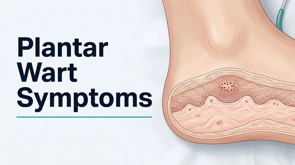
One of the most reliable visual features is the interruption of dermatoglyphs, the ridge and furrow pattern of the skin surface (think fingerprint lines, but on the sole of the foot). In normal skin, these lines flow continuously across the surface. A plantar wart interrupts this pattern. The lines run up to the wart’s edge and stop or deflect around it. A corn or callus, by contrast, preserves and may even exaggerate the underlying skin line pattern.
The lesion is typically flat with surrounding skin or slightly sunken due to inward growth pressure. It may appear to have a central “core” surrounded by a slightly raised border, particularly in well-established lesions. Surrounding the lesion, there is often a ring of thickened callus skin that builds up as the body responds to abnormal pressure distribution.
| Visual Feature | Plantar Wart | Corn | Callus |
|---|---|---|---|
| Skin line pattern | Interrupted, deflected | Preserved | Preserved |
| Black dots present | Yes (thrombosed capillaries) | No | No |
| Central hard core | Sometimes | Yes, translucent | No distinct core |
| Pain pattern | Worse with direct pressure (vertical) | Worse with direct pressure | Diffuse ache, less sharp |
| Surface texture | Rough, granular | Smooth, hard | Rough but uniform |
| Shape | Round to oval | Round | Irregular |
For children under 10, plantar warts may appear slightly flatter and smaller than in adolescents or adults, because the shorter time since infection means the lesion has not had as long to develop the thickened hyperkeratotic surface layer.
Plantar Wart Pain Symptoms
Plantar wart pain is produced by the downward compression of a growing endophytic lesion against sensory nerve fibers and thrombosed capillary loops in the papillary dermis, generating a sharp, aching, or burning pain that is consistently worse with direct vertical pressure and typically reduced when the lesion is pinched from the sides.
The pain is the primary functional symptom. Most people with a plantar wart do not notice the lesion visually first: they notice that a particular spot on the bottom of their foot hurts when they walk or stand. The pain is often described as similar to walking on a small pebble or a piece of gravel that seems to be embedded in the foot, because that is anatomically what is happening: the lesion is pressing against the underlying dermis from above with every step.
The directional pain test is one of the most clinically useful self-assessment tools. Press directly down on the lesion (vertical compression): plantar warts produce pain. Now pinch the lesion from both sides (horizontal compression): plantar warts produce less or no pain from this direction. Corns and calluses typically produce the reverse pattern, hurting more with lateral pressure across the corn’s central core. This difference exists because the wart’s pain is generated in the dermis below the lesion, while a corn’s pain comes from the central keratin plug pressing into the skin laterally.
Pain varies by location on the plantar surface. Warts on the metatarsal heads (the balls of the foot behind each toe) are typically more painful than those on the calcaneus (heel), because the metatarsal head area bears higher dynamic loading during the push-off phase of walking. A wart directly under the first or fifth metatarsal head may produce enough pain to alter gait mechanics, and long-standing gait changes can secondarily produce knee, hip, or lower back discomfort from compensatory posture changes.
Quick Tip:
- If the pain from a foot lesion is causing you to limp or alter your walking pattern, this warrants evaluation by a primary care physician or board-certified podiatrist regardless of how the lesion looks visually.
- People with rheumatoid arthritis on methotrexate or biologic therapies may have plantar warts with less pain than expected because both the wart burden and the immune-inflammatory response are modified by their medications, which can lead to underrecognition of the lesion.
Plantar Wart Black Dots Explained
The black or dark red dots visible in a plantar wart are thrombosed (clotted) capillary loops within the papillary dermis that have been disrupted by the wart’s inward growth and subsequently occluded, producing small pools of coagulated blood visible through the overlying skin.
These dots are often called “wart seeds” colloquially, but they are not seeds, virus particles, or any foreign material. They are a vascular sign. As the wart grows inward and the abnormal keratinocyte architecture distorts the dermal papillae, the small blood vessels that normally supply oxygen to the epidermis through the dermal papillae become stretched, compressed, and eventually thrombosed. The resulting clotted blood appears dark because it is deoxygenated hemoglobin viewed through a thin overlying layer of skin.
The presence of black dots within a plantar lesion is one of the most diagnostically useful features for distinguishing a plantar wart from a callus or corn. Neither calluses nor corns produce thrombosed capillaries, because neither involves viral disruption of the dermal architecture. If you pare away the surface layer of a plantar wart with a pumice stone or nail file, the black dots become more visible and may produce small pinpoint bleeding where the thrombosed loops have been opened. This is called the capillary bleeding sign or paring test, and it is a standard clinical diagnostic maneuver.
According to research reviewed in JAMA Dermatology, dermoscopy (magnified examination of the skin with a handheld dermoscope) reveals the thrombosed capillary loops as a characteristic pattern described as “black or red dots arranged in a mosaic or scattered pattern” within the lesion. This pattern is sufficiently distinctive that experienced dermatologists can distinguish plantar warts from other plantar lesions with high accuracy using dermoscopy alone, before any tissue sampling.
In children, the black dots may be smaller and less numerous than in adults because the lesion is typically at an earlier stage of development. Older, larger, or more deeply invasive warts typically show more numerous and more darkly pigmented thrombosed capillary loops.
Key Takeaway: The combination of pressure-specific pain (worse with vertical compression, less with lateral pinching) and visible black or dark red dots within a plantar skin lesion is the most diagnostically specific feature pattern for plantar warts that a non-clinician can assess reliably.
Early Stage Plantar Wart Symptoms
Early stage plantar wart symptoms are typically subtle and easily mistaken for a minor callus or skin thickening: a small area of slightly roughened, hard skin on the plantar surface, often no larger than a few millimeters, with mild tenderness when pressed and no visible black dots yet.
The earliest recognizable change is a localized area of skin that feels slightly harder or rougher than the surrounding plantar skin. At this point, the HPV infection has established itself in the stratum basale and begun disrupting normal keratinocyte turnover, but the lesion has not yet grown large enough to distort the dermal capillary architecture and produce thrombosed capillaries. There may be mild tenderness when the area is pressed firmly, but it is not yet the sharp, pebble-in-the-shoe pain of an established lesion.
Over the next several weeks, as abnormal keratinocyte proliferation accumulates, the surface becomes more clearly roughened and granular. The skin line disruption becomes visible to close inspection. The lesion develops a more defined boundary distinguishing it from surrounding normal skin. Tenderness increases as the inward growth begins to compress dermal nerve fibers.
The early stage window is the best time to intervene with over-the-counter treatments if you have confirmed or strongly suspected a plantar wart, because the lesion is smaller, the hyperkeratotic tissue layer is thinner, and the penetration depth of topical treatments like salicylic acid is more effective. According to a Cochrane Review on wart treatments, salicylic acid preparations show their best evidence base in smaller, early-to-moderate stage warts compared to large, deeply embedded, or mosaic lesions.
Early stage detection is particularly important in people with type 2 diabetes mellitus, because the concurrent reduction in plantar pain sensitivity from peripheral neuropathy means they may not notice the mild tenderness that serves as the primary early symptom in people with intact sensory function. For people with diabetes, visual inspection of the plantar surface as part of a regular foot self-examination protocol becomes the primary detection tool.
Mosaic Plantar Wart Symptoms
Mosaic plantar wart symptoms include a large, flat, tightly clustered group of individual warts that have merged into a single tile-like or cobblestone-pattern plaque covering a broad area of the plantar surface, most commonly across the ball of the foot or heel, and which is typically less painful per unit area than a single deep plantar wart.
Mosaic warts represent a distinct clinical presentation pattern caused primarily by HPV types 2, 4, and 27. Rather than a single deep wart growing inward, mosaic warts grow as a broad, relatively superficial cluster of individual lesions that coalesce. The resulting plaque can cover several centimeters of the plantar surface. The individual lesions within the mosaic each contain their own thrombosed capillary loops, giving the overall plaque a stippled, black-dotted surface when examined closely.
The relative shallowness of each individual lesion within a mosaic means the direct compression pain is often less severe than that of a single, deeply invasive HPV-1 myrmecia wart of equivalent area. However, the broad surface coverage of a mosaic plaque distributes the discomfort more diffusely, which can make walking uncomfortable across a wider contact area.
Mosaic warts are notoriously resistant to standard treatments. The AAD notes in its clinical guidance that mosaic warts typically require multiple treatment sessions, often combining modalities such as cryotherapy with topical keratolytic agents, because of their broad surface area and the presence of multiple viral foci rather than a single lesion core.
Mosaic warts are more common in people who are immunosuppressed, including organ transplant recipients on calcineurin inhibitors (such as tacrolimus or cyclosporine) and HIV-positive individuals with low CD4+ T-cell counts. In these populations, mosaic warts may cover large areas of both feet and prove highly refractory to standard treatment, sometimes requiring referral to a specialist in infectious disease dermatology. Pregnant women, who experience a physiological shift in immune tolerance during the second and third trimesters, may also develop new or enlarging wart clusters during pregnancy.
Plantar Wart Spreading Symptoms
Plantar wart spreading symptoms include the appearance of new, smaller satellite lesions adjacent to or near an existing plantar wart, a pattern caused by autoinoculation of HPV from an established lesion to nearby plantar skin through direct contact or shared footwear and flooring.
Autoinoculation is the primary spreading mechanism. When an established plantar wart sheds viral particles (through walking, scratching, picking at the lesion, or mechanical disruption during paring), those particles can contaminate adjacent areas of the same plantar surface. If those areas have microabrasions in the stratum corneum, new infections can establish. The new lesions initially appear as tiny, firm papules adjacent to the original wart, then develop the full symptom pattern over weeks.
A second spreading pathway is person-to-person transmission through fomites: shared surfaces such as shower floors, pool decks, locker room tiles, yoga mats, and shared footwear. HPV can survive on moist environmental surfaces for at least 24 hours under typical bathroom and pool-deck conditions, according to virological research reviewed by the CDC.
Spreading is distinguishable from a single wart enlarging: spreading produces multiple distinct lesions, each with their own boundary, their own skin-line interruption pattern, and their own set of black dots. A single wart that is merely growing larger remains a single lesion with a single continuous boundary.
Key signs that a plantar wart is spreading include:
- New rough, firm spots appearing on the plantar surface near the original lesion
- Similar spots appearing on the plantar surface of the other foot
- Periungual (around the toenail) lesions developing alongside plantar lesions, suggesting autoinoculation during bare-foot walking
- Multiple lesions appearing within days to weeks of first noticing the original lesion
People who habitually pick at or pare their plantar warts without clean technique are at highest risk for autoinoculation. Using a dedicated nail file or pumice stone that is not shared with other household members and thoroughly disinfecting any tools used to work on the wart reduces autoinoculation risk.
Key Takeaway: Mosaic plantar warts and spreading warts both signal that the immune system has not yet mounted an effective response to the HPV infection, and both presentations warrant professional evaluation rather than prolonged home treatment trials.
Plantar Wart Symptoms in Children
Plantar wart symptoms in children follow the same core clinical pattern as adults, but children between the ages of 8 and 16 develop plantar warts at higher rates than any other age group, experience faster lesion development, and also have the highest rate of spontaneous resolution without treatment.
The British Journal of Dermatology has published observational data indicating that spontaneous resolution rates for cutaneous warts in children approach 65 to 78 percent within two years without any treatment, compared to significantly lower rates in adults. This is clinically important because it changes the decision about whether to pursue aggressive treatment in a child with a mild or moderately symptomatic plantar wart, particularly given that many treatments cause their own discomfort.
Children’s plantar warts are often discovered by a parent noticing the child limping, complaining of a “sore spot” on the foot, or refusing to walk barefoot. The lesion may be partially hidden by callus buildup, and children may describe the pain as a feeling of something stuck in the foot rather than localized skin pain.
The higher incidence in this age group reflects behavioral factors (greater frequency of barefoot activity in communal settings, including school gym showers, swimming pools, and sports facilities) combined with an immune system that is encountering certain HPV genotypes for the first time and has not yet generated specific CD4+ T-cell memory responses to plantar HPV strains.
A pediatrician or primary care physician is the appropriate first point of contact for a child with a suspected plantar wart. For warts that are painful enough to affect walking or that do not improve after four weeks of appropriate over-the-counter salicylic acid treatment, referral to a board-certified dermatologist or board-certified podiatrist is warranted. Parents should not apply cryotherapy home kits to children under 4 years of age, as the cold tolerance and tissue response differ from older children and adults.
Quick Tip:
- Children with a plantar wart should wear shower shoes or flip-flops in communal shower and pool areas to reduce both spreading to others and autoinoculation.
- Parents of children with plantar warts should not share towels, socks, or bath mats, as HPV can be transferred through direct contact with contaminated surfaces.
- For an immunocompetent child under 10 with a small, minimally symptomatic plantar wart, watchful waiting is a clinically acceptable approach supported by the AAD’s guidance on pediatric wart management.
Plantar Wart Symptoms in Diabetics and Immunocompromised Individuals
People with diabetes mellitus, HIV infection, organ transplant status, or immunosuppressive therapy experience plantar wart symptoms differently and are at elevated risk for complications because impaired immune clearance allows greater viral burden, while peripheral neuropathy in diabetic patients removes the primary symptom signal that triggers care-seeking.
In people with type 1 or type 2 diabetes mellitus who have developed peripheral neuropathy, the sensory nerve fibers of the plantar dermis, specifically the small-diameter A-delta and C-fiber nociceptors responsible for sharp and burning pain respectively, are functionally impaired. This means the pressure pain that serves as the primary early warning symptom of a plantar wart may be absent or significantly reduced. A diabetic patient may develop a wart, have it grow for months, and never experience the pain that would prompt a person with intact sensation to seek care.
The clinical danger compounds because peripheral arterial disease, which occurs at higher rates in people with long-standing diabetes, impairs plantar microcirculation. Any foot lesion in a person with compromised peripheral blood flow, including a seemingly benign plantar wart, carries elevated risk of impaired healing if the skin is disrupted by self-treatment attempts. The American Academy of Dermatology advises that people with diabetes should not self-treat any plantar lesion with over-the-counter keratolytic agents, razors, or cryotherapy products without prior evaluation by a primary care physician, board-certified endocrinologist, or board-certified podiatrist.
| Population | Primary Risk Modification | Clinical Significance |
|---|---|---|
| Type 2 diabetes with neuropathy | Absent or reduced pain signal | Wart may grow undetected for months |
| Type 2 diabetes with peripheral arterial disease | Impaired plantar microcirculation | Higher risk of complications from any foot disruption |
| HIV with low CD4+ count (below 200 cells/mm³) | Severely impaired cell-mediated immunity | Mosaic or extensive warts, high treatment failure rate |
| Organ transplant on calcineurin inhibitors | Sustained immunosuppression | Higher wart burden, malignant transformation risk higher |
| Biologic therapy (anti-TNF agents) | Impaired innate immune surveillance | More rapid wart growth, reduced spontaneous resolution |
| Pregnancy (second and third trimester) | Physiological immune tolerance shift | New or enlarging wart clusters, generally resolves postpartum |
For organ transplant recipients specifically, the risk of cutaneous squamous cell carcinoma developing within a long-standing HPV-associated skin lesion is substantially higher than in the general population. Research published in the Journal of the American Academy of Dermatology has documented this elevated malignant transformation risk in transplant patients. Any plantar lesion in a transplant recipient that changes in color, bleeds, or does not respond to treatment requires biopsy rather than continued empiric treatment.
Plantar Wart vs Corn vs Callus: How to Tell the Difference
The three most reliably distinguishing features between a plantar wart and a corn or callus are the presence of thrombosed capillary dots within the lesion, the disruption of skin line continuity across the lesion surface, and the directional pain pattern: warts hurt with vertical compression, while corns hurt with lateral compression.
A corn (clavus) is a localized area of hyperkeratosis produced by repetitive mechanical friction against a bony prominence, most often at the fifth metatarsal head (fifth toe base) or over an interphalangeal joint. At its center is a translucent, conical keratin plug that points inward toward the dermis. When pared away, this plug has a glossy, almost glassy appearance. There are no black dots and no skin line disruption.
A callus (tyloma) is a broader, more diffuse area of thickened skin produced by repetitive pressure across a weight-bearing surface. Calluses preserve and may even amplify the underlying skin line pattern. They have no central core, no black dots, and typically produce a dull diffuse ache or no pain at all rather than a sharp localized tenderness.
The paring test is the single most useful clinical maneuver for distinguishing these three conditions. When a plantar wart is pared down with a blade or sharp instrument by a clinician, it reveals a whitish, granular core with pinpoint bleeding from thrombosed capillary loops when disrupted. When a callus is pared, it reveals smooth, homogeneous dead skin with no vascular component and no bleeding. When a corn is pared, it reveals the translucent conical plug, also with no bleeding.
According to clinical guidelines referenced by the Cleveland Clinic, the pain direction test is reliable enough for self-assessment in most cases: apply direct downward pressure with a fingertip (plantar warts hurt more), then squeeze the lesion from its sides (corns hurt more, warts hurt less). The combination of this test with visual inspection for black dots and skin line disruption provides reasonable preliminary self-assessment accuracy, though clinical confirmation by a healthcare provider is always more reliable.
HPV Symptoms on Feet
HPV symptoms on feet vary by the location of infection: plantar warts on the weight-bearing surface present with inward-growing painful lesions, while HPV-related common warts on the top (dorsal) surface of the foot and toes present with raised, outward-growing rough papules that are typically painless.
HPV infects the feet through two primary routes: direct contact with contaminated wet surfaces (pool decks, shower tiles, locker rooms), and skin-to-skin contact with an infected individual. The CDC notes that HPV can survive in moist environments on non-living surfaces for extended periods, which is why communal barefoot areas carry a measurably higher transmission risk than dry surfaces.
On the plantar surface specifically, the HPV genotypes with a preference for this location, primarily types 1, 2, 4, 27, and 57, produce symptoms that are mechanically driven by the contact pressure of walking. Elsewhere on the foot, the same or related HPV types produce common warts (verruca vulgaris) that look and behave differently because they are not under compression: they grow outward, appear as raised cauliflower-like papules, and are generally painless.
Around the toenails and nail folds, HPV can produce periungual warts, which may disrupt nail growth and appear as rough, irregular thickening of the skin at the nail margins. Periungual warts are harder to treat than standard plantar warts because the nail plate interferes with topical medication penetration and creates a protected microenvironment for the virus.
For people who have had genital HPV infections from high-risk HPV types (types 16 and 31, primarily), it is important to understand that plantar and common warts are caused by entirely different, low-risk HPV genotypes that do not cause cervical or other anogenital cancers. The two HPV categories are clinically and virologically distinct, though they share the family name.
Key Takeaway: HPV symptoms on feet are location-dependent: plantar surface HPV produces painful, inward-growing lesions with black dots, while dorsal foot HPV produces painless, outward-growing rough papules. The distinction matters for both treatment selection and communication with a healthcare provider.
Plantar Wart Symptoms That Require a Doctor Visit
Plantar wart symptoms that require professional evaluation include any lesion that bleeds spontaneously or with minimal contact, any lesion that changes in color, border, or texture over weeks, any lesion in a person with diabetes or compromised immunity, any lesion that does not improve after eight weeks of appropriate self-treatment, and any lesion that cannot be clearly distinguished from other possible diagnoses.
The most important clinical decision threshold for seeking professional evaluation is the failure of the lesion to behave like a typical plantar wart. Standard plantar warts are stable in their basic appearance, grow slowly, do not bleed spontaneously, and do not have irregular or changing borders. A lesion that violates any of these characteristics requires evaluation by a board-certified dermatologist before any additional home treatment is attempted.
Symptoms requiring a same-day or next-day evaluation appointment:
- Any new foot lesion in a person with diabetes mellitus, peripheral neuropathy, or peripheral arterial disease
- A foot lesion accompanied by surrounding redness, warmth, swelling, or any discharge, suggesting secondary bacterial infection
- A lesion that is growing rapidly (measurably larger week to week)
- Spontaneous bleeding from the lesion without trauma
- A lesion in a person with HIV, organ transplant status, or active cancer treatment who develops any new or enlarging foot lesion
Symptoms requiring evaluation within one to two weeks:
- A plantar lesion that has not responded to eight weeks of over-the-counter salicylic acid treatment applied correctly
- Multiple new lesions appearing in a short time frame
- A lesion with any unusual pigmentation (brown, black, or blue color within or around the lesion)
- Pain severe enough to alter walking pattern or prevent normal activity
The AAD recommends that patients with warts who are pregnant consult their obstetrician or primary care physician before using any wart treatment, including over-the-counter salicylic acid, because the systemic absorption risk in pregnancy, while low, has not been definitively established as safe for all formulations.
How Plantar Warts Are Diagnosed
Plantar warts are diagnosed primarily through clinical examination, with the combination of characteristic visual appearance, the paring test revealing thrombosed capillaries and granular tissue, and the directional pain pattern providing sufficient diagnostic accuracy for most uncomplicated cases without the need for biopsy.
To describe a plantar foot lesion to a healthcare provider for the most efficient evaluation:
- Note the exact location on the plantar surface: heel (calcaneal), ball of the foot (metatarsal head), toe base, or mid-arch.
- Describe how long the lesion has been present and whether it has grown, remained stable, or appeared to shrink.
- Note the pain characteristics: whether it hurts more with direct stepping pressure or with side-to-side pinching.
- Describe any visible black dots or dark speckling within the lesion surface.
- Note whether any other similar lesions have appeared elsewhere on either foot.
- Inform the provider of any relevant medical history: diabetes, HIV status, organ transplant status, current medications including immunosuppressants or biologics.
- Describe any previous treatment attempts and their results.
The clinical diagnostic tools a dermatologist or podiatrist may use include:
- Dermoscopy (dermatoscopy): A handheld instrument that magnifies the skin surface 10 to 20 times and illuminates it from the side, revealing the thrombosed capillary loop pattern characteristic of warts. Research published in JAMA Dermatology has shown that dermoscopy increases diagnostic accuracy for plantar lesions compared to unaided clinical examination.
- Paring test: The clinician uses a scalpel or blade to shave the surface of the lesion layer by layer. Plantar warts reveal a granular, white tissue with pinpoint bleeding. Calluses reveal smooth, homogeneous dead skin.
- Skin biopsy (shave or punch biopsy): Reserved for lesions that are atypical in appearance, fail to respond to appropriate treatment, bleed spontaneously, or in patients with elevated risk for malignant lesions. A biopsy provides tissue for histopathological examination, which can confirm the presence of koilocytes (HPV-infected keratinocytes with the characteristic perinuclear clearing and nuclear atypia) and rule out malignancy.
Plantar Wart vs Melanoma and Other Serious Mimics
A plantar wart can be visually confused with amelanotic melanoma, squamous cell carcinoma in situ (Bowen’s disease of the plantar surface), and porokeratosis plantaris: conditions that require urgent dermatological evaluation and, in the case of melanoma, can be life-threatening if mistaken for a benign wart and treated at home.
This is the content gap that competitors uniformly fail to address, and it is the most clinically consequential gap in the existing plantar wart information available to the public. Most people with a painful, thickened, blackish lesion on the bottom of their foot have a plantar wart. But some do not, and the conditions they might actually have are serious enough that the distinction matters.
Amelanotic melanoma on the plantar surface is a variant of malignant melanoma that lacks the typical brown or black pigmentation of most melanomas. It may appear as a skin-colored, pink, or reddish lesion that resembles a wart, callus, or granuloma. It does not have thrombosed capillary dots in the wart pattern. It may bleed easily. It may have an irregular, asymmetric border. It grows more rapidly than a typical plantar wart. Plantar melanoma, including the subungual (under the toenail) and plantar acral lentiginous subtypes, accounts for a disproportionate share of melanoma deaths because it is frequently diagnosed late, partly because it is mistaken for benign foot lesions.
Squamous cell carcinoma in situ (Bowen’s disease) on the plantar surface can appear as a persistent, scaly, flat or slightly raised lesion that does not respond to wart treatments. It is directly associated with certain HPV types in some cases, creating a particularly deceptive presentation where the clinician and patient assume ongoing wart infection.
| Lesion | Key Distinguishing Features | Urgency |
|---|---|---|
| Plantar wart | Black dots, skin line disruption, vertical pressure pain | Routine evaluation |
| Amelanotic melanoma | Rapid growth, bleeds easily, irregular border, no dots | Same-day dermatology evaluation |
| Squamous cell carcinoma in situ | Persistent, scaly, flat, no dots, no response to treatment | Prompt dermatology evaluation |
| Porokeratosis plantaris | Thin, raised rim, central atrophic center, no dots | Dermatology evaluation |
| Corn (clavus) | Translucent central plug, no dots, skin lines preserved | Routine, self-care often adequate |
Any plantar lesion that does not conform to the typical plantar wart presentation pattern, particularly one that bleeds spontaneously, grows rapidly, has an irregular or asymmetric border, or fails to respond to eight weeks of appropriate treatment, requires evaluation by a board-certified dermatologist who can perform dermoscopy and, if needed, order a biopsy.
When to See a Dermatologist or Podiatrist for Plantar Warts
A board-certified dermatologist is the appropriate specialist for any plantar lesion with atypical features suggesting a serious mimic, for warts in immunocompromised individuals, and for warts that require biopsy; a board-certified podiatrist is appropriate for mechanically troublesome warts affecting gait or arising in the context of structural foot problems.
The distinction between these two specialist types is practical. A board-certified dermatologist has specific training in skin lesion differential diagnosis, dermoscopy, skin biopsy technique, and the management of complex wart presentations including those in immunocompromised patients. A board-certified podiatrist has specific training in the mechanical function of the foot, which means they are particularly well-suited for managing warts that affect weight-bearing mechanics, cause gait alteration, or occur alongside other structural foot conditions such as bunions, hammer toes, or plantar fasciitis.
For most people with a straightforward plantar wart that does not respond to two months of over-the-counter salicylic acid applied correctly, the primary care physician is the appropriate first contact for a professional evaluation. The primary care physician can confirm the diagnosis, initiate a cryotherapy treatment series, and refer to a board-certified dermatologist or board-certified podiatrist if the wart does not respond or if atypical features emerge.
To prepare for a specialist appointment for a plantar wart:
- Take a clear photograph of the lesion in natural daylight before the appointment.
- Write down when you first noticed the lesion and whether it has changed.
- Note all previous treatments attempted and their outcomes.
- Bring a list of all current medications, including immunosuppressants, biologics, corticosteroids, and HIV antiretroviral medications.
- Be prepared to describe the pain pattern in detail: when it occurs, how severe it is, and whether it has changed your gait.
- Ask specifically about dermoscopy examination and whether the provider feels the lesion needs a biopsy.
People with rheumatoid arthritis on biologic disease-modifying antirheumatic drugs (DMARDs) such as adalimumab, etanercept, or tofacitinib should inform their dermatologist or podiatrist about their biologic therapy before starting any wart treatment, because the treatment approach and monitoring frequency for warts in biologic-treated patients differs from immunocompetent management.
Key Takeaway: Any plantar lesion that bleeds spontaneously, changes color or border over weeks, fails to respond to eight weeks of appropriate treatment, or occurs in a person with diabetes or immunosuppression requires professional evaluation by a board-certified dermatologist, not continued home treatment.
When to Seek Urgent Evaluation for a Foot Lesion
Plantar warts in immunocompetent individuals are not medical emergencies. However, certain presentations of foot lesions that may initially appear to be plantar warts require prompt professional evaluation because the underlying condition may be serious.
Seek evaluation by a board-certified dermatologist within 24 to 48 hours if you notice any of the following:
- A plantar lesion that is bleeding without any trauma or provocation
- Rapid visible enlargement of a foot lesion over days to weeks
- A lesion with irregular, asymmetric borders or uneven color, including any pigmented areas within or around it
- Any new foot lesion accompanied by systemic symptoms: fever, chills, swollen lymph nodes in the groin (inguinal lymph nodes), or unexplained fatigue
Seek same-day evaluation by a primary care physician or go to an urgent care facility if you have diabetes mellitus and notice any of the following alongside a foot lesion:
- Surrounding redness, warmth, or swelling of the skin around the lesion
- Any discharge or crusting from the lesion
- A lesion that appears to be enlarging with any break in the overlying skin surface
- Increased pain, redness extending beyond the lesion, or any foul odor from the foot
Diabetic foot complications can progress rapidly from a localized skin lesion to a serious soft tissue infection. These presentations require same-day medical evaluation rather than watchful waiting or home care. A primary care physician, board-certified endocrinologist, or board-certified podiatrist should be contacted immediately under these circumstances.
Frequently Asked Questions About Plantar Wart Symptoms
What does a plantar wart feel like when you press on it?
A plantar wart feels like a small, hard pebble or stone embedded just under the skin when you press directly downward on it.
The pain is sharp or aching with vertical pressure and typically much less severe when the lesion is pinched from its sides, which distinguishes it from a corn that hurts more with lateral compression.
The surrounding skin may feel slightly tender as well due to the callus that builds up around the wart from repeated pressure redistribution.
Can a plantar wart go away on its own without treatment?
Plantar warts can resolve without treatment, particularly in children, where the British Journal of Dermatology has documented spontaneous resolution rates of 65 to 78 percent within two years.
In adults, spontaneous resolution occurs but at lower rates and over longer timeframes, sometimes requiring three to five years or more without treatment.
Immunocompromised individuals, including people with HIV or on immunosuppressive medications, have substantially lower rates of spontaneous resolution and typically require professional treatment.
How do I know if I have a plantar wart or just a callus?
A plantar wart shows interrupted skin line patterns across its surface and contains visible black or dark red dots (thrombosed capillary loops), while a callus preserves normal skin ridge lines and contains no dots.
Press directly down on the lesion: warts produce sharp, localized pain with vertical pressure, while calluses produce a more diffuse ache without a distinct pain center.
If paring the surface with a clean nail file reveals tiny black dots or pinpoint bleeding, the lesion is almost certainly a plantar wart rather than a callus.
Are plantar warts contagious and how do they spread?
Plantar warts are contagious and spread through direct contact with HPV shed from an infected person’s plantar skin or through contact with contaminated moist surfaces such as pool decks, shower floors, and locker room tiles.
The virus requires a break or microabrasion in the stratum corneum to establish infection, which is why wet, softened plantar skin in communal barefoot environments carries the highest transmission risk.
Person-to-person direct skin contact is a less common but real transmission route, and autoinoculation from an existing wart to adjacent plantar skin on the same foot is a recognized mechanism for wart cluster development.
What do the black dots in a plantar wart mean?
The black or dark red dots in a plantar wart are thrombosed (clotted) capillary loops in the papillary dermis that have been occluded by the wart’s inward growth disrupting normal dermal capillary architecture.
These dots are sometimes called “wart seeds” colloquially but they contain no viral particles and are a vascular sign, not a biological seed structure.
Their presence within a plantar lesion is one of the most diagnostically reliable features for distinguishing a plantar wart from a corn or callus, both of which lack this vascular disruption pattern.
When should I stop treating a plantar wart at home and see a doctor?
Stop home treatment and schedule an evaluation with a primary care physician or board-certified dermatologist if the wart has not shown any improvement after eight weeks of correctly applied over-the-counter salicylic acid treatment.
Seek evaluation sooner if the lesion bleeds spontaneously, changes in color or border, grows rapidly, or if you have diabetes mellitus, HIV, are on immunosuppressive medications, or are pregnant.
Any lesion you cannot confidently identify as a plantar wart based on the characteristic features (black dots, skin line disruption, pressure-specific pain) should be evaluated by a clinician before any home treatment is attempted.
Closing
Recognizing plantar wart symptoms accurately depends on knowing the specific combination of features that makes this condition distinctive: the interrupted skin lines, the thrombosed capillary dots, and the pain that is sharp with vertical pressure but reduced with lateral pinching. These are not vague or subjective signs. They are mechanically and anatomically specific, and they allow for reasonable self-assessment in most straightforward cases.
The most important practical step this article can prompt is knowing when to stop self-assessing. A lesion with changing color or borders, spontaneous bleeding, or rapid growth in an adult is not a plantar wart until a board-certified dermatologist confirms it is one. For anyone with diabetes, HIV, organ transplant status, or immunosuppressive therapy, the threshold for professional evaluation is any new foot lesion, not just one that has already failed home treatment.
If your lesion matches the typical presentation, you are managing it correctly with the right over-the-counter preparation, and it is not worsening, monitoring for eight weeks is clinically reasonable. If it is not improving, or if anything about it seems atypical, book the appointment with a primary care physician or board-certified dermatologist and bring a clear photograph of the lesion taken in natural light.