Early Stage DVT Symptoms in the Leg: What to Know
Early stage DVT symptoms in the leg often appear subtly: a nagging ache in the calf, mild swelling in one ankle, or a heaviness that was not there before. These early signs matter because deep vein thrombosis (DVT) is a blood clot forming inside the deep venous system of the leg, and a clot that goes unrecognized can break free and travel to the lungs.
The National Heart Lung and Blood Institute (NHLBI) reports that venous thromboembolism, the category that includes both DVT and pulmonary embolism, affects an estimated 900,000 people in the United States every year, and that as many as 100,000 of those cases result in death. That figure reflects, in large part, cases where DVT was silent or mistaken for a muscle strain until a pulmonary embolism occurred.
This article covers every clinically recognized early-stage and progressive DVT symptom with the specific physiological reason each one occurs, how to distinguish a DVT from more benign causes, which populations carry a harder-to-read clinical picture, and the precise threshold that makes a leg symptom a same-day emergency rather than a next-week appointment.
What Is Deep Vein Thrombosis and How Does It Form in the Leg
Deep vein thrombosis is a condition in which a solid blood clot (thrombus) forms inside one of the major deep veins of the lower extremity, most commonly the femoral vein, the popliteal vein behind the knee, or the deep veins of the calf including the tibial veins and peroneal veins.
The mechanism of clot formation follows Virchow’s triad, the three-factor model first described in the 19th century and validated by more than a century of clinical and laboratory research. Those three factors are: venous stasis (blood moving too slowly through a vein), endothelial injury (damage to the inner lining of the vein wall, the tunica intima), and hypercoagulability (a state in which the blood’s tendency to clot is elevated above normal).
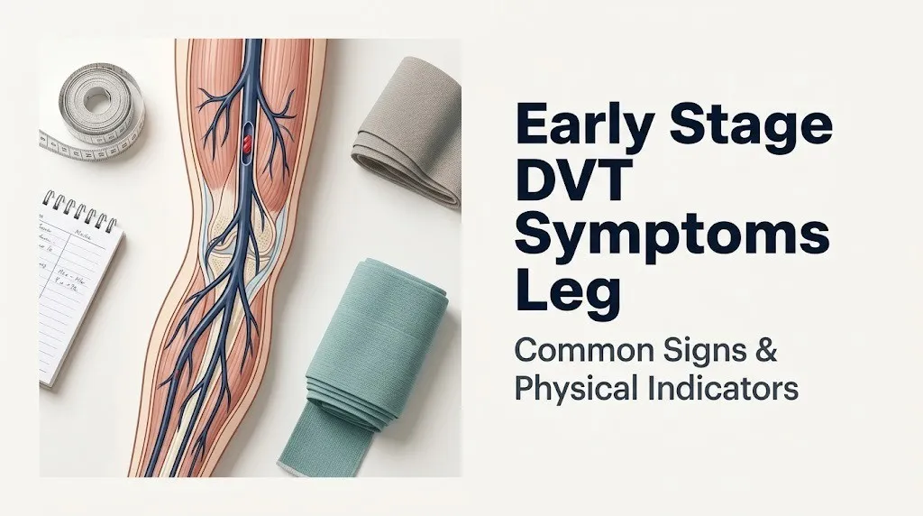
When any combination of these three conditions is present, the extrinsic coagulation cascade activates through tissue factor (Factor III) expressed on damaged endothelial cells. This cascade triggers Factor VIIa, then Factor Xa, which converts prothrombin to thrombin. Thrombin then cleaves fibrinogen into fibrin strands. Think of fibrin as the structural steel framework of a clot: it cross-links and traps red blood cells and platelets into an increasingly solid mass inside the vein.
Once that thrombus grows large enough to obstruct venous outflow, the physiology downstream from the clot changes immediately. Pressure rises in the venous segment below the obstruction, fluid shifts out of the vein and into surrounding tissue, and the tissue surrounding the clot becomes inflamed.
Key facts about DVT location:
- Approximately 90% of lower extremity DVTs originate in the calf veins
- Roughly 20 to 25% of untreated calf DVTs propagate proximally into the popliteal or femoral vein
- Proximal DVTs carry a substantially higher risk of pulmonary embolism than isolated distal calf DVTs
- The left leg is affected more frequently than the right, in part due to anatomical compression of the left common iliac vein by the right common iliac artery (a configuration called May-Thurner syndrome anatomy)
Early Stage DVT Symptoms Leg: The Core Clinical Picture
Early stage DVT symptoms in the leg are frequently subtle, often unilateral, and can be absent entirely in 30 to 50% of confirmed cases. The most common early symptoms are aching or cramping pain in the calf or thigh, mild swelling of the ankle or lower leg on the affected side, and a low-grade feeling of heaviness or tightness that tends to worsen with standing or walking.
What makes early DVT difficult to recognize is that none of these symptoms are specific to the condition on their own. A pulled calf muscle feels similar. So does moderate dehydration combined with prolonged sitting. The clinical red flags that distinguish early DVT from those causes are one-sidedness, a gradual and persistent worsening rather than improvement over 24 to 48 hours, and onset in the context of a known risk factor such as recent surgery, immobility, or hormone use.
The Centers for Disease Control and Prevention identifies the following as the primary recognized symptoms of DVT in the leg:
| Symptom | Typical Early Stage Appearance | Physiological Cause |
|---|---|---|
| Calf or thigh pain | Dull ache or cramping, may worsen with dorsiflexion | Prostaglandin E2 and bradykinin sensitizing C-fiber nociceptors near the thrombus |
| Leg swelling | Mild to moderate pitting edema, often unilateral | Venous outflow obstruction raising hydrostatic pressure, driving interstitial fluid accumulation via Starling forces |
| Skin warmth | Increased warmth to touch over affected area | Local inflammatory response including histamine and IL-6 release increasing local blood flow |
| Skin redness or discoloration | Mild erythema or blueish discoloration (cyanosis) | Venous stasis causing deoxygenated blood to pool in superficial vessels |
| Heaviness or fatigue in the leg | Persistent sense of leg being heavier than usual | Increased interstitial fluid volume and edema increasing limb mass and tension on surrounding fascial compartments |
One underappreciated early feature: the pain is often described as a tension or pressure rather than the sharp stabbing quality many people associate with a blood clot. That cramping, pressure-like quality in the calf after a long plane ride is often dismissed as stiffness. When it persists beyond 24 hours and is one-sided, it should not be dismissed.
What Does a Blood Clot in the Leg Feel Like
A blood clot in the leg typically produces a deep, persistent aching sensation located in the calf, behind the knee, or along the inner thigh, depending on where the thrombus is located within the deep venous system. Most people describe it as a cramp that will not go away, or a pulled muscle that is not getting better.
The quality of the sensation is important. Unlike a true muscle strain from overexertion, DVT pain does not typically sharpen immediately with physical activity or improve dramatically with rest and ice. It tends to be constant at a low level and worsens when the leg is in a dependent position (hanging down) rather than when elevated. That positional behavior reflects the underlying venous pressure dynamics: gravity increases venous hydrostatic pressure in the dependent leg, worsening both edema and pain.
Tenderness to firm touch along the inner calf or the popliteal fossa (the hollow behind the knee) is a frequently reported early sign. Some patients describe the sensation as a “bruise-like” ache in tissue that looks completely normal on the surface. That disconnect between the severity of the internal symptom and the lack of visible surface change is characteristic of early DVT, when the thrombus is not yet large enough to produce significant external swelling or skin color change.
How to describe your leg symptoms accurately to a healthcare provider:
- Note which leg is affected and whether the symptoms are strictly one-sided or present in both legs
- Record when the symptoms started and whether they have been getting worse, staying the same, or improving over the past 24 to 72 hours
- Note any associated activities, positions, or movements that make the sensation worse or better
- Recall any recent events in the past 4 to 8 weeks: surgery, hospitalization, injury, long travel, bed rest, change in medications including hormonal contraceptives
- Note whether any other symptoms have appeared alongside the leg symptoms: shortness of breath, chest pain, heart racing, or coughing up blood
- Measure and compare the circumference of both calves at the same level if possible, as a difference of more than 3 cm is a clinically noted asymmetry
Key Takeaway: Early stage DVT pain in the leg feels like a persistent, dull, cramping ache that does not improve with rest the way a muscle strain typically would, and it almost always affects only one leg at a time.
DVT Leg Swelling: Why It Happens and What It Looks Like
DVT-related leg swelling occurs because a thrombus blocking venous outflow forces fluid out of the vascular compartment and into the surrounding interstitial tissue through a mechanism governed by Starling forces: the balance between hydrostatic pressure pushing fluid out of vessels and oncotic pressure drawing fluid back in.
When the femoral or popliteal vein is blocked, venous pressure rises in everything downstream from the clot. That rising hydrostatic pressure overwhelms the normal oncotic gradient and pushes fluid into the tissue of the calf, ankle, and foot. The result is pitting edema: swelling that retains the indentation of finger pressure for several seconds before slowly refilling.
In early-stage DVT, the swelling may be subtle enough that the only way to detect it is by measuring both legs at the same point. A calf circumference difference of 3 centimeters or more between the affected and unaffected leg is a criterion recognized in the Wells DVT clinical prediction score, the validated clinical tool used by healthcare providers to stratify DVT probability before diagnostic testing.
The swelling in DVT has specific characteristics that help distinguish it from other causes:
- It is typically unilateral, affecting only the leg where the clot is present
- It is pitting in quality (leaves a temporary dimple when pressed)
- It tends to worsen throughout the day as the patient stands or walks, reflecting worsening venous hydrostatic pressure
- It improves somewhat with leg elevation, but does not fully resolve with elevation the way dependent edema from heart failure or prolonged sitting might
- In pregnancy-associated DVT, the swelling is often more pronounced and may extend higher into the thigh due to the elevated clot burden affecting iliac vein segments
Women taking oral contraceptives may notice leg swelling they attribute to the medication itself, as both estrogen-containing contraceptives and early DVT can produce mild leg edema. Any swelling that is unilateral, persistent, or associated with calf pain warrants same-day medical evaluation rather than self-monitoring.
DVT Leg Pain: How to Distinguish It from Muscle Pain or Cramping
DVT leg pain and muscle cramp pain share enough surface characteristics that the distinction matters enormously in the clinical decision about whether to get evaluated today or wait. The key distinction is mechanism: muscle pain originates from myofascial tissue damage, while DVT pain originates from venous obstruction and the inflammatory response to a thrombus.
Muscle cramp or strain pain typically has a clear precipitating event (overexertion, sudden movement, dehydration, electrolyte imbalance), peaks within the first 24 to 48 hours, and begins improving with gentle stretching, heat, and rest. It responds to over-the-counter anti-inflammatory medications. The affected area is tender at the muscle belly, not along the course of a deep vein.
DVT pain has no necessary preceding injury, peaks and holds rather than improving over 48 hours, and does not meaningfully respond to rest, stretch, or anti-inflammatory medication. Tenderness in DVT follows the path of the deep venous system, particularly along the medial calf (where the posterior tibial and peroneal veins run), the popliteal fossa, or the medial thigh along the course of the femoral vein. One older clinical sign, called Homans sign (pain in the calf with passive dorsiflexion of the foot), was historically used but is now considered unreliable: it has both low sensitivity and low specificity for DVT and should not be used to confirm or rule out the diagnosis.
| Feature | DVT Pain | Muscle Strain Pain |
|---|---|---|
| Onset | Gradual, no clear precipitating event required | Often after exertion, sudden movement, or dehydration |
| Quality | Dull, aching, cramping, pressure-like | Sharp initially, may transition to aching soreness |
| Behavior over 48 hours | Persists or worsens | Typically begins improving |
| Response to rest | Minimal improvement | Significant improvement |
| Tenderness location | Along venous anatomy (medial calf, popliteal fossa, inner thigh) | At muscle belly, triggered by palpation or contraction |
| Associated swelling | Frequently unilateral, pitting | Minimal, if any, localized to muscle belly |
| Associated warmth | Yes, diffuse over involved limb | Localized to the area of muscle injury |
Older adults and patients with pre-existing peripheral arterial disease may experience a mixed picture where DVT-related pain overlaps with ischemic leg pain, making clinical assessment particularly challenging. In these cases, professional evaluation is the only appropriate path.
DVT Redness, Warmth, and Skin Changes in the Affected Leg
Skin redness and warmth over the affected leg reflect the local inflammatory response that accompanies thrombus formation in the deep venous system. When a clot activates the vascular endothelium, inflammatory cytokines including interleukin-6 (IL-6) and histamine are released. These mediators cause local vasodilation in the superficial venous and capillary network overlying the deep veins, increasing blood flow to the area, which raises local skin temperature and produces visible erythema (redness).
The warmth in DVT is typically diffuse rather than sharply localized. In early stages, it may be noticeable only by comparing the temperature of both legs, and it tends to affect the calf most prominently in distal DVT or the thigh in proximal DVT. This diffuse warmth differs from the intense, sharply demarcated warmth of cellulitis, a bacterial skin infection that is frequently confused with DVT in clinical practice.
Skin discoloration in DVT varies by stage and severity. In early stages, mild erythema (pink to red flush) may appear over the inner calf. As venous obstruction progresses and deoxygenated blood pools in the superficial venous network, the skin may take on a bluish or mottled (patchy) discoloration called cyanosis or livedo reticularis. This is more common with extensive proximal DVT than with isolated distal calf DVT.
According to a review published in Thrombosis and Haemostasis, skin color change and warmth are present in only 40 to 50% of confirmed DVT cases, meaning their absence does not rule out the diagnosis. Physicians use skin findings as supporting evidence alongside other clinical signs, not as a standalone indicator.
Quick Tip:
- Compare both legs by touch in a warm room: place the back of your hand against the inner calf of each leg and note any asymmetric warmth
- Skin that is both warm and tender along the calf, especially with any associated swelling, is a combination that warrants same-day medical evaluation
- Dark skin tones may show DVT-associated discoloration as deepening of baseline pigmentation rather than visible redness, making visual assessment unreliable without palpation
Key Takeaway: Skin warmth and redness in DVT are caused by an inflammatory cytokine response from the activated venous endothelium, but these signs are absent in roughly half of confirmed cases, so their absence does not mean the vein is clear.
Silent DVT: When a Blood Clot Has No Symptoms at All
Silent DVT refers to the clinical reality that a substantial proportion of confirmed deep vein thromboses in the leg produce no recognizable symptoms at the time of formation. Research published in prospective imaging studies has consistently demonstrated that between 30 and 50% of DVTs are asymptomatic at their time of detection, meaning the person had no pain, swelling, warmth, or skin change they were aware of.
This happens for a specific physiological reason. A thrombus forming in a small-caliber distal calf vein may not obstruct enough venous outflow to raise hydrostatic pressure to the threshold that drives significant edema formation. The inflammatory mediator release in these small thrombi may also be insufficient to sensitize local nociceptors to the point of producing pain. The clot can remain entirely contained within the distal calf venous network, without symptoms, while either slowly resolving through endogenous fibrinolysis or propagating proximally into larger venous segments.
The danger of silent DVT is not the absence of symptoms: it is that the first symptom a person experiences may be a pulmonary embolism rather than leg pain. A thrombus that propagates into the popliteal, femoral, or iliac vein becomes large enough and floats freely enough in the venous lumen to fragment and embolize to the pulmonary arteries. When this happens, the lung symptom (sudden shortness of breath, pleuritic chest pain, rapid heart rate) is often the presenting complaint, with the leg DVT identified only on imaging done as part of the PE workup.
For this reason, the American Society of Hematology and the CHEST antithrombotic guidelines recommend routine DVT screening imaging (typically compression duplex ultrasonography) in certain high-risk populations after major surgery, even in the absence of leg symptoms.
People with Factor V Leiden mutation, prothrombin G20210A variant, antiphospholipid syndrome, active malignancy, or a prior episode of DVT face a higher probability that any given thrombus will be clinically silent, making periodic monitoring discussions with a vascular medicine specialist or hematologist clinically appropriate.
Proximal DVT vs Distal DVT: How Symptoms Differ by Clot Location
The location of a DVT within the deep venous system of the leg directly determines the severity, pattern, and urgency of early symptoms. Clinicians classify lower extremity DVT as either distal (involving calf veins below the knee) or proximal (involving the popliteal, femoral, or iliac veins above or at the level of the knee).
Distal DVT (confined to the tibial and peroneal veins and their tributaries) tends to produce more localized symptoms. The pain is typically centered in the calf belly rather than the thigh, swelling is modest if present, and the risk of immediate pulmonary embolism is substantially lower. The CHEST antithrombotic guidelines note that isolated distal DVT carries a lower short-term PE risk than proximal DVT, though they note the risk is not zero, particularly in patients with cancer or inherited thrombophilia.
Proximal DVT (involving the popliteal vein, the femoral vein in the medial thigh, or the iliac vein in the pelvis) typically produces more pronounced symptoms because a larger venous segment is blocked, preventing outflow from a greater volume of the lower extremity. Swelling in proximal DVT often involves the entire leg from the foot to the groin rather than just the calf and ankle. Thigh pain or groin aching is more common. Iliac vein DVT, in particular, can present primarily as groin or pelvic discomfort with leg swelling rather than calf-centered symptoms, which contributes to delays in recognition.
| DVT Location | Typical Pain Location | Swelling Extent | PE Risk (short-term) |
|---|---|---|---|
| Distal (tibial, peroneal veins) | Calf belly, behind ankle | Calf and ankle only | Lower (but not zero) |
| Popliteal vein | Behind knee, calf | Calf to below knee | Moderate to high |
| Femoral vein | Inner thigh, medial knee | Entire lower leg and thigh | High |
| Iliac vein | Groin, hip, lower abdomen | Entire leg, may include buttock | High |
| Bilateral iliac or IVC involvement | Both legs, lower back | Bilateral leg swelling | Very high |
Patients who have had prior DVT in a specific venous segment may develop chronic venous insufficiency in that segment, which can mask the early swelling and pain of a new DVT by blending it into a pre-existing baseline of leg discomfort and edema. This makes recurrent DVT recognition particularly difficult.
Key Takeaway: Proximal DVT above the knee typically causes more extensive swelling and carries significantly higher pulmonary embolism risk than distal calf DVT, and iliac vein clots can present primarily as groin or hip pain rather than classic calf symptoms.
Early Stage Blood Clot Symptoms in Leg vs Other Conditions
Distinguishing early stage blood clot symptoms in the leg from other common conditions is one of the most clinically challenging aspects of DVT recognition, because several high-frequency conditions produce almost identical surface symptoms. The most important competing diagnoses are: muscle strain or tear, cellulitis (bacterial skin infection), superficial thrombophlebitis (clot in a surface vein rather than a deep vein), chronic venous insufficiency, and ruptured Baker’s cyst behind the knee.
Cellulitis is a bacterial infection of the subcutaneous tissue that produces warmth, redness, tenderness, and swelling in a limb segment. It differs from DVT in that the redness is sharply demarcated (has a clear border), the skin surface may be visibly inflamed with streaking (lymphangitic spread), and the patient typically has a fever and elevated inflammatory markers. However, cellulitis and DVT can and do coexist, as venous stasis and skin barrier breakdown sometimes occur together.
Superficial thrombophlebitis is a clot in the greater or lesser saphenous vein or its tributaries, visible just under the skin surface. It presents as a palpable, tender cord under the skin, often with overlying redness. Unlike DVT, it is palpable at the skin surface and the vein can often be felt as a firm, inflamed rope. However, when superficial thrombophlebitis extends into the deep venous system through the saphenofemoral junction, it becomes DVT.
A ruptured Baker’s cyst can produce sudden calf pain and swelling that precisely mimics acute DVT. The popliteal cyst (Baker’s cyst) forms in the popliteal fossa behind the knee from joint fluid accumulation in conditions like osteoarthritis or rheumatoid arthritis. When it ruptures, synovial fluid tracks down into the calf, causing rapid pain, warmth, and swelling. Compression duplex ultrasonography can distinguish the two because a ruptured Baker’s cyst shows fluid tracking between muscle planes rather than thrombus within a deep vein.
None of these competing diagnoses can be reliably distinguished from DVT by symptoms alone. Compression duplex ultrasonography is the only clinically appropriate method for ruling in or ruling out deep vein thrombosis. A healthcare provider who suspects DVT should not manage the symptom complex as a muscle strain or cellulitis without imaging confirmation.
DVT Symptoms in Women, Pregnancy, and Hormonal Risk Groups
Women represent a particularly important population in DVT symptom recognition because several common hormonal exposures and biological states substantially elevate DVT risk while also altering the clinical presentation pattern. The National Heart Lung and Blood Institute reports that venous thromboembolism risk is 3 to 5 times higher during pregnancy and the postpartum period compared to non-pregnant women of the same age.
Estrogen-containing medications including combined oral contraceptives, the contraceptive patch, the vaginal ring, and postmenopausal hormone replacement therapy all elevate DVT risk by increasing hepatic production of several coagulation factors (Factors II, VII, VIII, X, and fibrinogen) while reducing levels of natural anticoagulants including protein S. The resulting hypercoagulable state directly addresses the third arm of Virchow’s triad.
In pregnancy, the specific anatomical pattern of DVT has a left-leg predominance because the gravid uterus compresses the left common iliac vein against the spine, reducing venous return from the left lower extremity. This creates both venous stasis and mild venous compression injury in the left iliac system, two of Virchow’s three factors. DVT in pregnancy often presents higher in the venous system (iliac and femoral veins rather than calf veins), which means the initial symptoms may be groin pressure, hip pain, or diffuse leg swelling rather than the classic calf cramp pattern.
Important considerations specific to this group:
- Leg swelling in pregnancy is extremely common and expected, making DVT-related swelling harder to identify; unilateral swelling that is dramatically worse in one leg than the other, or swelling that increases rapidly over hours rather than gradually over weeks, warrants same-day evaluation by an obstetrician-gynecologist or emergency medicine physician
- Postpartum women (in the first 6 weeks after delivery) carry the highest absolute VTE risk of any period in reproductive life and should treat any new leg symptoms in this period as DVT until imaging proves otherwise
- Women with an inherited thrombophilia who use combined hormonal contraceptives have a synergistically elevated DVT risk: Factor V Leiden carriers using estrogen-containing contraceptives have an approximately 35-fold elevated DVT risk compared to non-carrier, non-user controls, according to data published in the New England Journal of Medicine
DVT Symptoms in Older Adults and Post-Surgical Patients
Older adults and patients recovering from surgery represent two of the highest-risk groups for DVT, and both groups are more likely to have DVT symptoms that are masked, misattributed, or blunted by pre-existing conditions. The CDC reports that DVT incidence roughly doubles with each decade of life after age 40.
In older adults, the baseline state of the venous system differs from younger adults in ways that directly affect symptom recognition. Venous valve function declines with age, producing mild chronic venous insufficiency that causes low-level baseline leg swelling, heaviness, and skin changes in many people over 65. When an acute DVT develops on top of this baseline, the incremental swelling may not be dramatic enough to trigger recognition. An older adult who notices their usual “heavy legs” are more swollen than normal, or that their ankle edema is worse on one side and associated with new calf aching, should not dismiss this as just the usual venous insufficiency without evaluation.
Post-surgical DVT deserves particular emphasis because it is common, predictable, and often underestimated by patients in recovery. Orthopedic surgery (particularly total knee replacement, total hip replacement, and hip fracture surgery) carries the highest DVT risk of any surgical category. Abdominal and pelvic surgery, particularly for cancer, also carries very high post-operative DVT risk. According to CHEST antithrombotic guidelines, without pharmacological prophylaxis, the rate of DVT after major orthopedic surgery exceeds 40 to 60% detected by venographic imaging.
Post-surgical patients face a compound identification problem. The operated leg is already painful, swollen, and warm from surgical trauma. A DVT developing in the same extremity is not distinguishable from surgical inflammation by external examination alone. Any calf or thigh swelling or pain in a post-surgical patient that is disproportionate to what was present the day before, or that is worsening rather than improving on an expected post-operative recovery trajectory, warrants imaging.
Older adults or post-surgical patients taking anticoagulants for other conditions (atrial fibrillation, for example) may develop DVT even on therapeutic anticoagulation, particularly if their dosing is at the lower end of the therapeutic range or if they have cancer-associated hypercoagulability that exceeds standard anticoagulant coverage.
Key Takeaway: Older adults with pre-existing venous insufficiency and post-surgical patients in the operated limb are the two groups most likely to have DVT symptoms masked by their baseline condition, making imaging the only reliable way to clarify the diagnosis.
DVT Risk Factors That Make Early Recognition Harder
Recognizing early stage DVT symptoms is consistently harder in people who have multiple concurrent DVT risk factors, because those same factors often produce their own leg symptoms that overlap with DVT presentation. Understanding your specific risk factor profile is clinically useful because it determines the pre-test probability that a healthcare provider will assign to your symptom cluster before ordering diagnostic imaging.
The Wells DVT score formally quantifies this pre-test probability. It assigns points for: active cancer, paralysis or recent plaster cast immobilizing the leg, recent bed rest exceeding 3 days or major surgery within 12 weeks, localized tenderness along the deep venous system, entire leg swelling, calf swelling more than 3 cm larger than the asymptomatic side, pitting edema confined to the symptomatic leg, dilated collateral surface veins, and prior confirmed DVT. A score of 2 or higher places a patient in the high-probability category warranting immediate imaging.
DVT risk factors by category:
Acquired risk factors:
- Immobility exceeding 4 to 8 hours (including long-haul flights, bed rest, hospitalization)
- Recent surgery, particularly orthopedic, abdominal, or pelvic procedures
- Active malignancy (especially solid tumors producing procoagulant tissue factor)
- Obesity (BMI above 30: associated with impaired venous return and a pro-inflammatory state)
- Prior DVT or pulmonary embolism
- Estrogen-containing medications (oral contraceptives, hormone replacement therapy)
- Pregnancy and the 6 weeks postpartum
- Trauma or fracture of the lower extremity
- Central venous catheter placement
Inherited (congenital) risk factors:
- Factor V Leiden mutation (most common inherited thrombophilia in populations of European descent, present in approximately 5% of the general population)
- Prothrombin G20210A gene variant (second most common inherited thrombophilia)
- Protein C deficiency
- Protein S deficiency
- Antithrombin III deficiency
- Antiphospholipid syndrome (an acquired autoimmune condition that mimics inherited thrombophilia)
People with two or more concurrent risk factors face a synergistically elevated DVT probability, not merely an additive one. A 35-year-old woman who is 6 weeks post-cesarean section, has Factor V Leiden, and has been relatively immobile during postpartum recovery carries a DVT risk profile that warrants a much lower clinical threshold for imaging than any one of those factors alone.
The DVT-Pulmonary Embolism Connection: When a Leg Clot Becomes a Lung Emergency
A DVT in the deep venous system of the leg becomes a pulmonary embolism when part or all of the thrombus breaks free, travels through the inferior vena cava and right heart chambers, and lodges in a pulmonary artery or one of its branches, obstructing blood flow through the lungs. This progression from leg clot to lung emergency can happen within hours of DVT formation, or it can occur days to weeks later.
The risk of pulmonary embolism from an untreated proximal DVT (above the knee) is substantial. Research published in the New England Journal of Medicine has demonstrated that clinically apparent PE occurs in approximately 40 to 50% of untreated proximal DVT cases. That figure underscores why a suspected DVT is not a condition to monitor at home with a plan to call a doctor “if it gets worse.”
The physiological pathway from DVT to PE involves thrombus fragmentation. The deep venous thrombus, attached to the vein wall at its tail and floating freely at its proximal end (the “red clot” or propagating head of the thrombus), is vulnerable to fragmentation with any sharp increase in venous blood flow velocity: sudden standing after prolonged sitting, Valsalva maneuvers, or physical exertion. Once a fragment embolizes to the pulmonary vasculature, it obstructs blood flow and forces the right ventricle to pump against higher pulmonary vascular resistance, a condition called acute cor pulmonale in severe cases.
Symptoms that indicate a DVT has progressed to pulmonary embolism include:
- Sudden onset of shortness of breath at rest, not explained by exertion
- Sharp, stabbing chest pain that worsens with inhalation (pleuritic chest pain), caused by infarction of the pleural surface near the embolus
- Rapid heart rate (tachycardia) above 100 beats per minute
- Lightheadedness, near-fainting, or fainting
- Coughing up blood (hemoptysis)
- A sense of impending doom (this is a real and recognized clinical symptom of massive PE)
- Oxygen saturation below 95% if measured by pulse oximeter
Call 911 or go to the nearest emergency room immediately if any of these symptoms appear alongside or after leg symptoms consistent with DVT. Time is the critical variable in PE management.
Emergency Symptoms: When to Call 911 or Go to the ER
Certain symptoms associated with DVT and its complications require immediate emergency evaluation. Do not wait to see if these resolve on their own.
Call 911 or go to the nearest emergency room immediately if you experience:
- Sudden shortness of breath at rest, particularly if it comes on without an obvious explanation like exertion or respiratory illness
- Chest pain that is sharp, stabbing, or worsens when you breathe in deeply
- Rapid heart rate, heart pounding, or a sense that your heart is racing without physical exertion
- Lightheadedness, dizziness, or fainting
- Coughing up blood (hemoptysis), even a small amount
- Bluish discoloration of the lips or fingertips (cyanosis), indicating low oxygen levels
- Sudden leg swelling that is severe, spreading rapidly up the entire leg, associated with skin that is turning dusky blue or showing signs of tissue compromise
- Any combination of leg pain or swelling with chest symptoms: this combination makes pulmonary embolism highly probable until imaging proves otherwise
These presentations can indicate a pulmonary embolism or, in the most severe cases, massive PE leading to cardiovascular collapse, and they require emergency medical assessment, not an urgent care appointment or a phone call to a nurse line.
Key Takeaway: The progression from DVT to pulmonary embolism can happen within hours, and the first symptom of PE is often sudden shortness of breath or chest pain rather than worsening leg symptoms, which is why any respiratory symptom alongside a swollen painful leg is a 911 call.
How DVT Is Diagnosed: D-Dimer, Ultrasound, and the Wells Score
DVT diagnosis in clinical practice follows a structured two-step process: first, stratify the pre-test probability using a validated clinical prediction tool, then apply the appropriate imaging or laboratory test based on that probability assessment.
The Wells DVT clinical prediction score is the validated tool used most widely in this first step. As described in the risk factors section, it assigns points for specific clinical features and classifies patients as low probability (score 0 or below), moderate probability (score 1 to 2), or high probability (score 3 or above). A high-probability score warrants direct referral for imaging without waiting for laboratory results.
The D-dimer blood test measures fibrin degradation products in the bloodstream, specifically the D-dimer fragment released when plasmin dissolves fibrin cross-links. Because every DVT involves active fibrin formation and some degree of simultaneous endogenous fibrinolysis, D-dimer is elevated in virtually all active DVTs. Its strength is its high negative predictive value: a normal D-dimer in a low-probability patient essentially rules out clinically relevant DVT. Its weakness is low specificity: D-dimer is elevated by many other conditions including pregnancy, infection, inflammation, recent surgery, and malignancy, making a positive result uninterpretable without imaging.
Compression duplex ultrasonography is the first-line imaging test for suspected lower extremity DVT. It combines two-dimensional B-mode ultrasound imaging of the deep venous anatomy with Doppler flow measurement to assess venous compressibility and blood flow velocity. A vein containing thrombus does not compress fully under probe pressure, which is the definitive imaging sign of DVT. The test is non-invasive, does not involve radiation, and has a sensitivity exceeding 95% for proximal DVT according to data published in JAMA Internal Medicine. Its sensitivity for isolated distal calf DVT is somewhat lower (around 75 to 80%), which is why repeat ultrasound at 5 to 7 days may be recommended in high-probability patients with an initially negative distal study.
Computed tomography pulmonary angiography (CTPA) is the imaging gold standard for pulmonary embolism diagnosis. If a patient presents with both DVT symptoms and PE symptoms, CTPA is the priority imaging study because it directly evaluates the pulmonary vasculature and establishes the diagnosis that determines the most urgent treatment decisions.
What to bring to your DVT evaluation appointment:
- A written timeline of when your leg symptoms started and how they have changed
- A list of all medications, supplements, and hormonal contraceptives including start dates
- A list of any surgeries, hospitalizations, or immobility periods within the past 3 months
- Information about any personal or family history of blood clots, DVT, or PE
- Results of any prior ultrasound or vascular imaging if available
Post-Thrombotic Syndrome: Long-Term Leg Symptoms After DVT
Post-thrombotic syndrome (PTS) is a chronic venous condition that develops in a subset of patients after a DVT episode, producing persistent leg symptoms that continue after the acute clot has resolved or been treated. According to research published in Thrombosis and Haemostasis, PTS develops in approximately 20 to 50% of patients who have had a proximal DVT, typically appearing within 2 years of the initial event.
The mechanism of PTS involves two pathological changes to the venous system caused by the original DVT. First, the thrombus and the inflammatory response it triggers damage the delicate one-way venous valves within the affected vein. When these valves are damaged or destroyed, blood refluxes backward with gravity in the standing position, a condition called venous reflux. Second, in some cases the thrombus does not fully dissolve and the vein remains partially obstructed, reducing the total venous outflow capacity of the limb permanently.
Both of these changes produce chronic venous hypertension: persistently elevated venous pressure in the lower leg that forces fluid, inflammatory cells, and plasma proteins into the interstitial tissue. Over months and years, this produces a progressive clinical picture: chronic leg swelling that worsens through the day, a persistent aching or heaviness in the affected leg, skin thickening and pigmentation change (brownish or reddish discoloration called lipodermatosclerosis), and in severe cases, venous stasis ulcers near the medial ankle.
PTS symptoms can be difficult to distinguish from a new DVT episode, which is clinically significant because the treatment differs. New DVT requires anticoagulation. PTS is managed with compression therapy, graded exercise, and in some cases endovascular procedures to relieve venous obstruction. Any patient with known prior DVT who develops new or acutely worsening leg symptoms should be re-evaluated with imaging rather than assumed to have PTS flare.
Patients with prior DVT should discuss PTS risk and long-term compression therapy options with a vascular medicine specialist or their managing hematologist, as there is evidence that consistent use of graduated compression stockings in the first 2 years after proximal DVT reduces PTS severity.
When to Go to the ER for Blood Clot Symptoms in the Leg
The decision threshold for emergency evaluation of suspected leg DVT is straightforward once the clinical picture is understood: any symptom combination consistent with DVT in the leg that is accompanied by respiratory symptoms, cardiovascular instability, or rapidly worsening limb changes is an emergency. The question of when to go to the ER versus when to call your primary care physician on the same day should be answered by one factor: whether you have any symptoms beyond the leg.
Go to the emergency room or call 911 immediately if your leg symptoms are accompanied by shortness of breath, chest pain, rapid or irregular heart rate, coughing up blood, lightheadedness, or fainting. This symptom combination warrants emergency evaluation for pulmonary embolism.
Seek same-day medical evaluation from a primary care physician or urgent care clinic if you have isolated leg symptoms (swelling, pain, warmth, redness limited to the leg) without any respiratory or cardiovascular accompaniment, particularly if those symptoms have been present for fewer than 72 hours and have not been improving.
Seek evaluation from a vascular medicine specialist or hematologist if you have had a prior confirmed DVT, have a known inherited thrombophilia, or have been told you are at high lifetime DVT risk, and you develop new symptoms consistent with DVT even if they appear mild.
| Symptom Picture | Urgency Level | Action |
|---|---|---|
| Leg swelling, pain, warmth only, no respiratory symptoms, mild to moderate | Same-day clinic | Primary care physician or urgent care; ask for DVT evaluation and imaging referral |
| Leg swelling plus any shortness of breath or chest pain | Emergency | Call 911 or go to the nearest ER immediately |
| Severe rapid leg swelling, skin turning dark or dusky | Emergency | Call 911 or go to the nearest ER immediately |
| Fainting, racing heart, coughing blood, low oxygen | Life-threatening emergency | Call 911; do not drive yourself |
| Mild leg aching only, no swelling, no warmth, high-risk person | Same-day to next-day | Primary care physician; mention risk factors explicitly when calling |
Do not attempt to self-manage suspected DVT with compression wrapping, elevation, or anti-inflammatory medications as a substitute for evaluation. Compression applied to an unstable thrombus carries a theoretical risk of fragment displacement, and anti-inflammatory pain relief may mask the symptom progression that would otherwise prompt timely medical evaluation.
Key Takeaway: Isolated leg symptoms without any shortness of breath or chest pain warrant same-day medical evaluation; any respiratory symptom appearing alongside leg DVT signs warrants calling 911 or going directly to the emergency room, not urgent care.
Frequently Asked Questions About Early Stage DVT Symptoms in the Leg
What are the first signs of a blood clot in the leg?
The first signs of a blood clot in the leg are typically a persistent dull aching or cramping in the calf that does not improve with rest, mild unilateral ankle or lower leg swelling, and subtle warmth over the affected area.
These signs often appear gradually over 24 to 48 hours and may be initially dismissed as a muscle strain, particularly when they follow immobility, a long flight, or surgery.
Any combination of unilateral leg pain, swelling, and warmth that persists beyond 48 hours without an obvious improving trajectory warrants evaluation by a primary care physician the same day.
Can a blood clot in the leg go away on its own?
Small distal DVTs in the calf veins do occasionally resolve through the body’s own fibrinolytic system without treatment, but this cannot be predicted or relied upon without professional monitoring.
Proximal DVTs in the femoral, popliteal, or iliac veins do not reliably resolve without anticoagulation and carry substantial pulmonary embolism risk if left untreated.
The American Society of Hematology recommends anticoagulation treatment for most confirmed DVTs; the decision about whether to treat a small isolated distal DVT with anticoagulation or serial imaging is one that requires individualized assessment by a physician.
How do I know if my leg pain is a blood clot or a muscle cramp?
DVT pain does not have a clear precipitating event, does not improve meaningfully with rest or stretching within 48 hours, and tends to be located along the path of the deep venous system rather than at the muscle belly.
Muscle cramp or strain pain typically has a clear cause (overexertion, sudden movement, dehydration), peaks quickly, and begins improving within 24 to 48 hours with rest, gentle stretching, and hydration.
When the pain is one-sided, persistent, and associated with any leg swelling or warmth, evaluation with compression duplex ultrasonography is the only way to rule out DVT.
What does early stage DVT swelling look like?
Early stage DVT swelling is typically mild to moderate pitting edema of the calf and ankle on the affected side only, often noticeable as a visible asymmetry between both legs when standing.
The swelling tends to worsen throughout the day with standing or walking and improves somewhat when the leg is elevated, but it does not fully resolve with elevation the way simple dependent edema from prolonged sitting does.
A calf circumference difference of 3 centimeters or more between both legs at the same measurement point is a clinically recognized criterion in the Wells DVT scoring system.
Can you have DVT without any symptoms?
Yes, between 30 and 50% of confirmed DVT cases are entirely asymptomatic at the time the clot is present, particularly when the thrombus is limited to small distal calf veins that do not obstruct enough venous outflow to produce recognizable edema or pain.
Silent DVT is clinically significant because the first symptom a person experiences may be a pulmonary embolism if the clot propagates proximally and embolizes before any leg symptoms develop.
People with known high DVT risk factors including recent major surgery, active malignancy, or inherited thrombophilia may warrant prophylactic anticoagulation or surveillance imaging even without leg symptoms, a decision to discuss with a vascular medicine specialist or hematologist.
How quickly does DVT need to be treated?
DVT requires treatment as soon as the diagnosis is confirmed, and in high-probability cases most clinical guidelines support initiating anticoagulation before imaging confirmation is complete if imaging will be significantly delayed.
The American Society of Hematology recommends starting anticoagulation within hours to a day of diagnosis for proximal DVT to reduce the risk of pulmonary embolism and thrombus propagation.
Delay in treatment is associated with higher rates of PE, larger ultimate thrombus burden, and greater risk of post-thrombotic syndrome developing after resolution.
What You Now Know and What to Do With It
Early stage DVT symptoms in the leg are subtle, variable, and frequently mistaken for muscle injury or routine swelling. The mechanism behind them is specific: a thrombus obstructing venous outflow raises hydrostatic pressure and drives inflammatory mediators that produce the characteristic combination of one-sided swelling, persistent aching, warmth, and skin change. Knowing these mechanisms helps you recognize the pattern even when each individual symptom seems unremarkable on its own.
The critical clinical action point is this: unilateral leg swelling or pain that persists beyond 48 hours, does not have a clear improving trajectory, and occurs in the context of any known DVT risk factor warrants same-day medical evaluation and imaging, not watchful waiting. Any respiratory symptom appearing alongside that leg picture means calling 911 is the correct decision.
If you have had a confirmed DVT in the past, carry an inherited thrombophilia, or are currently pregnant or postpartum, your threshold for seeking evaluation should be lower than average. A vascular medicine specialist or hematologist who knows your specific history is the right provider for ongoing DVT risk management. The leg symptom that feels like nothing today is the one that deserves a professional’s eyes before tomorrow.