MCTD Symptoms: What Mixed Connective Tissue Disease Feels Like
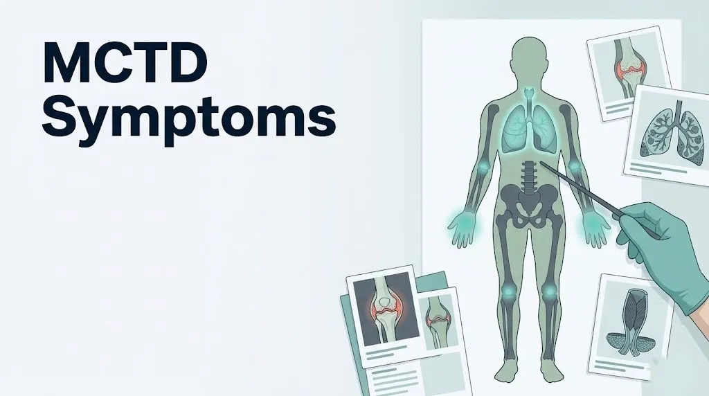
Mixed connective tissue disease (MCTD) symptoms span multiple organ systems simultaneously, including the joints, skin, muscles, lungs, esophagus, and cardiovascular system, because MCTD is a systemic autoimmune condition where the immune system attacks a specific nuclear protein complex across many tissue types. Understanding what this condition actually feels like, and which symptoms signal a progression that needs urgent attention, is the difference between managing MCTD well and missing a complication that changes the prognosis.
According to the National Institute of Arthritis and Musculoskeletal and Skin Diseases (NIAMS), MCTD is considered a distinct autoimmune overlap syndrome, meaning it carries features of lupus, scleroderma, polymyositis, and rheumatoid arthritis simultaneously or sequentially. Research published in the Journal of Rheumatology indicates that approximately 80% of MCTD patients are female, with peak onset occurring in the second through fourth decades of life.
This article covers every clinically recognized symptom category in MCTD, including the physiological mechanism driving each one, how symptoms evolve from early to advanced stages, which populations face the highest risk of severe complications, and exactly when certain symptoms require emergency evaluation rather than a routine appointment.
What Is MCTD (Mixed Connective Tissue Disease)?
Mixed connective tissue disease is a systemic autoimmune disorder defined by the presence of high-titer anti-U1 ribonucleoprotein (anti-U1 RNP) antibodies combined with clinical features overlapping at least two of the following conditions: systemic lupus erythematosus, systemic sclerosis, polymyositis, and rheumatoid arthritis.
The U1 ribonucleoprotein (U1 RNP) complex is a structure inside the cell nucleus that assists with pre-mRNA splicing, a process essential for protein production. In MCTD, the immune system misidentifies the U1 RNP complex as a foreign threat and produces IgG autoantibodies against it. These anti-U1 RNP antibodies circulate through the bloodstream, deposit in multiple tissues, and trigger chronic inflammatory injury across organ systems.
Think of the immune system in MCTD as a security alarm that has been miscalibrated. Instead of responding only to genuine threats, it fires continuously at a normal cellular component, and because that component is present in cells throughout the body, the inflammatory response spreads to joints, skin, muscles, blood vessels, lungs, and organs simultaneously.
The condition was first described by Gordon Sharp in 1972, and Sharp’s diagnostic criteria, now refined through subsequent work by Kasukawa and Alarcon-Segovia, remain the foundation of MCTD classification. MCTD carries the ICD-10 code M35.1.
What distinguishes MCTD from other autoimmune diseases is the specificity of the anti-U1 RNP antibody. Lupus patients may have anti-U1 RNP antibodies, but MCTD requires them at high titer as the dominant antibody finding, without the anti-double-stranded DNA (anti-dsDNA) or anti-Sm antibodies that more specifically define lupus. This distinction matters clinically because the symptom pattern and complication profile differ.
- MCTD has a relatively lower rate of kidney disease compared to lupus
- MCTD carries a higher rate of pulmonary arterial hypertension than lupus
- MCTD more commonly produces esophageal dysmotility than rheumatoid arthritis alone
- The overlapping nature means symptom presentation varies substantially between individuals
People with a first-degree relative with any autoimmune connective tissue disease carry a higher background risk, though MCTD itself does not follow a simple inheritance pattern.
MCTD Symptoms Overview
MCTD symptoms typically involve a combination of Raynaud’s phenomenon, puffy or swollen fingers, inflammatory arthritis, and constitutional fatigue in the early phase, with potential progression to lung, esophageal, cardiac, and other organ-system involvement over time.
No two people with MCTD present identically. The American College of Rheumatology recognizes MCTD’s variable presentation as one of its defining clinical challenges, particularly because early MCTD may be indistinguishable from early lupus, early scleroderma, or early inflammatory myopathy before the full symptom picture and laboratory findings clarify over months or years.
| Symptom Category | Approximate Frequency in MCTD | Clinical Significance |
|---|---|---|
| Raynaud’s phenomenon | 85 to 96% of patients | Early and persistent; indicates vascular involvement |
| Puffy or swollen fingers | 66 to 88% of patients | Often an early presenting feature |
| Arthritis or arthralgia | 60 to 90% of patients | May resemble rheumatoid arthritis |
| Muscle weakness or myositis | 40 to 72% of patients | Can be the dominant presenting feature |
| Esophageal dysmotility | 50 to 80% of patients | Often subclinical initially |
| Pulmonary involvement | 20 to 85% of patients (varies by study) | Most serious long-term complication |
| Skin changes | 30 to 70% of patients | Variable; includes malar rash, discoid lesions |
| Cardiac involvement | 10 to 30% of patients | Pericarditis most common; myocarditis less frequent |
| Renal disease | 10 to 25% of patients | Less common than in lupus |
| Neurological symptoms | 10 to 30% of patients | Trigeminal neuralgia characteristic |
Frequency data referenced from reviews published in Seminars in Arthritis and Rheumatism (2019) and Annals of the Rheumatic Diseases.
People who present initially with only one or two features and who lack the high-titer anti-U1 RNP antibody are often classified as having undifferentiated connective tissue disease (UCTD) rather than MCTD until the full clinical picture develops.
Raynaud’s Phenomenon in MCTD
Raynaud’s phenomenon is the most consistently reported symptom in MCTD, present in 85 to 96% of patients according to published clinical series, and it is frequently the first symptom to appear, often preceding other features by months to years.
Raynaud’s occurs when the small digital arteries and arterioles in the fingers and toes undergo pathological vasospasm in response to cold temperatures or emotional stress. In MCTD, this vasospasm is amplified by underlying vascular endothelial injury caused by the anti-U1 RNP autoimmune process. Normally, cold exposure causes modest vasoconstriction to preserve core body temperature. In MCTD, the damaged blood vessel lining responds with extreme, prolonged constriction.
The classic visible pattern involves three sequential color changes. Fingers turn white (pallor) as blood flow is abruptly cut off. They then turn blue or purple (cyanosis) as oxygen in the remaining blood is consumed. Finally, they turn red (rubor) as blood rushes back when the spasm resolves. Not every patient goes through all three phases. Some experience only pallor and cyanosis. The duration of each episode ranges from a few minutes to over an hour.
Beyond discomfort and numbness, severe or prolonged Raynaud’s in MCTD can progress to digital ischemia, where tissue damage occurs from sustained oxygen deprivation. In advanced cases, this can produce painful sores (digital ulcers) at the fingertips. A review published in Annals of the Rheumatic Diseases identified digital ulcers as a marker of more aggressive vascular disease in MCTD and scleroderma overlap presentations.
Cold avoidance, layered gloves, and hand warmers are appropriate home management for mild Raynaud’s. Persistent or severe episodes, digital ulcers, or any tissue color change lasting more than one hour requires evaluation by a board-certified rheumatologist, who may recommend calcium channel blockers such as nifedipine or vascular-directed therapy.
Raynaud’s severity in MCTD tends to be more pronounced than in primary Raynaud’s phenomenon (which occurs without autoimmune disease) and may be an early signal that warrants autoimmune workup, particularly an ANA and anti-U1 RNP antibody test.
Swollen Fingers and Hand Symptoms in MCTD
Puffy or sausage-like swelling of the fingers, particularly in the morning, is one of the hallmark early symptoms of MCTD, reported in approximately 66 to 88% of patients, and it results from inflammatory edema within the subcutaneous tissue of the digits.
Unlike the joint-centered swelling of rheumatoid arthritis, the finger swelling in MCTD often involves the entire digit, including the skin over the proximal interphalangeal (PIP) joints, the dorsal hand, and sometimes extending to the wrist. This pattern is sometimes called “puffy hands” or “sausage digits,” and it reflects diffuse soft tissue edema driven by localized inflammatory cytokine release and increased vascular permeability.
In the morning, this swelling is typically worse because fluid accumulates in dependent tissues during sleep and because inflammatory activity tends to peak in the early hours due to the normal overnight rise in pro-inflammatory cytokines including interleukin-6 (IL-6). The hands may feel stiff and difficult to close fully. Many patients describe it as trying to make a fist while wearing thick rubber gloves.
Over time, in patients with overlapping scleroderma features, the finger swelling can transition to sclerodactyly, where the skin over the fingers becomes progressively thickened and tightened. This is distinct from simple inflammatory edema and indicates fibrosis driven by transforming growth factor-beta (TGF-beta) pathways in addition to the autoimmune inflammation. Not all MCTD patients develop sclerodactyly; it is more likely in those with stronger scleroderma overlap features in their anti-U1 RNP antibody profile.
Older adults with MCTD may find it harder to distinguish MCTD finger swelling from osteoarthritis or carpal tunnel syndrome without a formal rheumatological evaluation. A rheumatologist can differentiate these through physical examination and targeted laboratory testing.
Quick Tip:
- Morning finger swelling that improves after an hour of gentle movement is consistent with inflammatory edema and warrants rheumatological evaluation
- Swelling that persists all day without improvement may indicate more active inflammation or a new complication
- People with diabetes should not assume foot or hand swelling is only metabolic without ruling out autoimmune causes, particularly if accompanied by Raynaud’s
Key Takeaway: Raynaud’s phenomenon that begins at a young age, particularly in women, combined with unexplained morning finger swelling, is a clinical pattern that warrants an ANA and anti-U1 RNP antibody blood test before attributing symptoms to stress, cold sensitivity, or lifestyle factors.
Joint Pain and Muscle Weakness in MCTD
Joint pain and inflammatory arthritis are present in 60 to 90% of MCTD patients, making them among the most common presenting features, and the pattern often resembles rheumatoid arthritis with symmetric involvement of the small joints of the hands, wrists, and knees.
The joint pain in MCTD is driven by synovitis, inflammation of the synovial membrane lining the joint space. Anti-U1 RNP immune complexes deposit in the synovium, activate complement proteins C3 and C4, and recruit neutrophils and macrophages that release inflammatory mediators including prostaglandins and interleukins. This produces joint warmth, swelling, tenderness, and stiffness. Unlike lupus arthritis, which rarely causes joint erosion on X-ray, MCTD arthritis can produce erosive changes in a subset of patients, more closely resembling seropositive rheumatoid arthritis.
Muscle weakness in MCTD, which affects 40 to 72% of patients, results from inflammatory myopathy, meaning the immune system also targets skeletal muscle fibers. The proximal muscle groups are primarily affected: the deltoid, iliopsoas, quadriceps femoris, and paraspinal muscles. Patients describe difficulty raising their arms above their head, climbing stairs, rising from a seated position, or lifting objects. This is distinct from the diffuse, global weakness of deconditioning. The inflammation elevates blood levels of creatine kinase (CK) and aldolase, which are measurable markers of active muscle injury.
A physical therapist working under rheumatologist supervision can help maintain muscle strength during low-disease-activity periods, but strengthening exercise during an active myositis flare can worsen muscle injury and should be avoided without physician guidance.
Pediatric patients with MCTD present more often with prominent arthritis and myositis than adults do, according to a clinical review published in the Journal of Rheumatology. Children may be misdiagnosed with juvenile idiopathic arthritis before the anti-U1 RNP antibody result clarifies the diagnosis.
| Joint or Muscle Feature | Description | Distinguishing Detail |
|---|---|---|
| Symmetric small joint arthritis | Fingers, wrists, MCPs, PIPs | Resembles RA; erosions possible |
| Proximal muscle weakness | Shoulders, hips, thighs | Elevated CK and aldolase confirm myositis |
| Morning stiffness | Worse after inactivity | Lasts more than one hour, unlike OA |
| Joint tenderness without swelling | Arthralgia only | Present in milder or early-stage disease |
MCTD Fatigue and Its Causes
Fatigue in MCTD is not general tiredness. It is a pathological, inflammatory-driven exhaustion that persists regardless of sleep duration and is driven by chronically elevated pro-inflammatory cytokines, particularly interleukin-6 (IL-6) and tumor necrosis factor-alpha (TNF-alpha), circulating in the blood.
This distinction matters because fatigue caused by elevated cytokines responds differently than sleep-deprivation fatigue. The cytokines produced during active MCTD inflammation signal the hypothalamic-pituitary-adrenal (HPA) axis and the central nervous system to reduce physical activity, increase perceived effort, and produce sleep disruption. The body is conserving resources for what the immune system has incorrectly identified as a systemic threat. No amount of coffee or improved sleep hygiene fully reverses this until the underlying inflammation is treated.
Fatigue in MCTD is typically worse during flares and correlates with other markers of disease activity including elevated ESR and CRP. Patients often describe it as a physical heaviness, a cognitive fog that makes sustained concentration difficult, and a recovery period after any moderate exertion that is disproportionate to the effort expended.
Several contributing factors amplify MCTD fatigue beyond cytokine activity alone. Inflammatory anemia, where chronic inflammation suppresses red blood cell production through hepcidin-mediated iron restriction, reduces oxygen delivery to tissues and compounds physical fatigue. Sleep disruption from joint pain, Raynaud’s episodes during the night, or esophageal reflux further degrades sleep quality. Hypothyroidism co-occurs with autoimmune conditions at a higher rate than in the general population, according to the Arthritis Foundation, and undiagnosed thyroid disease can dramatically worsen fatigue in MCTD patients.
For women in their third and fourth decade, which is the peak age of MCTD onset, fatigue severe enough to interfere with work or daily function and unexplained by thyroid disease, anemia, or sleep disorder should prompt rheumatological evaluation rather than attribution to stress or lifestyle.
Skin Symptoms of MCTD
Skin symptoms in MCTD include a malar rash resembling lupus, photosensitivity, livedo reticularis, telangiectasias, and in some patients with scleroderma overlap features, progressive skin thickening beginning at the fingers.
The malar rash of MCTD is a flat or slightly raised redness across the cheeks and bridge of the nose that spares the nasolabial folds, identical in appearance to the butterfly rash of systemic lupus erythematosus. It is produced by immune complex deposition in the dermal capillary walls with subsequent complement activation and local inflammatory injury to the skin microvasculature.
Photosensitivity affects a substantial proportion of MCTD patients and results from ultraviolet light triggering translocation of nuclear antigens, including the U1 RNP complex, to the surface of keratinocytes (skin cells), where circulating anti-U1 RNP antibodies can reach and attack them. Sun exposure in photosensitive MCTD patients can provoke not only a skin rash but also a broader systemic flare.
Livedo reticularis appears as a purplish, net-like mottling of the skin, most visible on the legs and arms, caused by sluggish blood flow through superficial skin capillaries due to underlying vascular inflammation and vasospasm. Telangiectasias, tiny dilated capillaries visible as red thread-like markings on the skin surface, particularly on the face and hands, reflect chronic vascular dilation from endothelial injury.
According to the National Institutes of Health, hair loss (alopecia) occurs in a subset of MCTD patients, particularly those with more lupus-like features, and results from inflammatory damage to hair follicles rather than androgenic hair loss. This distinction can be confirmed by scalp biopsy if needed.
- Malar rash: symmetric, cheeks and nose bridge, spares nasolabial folds
- Photosensitivity: new or worsened rash within 24 hours of sun exposure
- Livedo reticularis: purplish net-like skin mottling, usually legs
- Telangiectasias: visible small red vessel networks, face and hands
- Skin thickening at fingers (sclerodactyly): indicates scleroderma overlap
- Hair thinning or loss: inflammatory follicular injury, not androgenic pattern
Key Takeaway: A butterfly-shaped facial rash combined with cold-triggered finger color changes and persistent fatigue in a young woman is a clinical pattern that deserves anti-U1 RNP antibody testing, not just a topical skin treatment.
Lung Symptoms of MCTD
Lung involvement is the most serious long-term complication of MCTD and the leading cause of disease-related death, with pulmonary arterial hypertension (PAH) and interstitial lung disease (ILD) representing the two most clinically important forms.
Interstitial lung disease (ILD) in MCTD develops when immune-mediated inflammation damages the alveolar interstitium, the delicate scaffolding of connective tissue between the air sacs. Over time, this tissue becomes scarred (fibrotic), reducing the lung’s ability to expand and transfer oxygen into the bloodstream. The earliest symptom is exertional dyspnea, shortness of breath that occurs with activity levels previously tolerated without difficulty. A dry, nonproductive cough is also common. Some patients have bibasilar crackles on lung examination. Pulmonary function tests (PFTs) show a restrictive pattern with reduced total lung capacity (TLC) and reduced diffusing capacity (DLCO).
Pulmonary arterial hypertension (PAH) in MCTD results from a different mechanism. Progressive immune-mediated injury to the walls of small pulmonary arteries causes intimal proliferation, medial hypertrophy, and in-situ thrombosis, narrowing the vessel lumen and forcing the right ventricle of the heart to pump against dramatically elevated resistance. The right ventricle, which is not built for sustained high-pressure work, gradually fails. Early symptoms are insidious: mild exertional breathlessness, reduced exercise capacity, fatigue. Later symptoms include dyspnea at rest, leg edema from right heart failure, syncope with exertion, and palpitations.
A 2019 systematic review published in Seminars in Arthritis and Rheumatism found that pulmonary arterial hypertension occurs in approximately 20 to 35% of MCTD patients over the disease course and accounts for the majority of MCTD-related mortality. Early detection through annual echocardiography and pulmonary function testing is recommended by rheumatologists for all MCTD patients, regardless of current respiratory symptoms.
Any MCTD patient experiencing new or worsening shortness of breath should be evaluated by both a board-certified pulmonologist and a cardiologist rather than attributing breathlessness to deconditioning or anxiety.
Esophageal and Digestive Symptoms of MCTD
Esophageal dysmotility, meaning impaired movement of food and liquid through the esophagus, is present in 50 to 80% of MCTD patients and results from autoimmune injury to the smooth muscle of the esophageal wall and the lower esophageal sphincter.
The lower esophageal sphincter (LES) is a circular muscle band at the junction of the esophagus and stomach that prevents stomach acid from flowing backward. In MCTD, immune-mediated smooth muscle injury weakens both the propulsive contractions that move food downward (peristalsis) and the sphincter tone that keeps stomach acid contained. The result is a spectrum of symptoms including heartburn, acid reflux, regurgitation, and difficulty swallowing (dysphagia).
Many patients describe a sensation of food sticking in the chest or throat, particularly with solid foods or bread. This is a symptom of impaired peristalsis rather than a structural obstruction. If severe, it can lead to aspiration, where small amounts of esophageal or gastric contents enter the airway, potentially causing aspiration pneumonia and further complicating lung disease.
Gastroesophageal reflux in MCTD also damages the lower esophageal mucosa over time and can produce Barrett’s esophagus if untreated over years, a premalignant change in the esophageal lining associated with longstanding acid exposure.
Patients with MCTD should inform a gastroenterologist of their diagnosis before any upper endoscopy, as esophageal motility studies (esophageal manometry) and 24-hour pH monitoring are more informative than standard endoscopy for identifying the pattern of dysmotility specific to connective tissue disease. A standard endoscopy may appear normal even when significant motility impairment is present.
Elevating the head of the bed by six to eight inches and avoiding eating within three hours of lying down are practical home measures for acid reflux, but these do not address the underlying dysmotility and are not a substitute for formal gastroenterological evaluation.
Cardiac Symptoms of MCTD
Cardiac involvement in MCTD most commonly presents as pericarditis, inflammation of the pericardial sac surrounding the heart, and produces chest pain that worsens when lying flat and improves when sitting forward.
The pericardium is a double-layered fibrous sac encasing the heart. In MCTD, immune complex deposition in pericardial tissue activates complement and inflammatory pathways, causing the pericardial layers to become inflamed and sometimes fluid-filled (pericardial effusion). The characteristic chest pain of pericarditis is sharp, positional, and pleuritic, meaning it often worsens with deep breathing or lying supine and is eased by sitting upright and leaning forward.
Myocarditis, inflammation of the heart muscle itself, occurs less frequently but is more serious. It can reduce the heart’s pumping efficiency, cause arrhythmias, and in rare cases lead to heart failure. Symptoms include palpitations, unexplained fatigue, exercise intolerance, and chest discomfort. An elevated troponin level on blood testing and abnormalities on cardiac MRI or echocardiogram are diagnostic findings.
Mitral valve prolapse has been reported at a higher frequency in MCTD patients compared to the general population in observational studies published in the Journal of Rheumatology, though the clinical significance of this finding varies among individuals.
Cardiac symptoms in MCTD require evaluation by a board-certified cardiologist with experience in autoimmune-related cardiac disease. An echocardiogram is typically the first-line imaging tool. A 12-lead electrocardiogram (ECG) helps identify arrhythmias. If pericardial effusion is large or causing hemodynamic compromise, emergency evaluation is required.
Older MCTD patients and those with concurrent hypertension, diabetes, or a smoking history face compounded cardiovascular risk from both traditional risk factors and autoimmune-mediated vascular inflammation, and should discuss cardiovascular risk reduction specifically with both their rheumatologist and cardiologist.
Key Takeaway: Any MCTD patient with new chest pain, particularly pain that worsens lying flat or with deep breaths, needs same-day cardiac evaluation, not a watchful-wait approach, because pericarditis can progress to pericardial tamponade if left unassessed.
Kidney and Nervous System Symptoms of MCTD
Kidney involvement in MCTD, though less common than in systemic lupus erythematosus, affects approximately 10 to 25% of patients and most often presents as membranous nephropathy or mesangial nephritis, producing proteinuria (protein in the urine) that may initially cause no noticeable symptoms.
The kidneys filter blood through the glomeruli, tiny capillary clusters within each nephron. Immune complex deposition in the glomerular basement membrane triggers complement-mediated injury. Early kidney involvement in MCTD is largely asymptomatic, which is why annual urinalysis and serum creatinine measurement are standard monitoring tests for all MCTD patients. As kidney inflammation progresses, symptoms may include leg or ankle swelling from protein loss, foamy urine (a sign of heavy proteinuria), and eventually fatigue from declining kidney function.
A nephrologist should be involved in the care of any MCTD patient with documented proteinuria above 0.5 grams per 24 hours, rising creatinine, or urinary sediment showing red blood cell casts, which indicate active glomerular inflammation.
Nervous system involvement in MCTD is diverse and includes trigeminal neuralgia, a striking and often underrecognized symptom. The trigeminal nerve (cranial nerve V) innervates the face and is subject to immune-mediated injury in MCTD, producing brief but severely painful electric-shock sensations across one side of the face triggered by light touch, talking, eating, or wind. Trigeminal neuralgia in a young woman without a structural brain lesion should prompt evaluation for MCTD and other connective tissue diseases.
Other neurological symptoms in MCTD include sensorineural hearing loss in some patients, aseptic meningitis (a rare but recognized feature), and peripheral neuropathy manifesting as numbness or tingling in the hands and feet. Any new or progressive neurological symptom in a person with known or suspected MCTD warrants evaluation by a neurologist alongside the rheumatologist.
Early Versus Advanced MCTD Symptoms
Early MCTD typically presents with Raynaud’s phenomenon, morning finger swelling, fatigue, and mild joint pain, while advanced MCTD involves organ-system complications including pulmonary arterial hypertension, interstitial lung disease, and esophageal dysmotility that require specialist management.
The distinction between early and advanced MCTD matters practically because the disease can remain stable for years in some patients while progressing rapidly in others. Recognizing the transition from the inflammatory-dominant early phase to the fibrotic and vascular complication phase allows earlier intervention.
| Feature | Early MCTD | Advanced or Complicated MCTD |
|---|---|---|
| Raynaud’s phenomenon | Present, episodic, cold-triggered | Severe, may produce digital ulcers |
| Finger swelling | Inflammatory edema, morning predominance | May transition to sclerodactyly (skin tightening) |
| Arthritis | Inflammatory, symmetric small joints | May become erosive |
| Fatigue | Present, cytokine-driven | Worsened by cardiopulmonary compromise |
| Lung | Usually normal or mildly restricted | ILD, PAH, reduced DLCO |
| Esophageal | Mild reflux or subclinical dysmotility | Severe dysphagia, aspiration risk |
| Cardiac | Possible mild pericarditis | Right heart failure from PAH |
| Kidney | Normal or trace proteinuria | Membranous nephropathy, rising creatinine |
| Nervous system | Occasional trigeminal symptoms | Peripheral neuropathy, hearing loss |
According to the American College of Rheumatology, the prognosis of MCTD is more favorable than pure systemic sclerosis but more variable than initially described by Sharp. A minority of patients achieve sustained remission; others develop progressive organ-system complications over decades.
The most important prognostic factor appears to be the degree of pulmonary vascular involvement, which does not always correlate with how severe other symptoms feel day to day. A patient may feel reasonably functional and yet have developing PAH on echocardiogram, reinforcing why regular specialist monitoring remains necessary even during periods of low perceived disease activity.
MCTD Flare Symptoms
An MCTD flare is a period of increased disease activity in which previously controlled or mild symptoms worsen substantially, typically affecting multiple organ systems at once, and can be triggered by infection, sun exposure, physical or emotional stress, pregnancy, or medication changes.
During a flare, baseline symptoms intensify. Raynaud’s episodes become more frequent and prolonged. Joint pain and stiffness worsen significantly, often affecting joints that were previously not troublesome. Fatigue deepens to the point of impairing daily function. New symptoms not present in between flares can emerge, including pleuritis (inflammation of the lining around the lungs producing sharp chest pain with breathing), oral ulcers, and worsening skin rash.
Laboratory markers typically change during a flare. The erythrocyte sedimentation rate (ESR) and C-reactive protein (CRP) rise. Complement levels (C3, C4) may fall as complement is consumed by immune complex activity. Anti-U1 RNP antibody titers do not always correlate well with disease activity, which distinguishes MCTD from lupus where anti-dsDNA antibody titers often track flare activity more reliably.
Flare recognition is important because untreated flares can cause cumulative organ damage over time. The Arthritis Foundation advises patients with MCTD to maintain a symptom diary that tracks Raynaud’s frequency, joint counts, fatigue severity rated on a scale, and any new symptoms between rheumatology appointments. This documentation gives a rheumatologist a more accurate picture of flare frequency and severity than point-in-time appointment reporting.
Patients who notice two or more baseline symptoms worsening simultaneously over three to five days should contact their board-certified rheumatologist within 48 hours rather than waiting for a scheduled appointment.
- Track daily which symptoms are present and rate their severity on a 0 to 10 scale
- Note any potential trigger in the days before worsening (infection, unusual sun exposure, missed medication dose, major stressor)
- Photograph any skin changes including rashes, livedo reticularis, or finger color changes
- Record morning joint stiffness duration in minutes
- Bring this log to your rheumatology appointment or share it electronically in advance
Key Takeaway: MCTD flares can involve new symptoms not present at baseline. Any new chest pain, breathing difficulty, or neurological symptom during a suspected flare requires same-day professional evaluation rather than assuming it is simply a typical flare pattern.
MCTD in Women and During Pregnancy
Approximately 80% of MCTD cases occur in women, with peak onset between ages 15 and 45, a pattern that strongly implicates estrogen and other sex hormones in modulating the autoimmune activity driving this condition.
Estrogen influences B lymphocyte maturation, antibody production, and innate immune signaling. Higher estrogen states, including the reproductive years, appear to promote the production of autoantibodies including anti-U1 RNP in genetically susceptible individuals. Women with HLA-DR4 genetic haplotypes, which are associated with autoimmune susceptibility, carry a particularly elevated background risk. The condition can present in adolescent girls, in whom the onset of estrogen-driven puberty may be a triggering factor.
Pregnancy in women with MCTD carries meaningful risks that require co-management between a board-certified rheumatologist and a maternal-fetal medicine specialist (perinatologist). Flares can occur during pregnancy and in the postpartum period, particularly in the first trimester and after delivery when hormonal shifts are most pronounced. MCTD is associated with an increased risk of preeclampsia, preterm birth, fetal growth restriction, and pregnancy loss compared to the general population.
Anti-U1 RNP antibodies can cross the placenta and, in a small percentage of cases, produce neonatal lupus in the newborn, characterized by a transient skin rash, heart block (a cardiac rhythm problem), and blood count abnormalities. Neonatal lupus caused by anti-U1 RNP is typically less severe than that caused by anti-Ro or anti-La antibodies, but cardiac monitoring of the fetus during pregnancy is recommended.
Women planning pregnancy should discuss their MCTD medications with both specialists well in advance. Several immunosuppressive drugs used in MCTD, including methotrexate and mycophenolate mofetil, are contraindicated during pregnancy and require a medication transition period before conception.
Postmenopausal women with MCTD sometimes experience reduced autoimmune activity as estrogen levels fall, but this pattern is not universal and cardiopulmonary monitoring should continue regardless of menopausal status.
How MCTD Is Diagnosed
MCTD is diagnosed through a combination of clinical findings, specifically the presence of at least two features overlapping lupus, scleroderma, polymyositis, or rheumatoid arthritis, plus a high-titer anti-U1 RNP antibody on blood testing, using classification systems such as the Sharp criteria or Kasukawa criteria.
The first and most critical laboratory test in suspected MCTD is the antinuclear antibody (ANA) test, which screens for autoimmune nuclear targeting. MCTD virtually always produces a strongly positive ANA with a speckled immunofluorescence pattern, which reflects antibody binding to nuclear proteins rather than to DNA. A positive ANA alone is nonspecific and appears in many autoimmune conditions. The anti-U1 RNP antibody test is the confirmatory step. High-titer anti-U1 RNP positivity (typically above 1:1600 on dilution testing or elevated on ELISA) in the absence of high-titer anti-dsDNA or anti-Sm antibodies is the serological hallmark of MCTD.
| Diagnostic Test | What It Measures | Clinical Relevance in MCTD |
|---|---|---|
| ANA with speckled pattern | Nuclear antigen reactivity | Positive in nearly all MCTD; first screening step |
| Anti-U1 RNP antibody | Specific U1 RNP complex antibody | High titer is diagnostic hallmark |
| Anti-dsDNA antibody | DNA-directed autoantibody | Usually negative in MCTD; positive suggests lupus |
| Creatine kinase (CK) | Muscle cell injury marker | Elevated with inflammatory myopathy |
| Complete blood count (CBC) | Blood cell counts | Anemia, low white cell or platelet count in active disease |
| Complement C3 and C4 | Complement protein levels | Low levels indicate immune complex consumption |
| ESR and CRP | Inflammatory markers | Elevated during active disease or flare |
| Pulmonary function tests | Lung capacity and diffusing capacity | Detects ILD (restrictive pattern, reduced DLCO) |
| Echocardiogram | Heart and pulmonary artery pressure | Screens for PAH; repeated annually |
| High-resolution CT (HRCT) | Lung parenchymal detail | Defines ILD pattern and extent |
According to the National Institute of Arthritis and Musculoskeletal and Skin Diseases (NIAMS), there is no single definitive diagnostic test for MCTD. Diagnosis requires integrating the clinical picture with laboratory and imaging findings, which is why a board-certified rheumatologist is the appropriate specialist to establish and confirm the diagnosis. Primary care physicians may initiate the ANA and anti-U1 RNP workup, but rheumatological expertise is needed to interpret the full picture correctly.
Emergency Symptoms: When to Call 911 or Go to the ER
Certain symptoms associated with MCTD require immediate emergency evaluation. Do not wait to see if these resolve on their own.
Call 911 or go to the nearest emergency room immediately if you experience:
- Severe shortness of breath at rest or with minimal exertion, particularly if new or rapidly worsening. This may indicate acute pulmonary arterial hypertension decompensation, massive pleural effusion, or acute lung injury and requires emergency cardiopulmonary evaluation.
- Syncope (fainting) or near-syncope, especially with exertion. This pattern in MCTD carries high risk for severe PAH with right ventricular compromise and requires emergent cardiac and pulmonary assessment.
- Severe chest pain that does not improve with sitting forward, combined with difficulty breathing, hypotension, distended neck veins, or muffled heart sounds. These signs suggest pericardial tamponade, a life-threatening buildup of fluid compressing the heart requiring emergency drainage.
- Sudden severe facial pain with neurological changes, visual disturbance, limb weakness, or speech difficulty. Stroke-like presentations require immediate stroke evaluation.
- Fingers or toes that are cold, pale, and painful for more than one hour with no color recovery, combined with skin breakdown or open sores. Severe digital ischemia can progress to irreversible tissue loss.
- High fever with severe joint pain and confusion or neck stiffness, which may indicate aseptic meningitis or septic arthritis, both of which require urgent evaluation in immunosuppressed patients.
- Rapid or irregular heartbeat combined with chest pain and shortness of breath, suggesting acute myocarditis or arrhythmia from MCTD cardiac involvement.
These presentations can indicate life-threatening complications of MCTD including right heart failure, pericardial tamponade, acute lung injury, or cerebrovascular events, and require emergency medical assessment, not an urgent care appointment or a phone call to a rheumatologist’s office.
Frequently Asked Questions About MCTD Symptoms
What are the first signs of MCTD?
The first signs of MCTD are most often Raynaud’s phenomenon (cold-triggered color changes in the fingers), puffy or swollen fingers in the morning, and unexplained fatigue combined with joint pain.
These early features can appear months to years before the full clinical picture of MCTD develops, which is why a high-titer anti-U1 RNP antibody test is the key confirmatory step when these symptoms occur together in a young woman.
A board-certified rheumatologist should be involved in evaluation when these three features co-exist, particularly if the ANA blood test is strongly positive.
Is MCTD a serious disease?
MCTD ranges in severity from relatively mild, with manageable joint pain and Raynaud’s, to potentially life-threatening when pulmonary arterial hypertension or other organ-system complications develop.
According to a 2019 systematic review published in Seminars in Arthritis and Rheumatism, pulmonary arterial hypertension in MCTD is the leading cause of disease-related death and occurs in approximately 20 to 35% of patients over the disease course.
With appropriate specialist monitoring and treatment, many patients maintain good functional status for decades, but the condition requires lifelong management rather than a short treatment course.
What does MCTD fatigue feel like?
MCTD fatigue feels like a persistent physical heaviness and mental fog that does not improve significantly with sleep or rest, because it is driven by pro-inflammatory cytokines rather than by sleep deprivation.
Many patients describe it as disproportionate exhaustion after mild activity, a need for rest that their workload or activity level does not justify, and difficulty sustaining concentration for normal tasks.
This is clinically distinct from general tiredness and should be evaluated for contributing factors including inflammatory anemia, hypothyroidism, and sleep disruption from pain, all of which can be identified and treated by a rheumatologist and primary care physician working together.
Can MCTD symptoms come and go?
Yes, MCTD follows a relapsing-remitting course in many patients, with periods of relatively low disease activity alternating with flares in which symptoms intensify across multiple systems.
Flares can be triggered by infection, sun exposure, physical stress, missed medication doses, or hormonal changes including pregnancy and the postpartum period.
Symptom-free or low-activity periods do not mean the disease is cured or that monitoring can stop, because organ-system complications including pulmonary involvement can develop silently during periods when symptoms feel manageable.
How is MCTD different from lupus or scleroderma?
MCTD is defined by the specific presence of high-titer anti-U1 RNP antibodies as the dominant autoimmune marker, while lupus is primarily defined by anti-double-stranded DNA (anti-dsDNA) and anti-Sm antibodies, and scleroderma is defined by anti-topoisomerase I (anti-Scl-70) or anti-centromere antibodies.
Clinically, MCTD carries a higher rate of pulmonary arterial hypertension than lupus, a lower rate of severe kidney disease than lupus, and a lower rate of severe skin fibrosis than diffuse systemic sclerosis.
These distinctions matter for prognosis and monitoring strategy, which is why accurate antibody testing and rheumatological classification are necessary rather than treating all connective tissue diseases as interchangeable.
When should I see a rheumatologist for MCTD symptoms?
You should see a board-certified rheumatologist if you have Raynaud’s phenomenon plus any one of the following: morning finger swelling, unexplained joint inflammation in multiple joints, muscle weakness affecting the shoulders or hips, a strongly positive ANA blood test, or an anti-U1 RNP antibody result reported as positive or high-titer.
A rheumatologist is the specialist equipped to integrate the clinical picture with laboratory and imaging findings to confirm or exclude an MCTD diagnosis and to establish a monitoring and treatment plan.
Delaying specialist evaluation when multiple connective tissue disease features co-exist risks missing early organ-system complications that are more manageable when identified before they progress.
A Final Word on Understanding Your MCTD Symptoms
MCTD is a condition that reveals itself gradually across organ systems, which means no single symptom tells the full story. The combination of Raynaud’s phenomenon, puffy fingers, fatigue, and joint pain in a young woman is the clinical entry point. The pulmonary, cardiac, esophageal, and neurological complications are where the stakes rise, and where early detection through regular specialist monitoring changes outcomes.
If you are experiencing symptoms that fit this pattern, particularly a strongly positive ANA with cold-triggered finger color changes and morning hand swelling, the right next step is a blood test for anti-U1 RNP antibodies and a referral to a board-certified rheumatologist. You do not need to wait for a confirmed diagnosis to ask for this evaluation.
The reader who leaves this article knowing that MCTD’s most dangerous complication, pulmonary arterial hypertension, can develop silently while other symptoms feel manageable is a reader who will keep their monitoring appointments, report new breathlessness promptly, and arrive at a specialist visit with the right questions. That knowledge, acted on, is what changes the clinical course of this condition