Adenomyosis Cancer Symptoms: Risks and Warning Signs
Adenomyosis cancer symptoms are a genuinely complex topic because adenomyosis itself is not cancer, yet its core symptoms overlap meaningfully with the early warning signs of endometrial and ovarian cancer. Understanding exactly where that overlap exists, where the distinction lies, and which specific symptom patterns should prompt you to seek evaluation beyond routine gynecological management is the most practically useful thing you can take from this article.
According to the National Cancer Institute, endometrial cancer is the most common gynecological cancer in the United States, with approximately 67,000 new cases diagnosed annually. Research published in Gynecologic Oncology has identified shared molecular pathways between adenomyosis tissue and endometrial carcinoma, specifically involving the PI3K-AKT-mTOR signaling pathway and reduced PTEN tumor suppressor protein expression. These findings mean the cancer question is not trivial and the biological relationship between the two conditions deserves a precise, evidence-grounded explanation.
This article covers the full clinical picture: what adenomyosis is and how it works at the tissue level, the complete symptom profile, where symptoms overlap with cancer and where they diverge, which populations face elevated cancer risk alongside adenomyosis, how diagnosis works, and the specific warning signs that require you to move beyond watchful waiting.
Adenomyosis Cancer Symptoms: What You Need to Know First
Adenomyosis cancer symptoms refer to the clinical overlap between the symptoms of adenomyosis and those of uterine or ovarian cancer, which is a real and clinically meaningful concern that every woman with this diagnosis deserves a direct answer about.
Adenomyosis is not cancer, and it does not directly cause cancer. That is the clear starting point. However, the distinction matters precisely because the two conditions share several symptoms, because they share some underlying biological drivers, and because having adenomyosis does not make you immune to a concurrent or subsequent gynecological malignancy.
The symptom overlap is not coincidental. Both adenomyosis and endometrial cancer arise in estrogen-stimulated uterine tissue. Both produce heavy bleeding. Both can cause an enlarged uterus. Both generate pelvic pain and pressure. Without a clinician examining you and ordering the right imaging or tissue sampling, the symptoms alone cannot reliably separate the two conditions.
What changes the clinical picture is the pattern, the timing, and the presence of specific red flag symptoms. Postmenopausal bleeding, for example, is never explained by adenomyosis alone because adenomyosis depends on active ovarian estrogen production. Any bleeding that occurs after menopause warrants prompt evaluation for endometrial cancer regardless of a prior adenomyosis diagnosis.
Key symptoms to hold in mind from the outset:
- Heavy cyclical uterine bleeding (menorrhagia): present in both adenomyosis and endometrial cancer
- Pelvic pain and pressure: present in both, but with important pattern differences
- Uterine enlargement: present in both, but the specific characteristics differ
- Postmenopausal bleeding: a red flag that points toward cancer, not adenomyosis
- Abnormal bleeding between periods: more consistent with cancer than adenomyosis
What Is Adenomyosis and How Does It Affect the Uterus
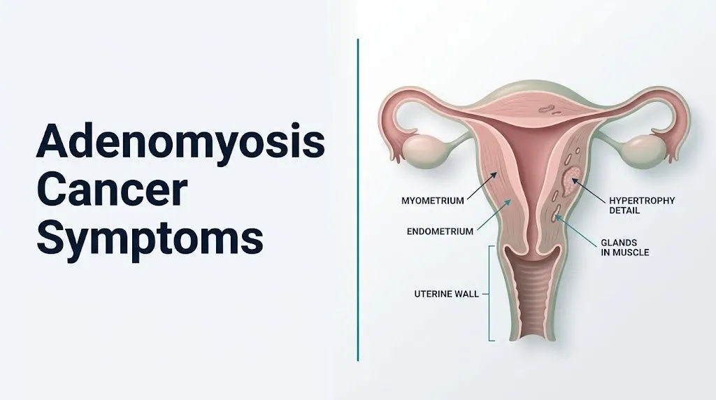
Adenomyosis occurs when endometrial glands and stroma, the tissue that normally lines the inner cavity of the uterus, invade through the junctional zone and embed themselves within the myometrium, the thick smooth muscle layer that forms the wall of the uterus.
Think of the uterus like a three-layered cylinder. The innermost layer, the endometrium, is designed to thicken and shed each month in response to hormonal signaling. The middle layer, the myometrium, is the muscle that contracts during menstruation to expel that shed tissue. Between these two layers lies the junctional zone, a thin transitional band of compacted smooth muscle. In adenomyosis, endometrial tissue breaks through the junctional zone and takes root inside the muscle itself. Those ectopic glands then respond to the same monthly hormonal signals as the uterine lining, bleeding and inflaming the surrounding muscle with each menstrual cycle.
According to the American College of Obstetricians and Gynecologists, adenomyosis affects an estimated 20 to 35 percent of people with a uterus, though prevalence estimates vary because definitive diagnosis historically required hysterectomy. Advances in pelvic MRI imaging have allowed more precise diagnosis without surgery.
The condition can be diffuse, spreading throughout the myometrium, or focal, concentrated in a discrete nodule called an adenomyoma. Diffuse adenomyosis tends to produce more severe bleeding and a uniformly enlarged, boggy-feeling uterus. Focal adenomyoma can occasionally be misidentified on imaging as a uterine fibroid, which is why imaging expertise and clinical correlation matter in diagnosis.
Women who have had prior uterine procedures including cesarean delivery, dilation and curettage, or myomectomy face higher adenomyosis prevalence, possibly because surgical disruption of the junctional zone allows endometrial tissue easier access to the myometrium.
Adenomyosis Symptoms: Heavy Bleeding, Pain, and Pelvic Pressure
The primary symptom of adenomyosis is menorrhagia, clinically defined as uterine bleeding heavy enough to soak through a pad or tampon in less than an hour or to last longer than seven days, occurring in the context of regular menstrual cycles.
The mechanism behind this heavy bleeding is direct. Ectopic endometrial glands embedded in the myometrium bleed during each menstrual cycle, adding blood volume to what the uterine cavity itself sheds. Simultaneously, those ectopic glands produce elevated levels of prostaglandin E2 through upregulation of the COX-2 enzyme, which impairs normal myometrial contractility. A uterine muscle that cannot contract efficiently cannot compress the spiral arterioles that supply the endometrium, so bleeding continues longer and more heavily than in an unaffected uterus.
Dysmenorrhea, severe menstrual cramping, is the second hallmark symptom. Prostaglandin F2-alpha, also synthesized in excess by COX-2-active ectopic glands, drives intense uterine contractions. The contractions in adenomyosis are not coordinated. They can be disorganized and prolonged, producing cramping that often begins before bleeding starts and continues through the first several days of the period.
Pelvic pressure and a sensation of heaviness or fullness in the lower pelvis arise from uterine enlargement and from the inflammatory reaction within the myometrium surrounding the ectopic glands. Macrophage infiltration and local cytokine release, including IL-6 and TNF-alpha, contribute to persistent pelvic inflammation between cycles.
| Symptom | Mechanism | Typical Timing |
|---|---|---|
| Heavy menstrual bleeding | Ectopic gland bleeding + impaired myometrial contraction | During menstrual days 1 to 7 |
| Severe cramping | Elevated prostaglandin F2-alpha, disorganized contractions | Day before bleeding through day 3 to 5 |
| Pelvic pressure/heaviness | Uterine enlargement, myometrial inflammation | Throughout cycle, worse with periods |
| Painful intercourse (dyspareunia) | Posterior uterine wall involvement, pelvic congestion | Variable, often positional |
| Bloating | Pelvic venous congestion, uterine enlargement | Premenstrual and menstrual phases |
Women in their 40s who experience a sudden worsening of previously manageable period pain should not assume this is simply a progression of existing adenomyosis. Hormonal fluctuation in perimenopause can amplify symptoms, but a clinician should rule out new pathology, including endometrial hyperplasia, before attributing the change to adenomyosis alone.
Is Adenomyosis a Cancer or Does It Cause Cancer
Adenomyosis is classified as a benign gynecological condition, meaning the ectopic endometrial tissue it involves does not possess the genetic alterations that define malignancy: uncontrolled cell division, local tissue invasion with metastatic potential, or the ability to spread to distant organs.
That said, the relationship between adenomyosis and cancer is more nuanced than the word “benign” implies, and research over the past decade has shifted how clinicians think about this relationship.
A 2021 large cohort study published in JAMA Oncology analyzed data from over 100,000 women and found that those with a diagnosis of adenomyosis had a statistically elevated risk of developing endometrial cancer compared to women without adenomyosis, with an adjusted hazard ratio of approximately 1.82. This does not mean adenomyosis causes endometrial cancer. It means the two conditions share a biological environment: excess unopposed estrogenic stimulation of uterine tissue.
The shared mechanism is the aromatase enzyme (CYP19A1). Ectopic endometrial glands in adenomyosis overexpress aromatase, synthesizing local estrogen independent of ovarian output. This local estrogen drives continuous proliferative signaling in adjacent endometrial tissue. Prolonged unopposed estrogen stimulation of the endometrium is the same primary driver behind endometrial hyperplasia, which is the established precursor lesion to endometrioid endometrial carcinoma.
Additionally, both adenomyosis tissue and endometrial carcinoma frequently show reduced expression of the PTEN tumor suppressor protein and activation of the PI3K-AKT-mTOR signaling cascade. These shared molecular features suggest that adenomyosis and endometrial carcinoma may arise in a common estrogenic microenvironment, not that one transforms into the other directly.
The practical clinical point: adenomyosis does not become cancer, but having adenomyosis means your uterine tissue is operating in a hormonal environment that also elevates endometrial cancer risk. That distinction matters for how often and how thoroughly you are monitored.
Key Takeaway: Adenomyosis is benign but shares molecular pathways with endometrial cancer, specifically aromatase-driven local estrogen excess and PI3K-AKT signaling activation, which is why women with adenomyosis warrant closer monitoring rather than simple reassurance.
Adenomyosis vs Endometrial Cancer: How to Tell the Difference
Distinguishing adenomyosis from endometrial cancer based on symptoms alone is genuinely difficult, and no clinician should attempt it without imaging and, when indicated, endometrial tissue sampling.
The symptom overlap is real and extensive. Both conditions produce heavy uterine bleeding, pelvic pain, and uterine enlargement. The differences that matter most are the pattern of bleeding, its relationship to the menstrual cycle and reproductive status, and the presence or absence of specific accompanying features.
Adenomyosis bleeding is cyclical. It occurs predictably with menstruation, is typically heaviest in the first few days of a period, and largely resolves between cycles. Endometrial cancer bleeding is often acyclical. It may occur between periods, it may appear as light spotting rather than heavy flow, and critically, it occurs after menopause when any uterine bleeding is, by clinical definition, abnormal and requires immediate evaluation.
| Feature | Adenomyosis | Endometrial Cancer |
|---|---|---|
| Bleeding pattern | Heavy cyclical menstrual bleeding | Acyclical, postmenopausal, or intermenstrual bleeding |
| Pain | Severe dysmenorrhea, cyclical cramping | Pelvic pain often without strong cyclical pattern |
| Uterine size | Diffusely enlarged, soft, boggy | May be enlarged; can be normal size in early cancer |
| Age of onset | Typically 35 to 50 (reproductive years) | More common after age 55, postmenopause |
| Relationship to periods | Symptoms tied to menstrual cycle | Symptoms not reliably tied to cycle |
| Associated risk factors | Multiparity, prior uterine surgery | Nulliparity, obesity, unopposed estrogen, Lynch syndrome |
| Imaging appearance (MRI) | Thickened junctional zone, diffuse myometrial change | Endometrial thickening, irregular endometrial lining |
| Definitive diagnosis | MRI, may require histopathology at hysterectomy | Endometrial biopsy, hysteroscopy with biopsy |
The differentiating feature that carries the most clinical weight is reproductive status. Adenomyosis is estrogen-dependent and typically resolves or regresses after menopause. Any woman who has a prior adenomyosis diagnosis and then develops uterine bleeding after menopause should not assume it is residual adenomyosis activity. A board-certified gynecologist or gynecologic oncologist should evaluate that bleeding with endometrial biopsy within one to two weeks.
Adenomyosis and Ovarian Cancer Risk
The relationship between adenomyosis and ovarian cancer is biologically distinct from its relationship to endometrial cancer, though both associations are estrogen-driven.
Research published in Obstetrics and Gynecology in 2020 found that women with adenomyosis had a modestly elevated risk of certain ovarian cancer subtypes, particularly endometrioid ovarian carcinoma and clear cell ovarian carcinoma, compared to the general population. The association was stronger in women who also had concurrent endometriosis, a related but distinct condition involving endometrial tissue outside the uterus rather than within the uterine wall.
The proposed biological mechanism connects through shared retrograde implantation pathways and the same estrogen-permissive environment. Endometrioid ovarian carcinoma and clear cell ovarian carcinoma are now understood by gynecologic oncologists to arise, in a subset of cases, from endometriosis and related endometrial tissue implants on or near the ovaries. Adenomyosis and endometriosis frequently co-exist: a 2019 meta-analysis published in Fertility and Sterility reported co-occurrence rates ranging from 25 to 50 percent depending on diagnostic criteria used.
The elevated ovarian cancer risk associated with adenomyosis is modest in absolute terms. The lifetime risk of ovarian cancer in the general population is approximately 1.3 percent according to the National Cancer Institute. Women with adenomyosis and concurrent endometriosis may face a risk closer to 1.5 to 2 percent based on available epidemiological data, which represents a relative increase but remains low in absolute risk terms.
What this means practically: women with adenomyosis should not assume that pelvic symptoms originate solely from their uterus. New or worsening ovarian pain, a palpable adnexal mass identified on examination, or a rising CA-125 serum level in the context of clinical symptoms warrants evaluation by a gynecologic oncologist, not simply reassurance based on the existing adenomyosis diagnosis.
Younger women in their 20s and 30s with adenomyosis are less likely to face ovarian cancer risk in the short term, but those with concurrent endometriosis and a family history of ovarian cancer should discuss surveillance with a gynecologist familiar with hereditary cancer risk.
Shared Symptoms of Adenomyosis and Uterine Cancer
The symptom overlap between adenomyosis and uterine cancer (a term that most commonly refers to endometrial carcinoma, which arises from the endometrial lining of the uterine cavity) creates genuine clinical ambiguity that cannot be resolved by symptom assessment alone.
Both conditions produce heavy uterine bleeding, and both cause pelvic pain. Both can enlarge the uterus to a degree visible or palpable on clinical examination. Both may cause bleeding that produces iron deficiency anemia with symptoms including fatigue, pallor, and exertional shortness of breath from reduced oxygen-carrying capacity in the bloodstream.
The shared symptoms, listed by clinical significance:
- Heavy uterine bleeding: the most common presenting symptom of both conditions
- Pelvic pain or pressure: present in both, with mechanism differences explained in the prior section
- Uterine enlargement: both can enlarge the uterus, detected on pelvic exam or imaging
- Iron deficiency anemia: a downstream consequence of chronic blood loss from either condition
- Pelvic fullness or bloating: associated with uterine size increase in both conditions
- Painful intercourse (dyspareunia): present in adenomyosis and in uterine cancer with posterior wall involvement
The symptom that does not overlap is postmenopausal bleeding. Adenomyosis does not produce postmenopausal bleeding in women who are not taking exogenous estrogen. Endometrial cancer frequently does. This is the single most reliable clinical differentiator in a postmenopausal woman.
According to the National Cancer Institute, postmenopausal bleeding is the presenting symptom in approximately 90 percent of endometrial cancer cases. That statistic is clinically powerful. Any postmenopausal bleeding, regardless of how light, brief, or isolated it appears, must be evaluated with endometrial biopsy.
For premenopausal women, differentiating adenomyosis from early endometrial cancer based on symptoms alone is not reliably possible, which is why imaging and tissue sampling protocols exist.
Key Takeaway: Both adenomyosis and endometrial cancer share heavy bleeding, pelvic pain, and uterine enlargement as presenting symptoms, but postmenopausal bleeding is not explained by adenomyosis and must be evaluated for endometrial cancer promptly.
Warning Signs That Adenomyosis Symptoms May Indicate Something More Serious
Several specific symptom features should prompt a woman with known or suspected adenomyosis to seek evaluation beyond her regular gynecological follow-up.
These are not vague concerns about “worsening symptoms.” These are specific clinical patterns that clinicians use to expand the differential diagnosis from adenomyosis to include endometrial cancer, ovarian cancer, or atypical endometrial hyperplasia, which is a premalignant lesion requiring treatment.
Warning signs that warrant prompt gynecological evaluation:
- Postmenopausal uterine bleeding of any volume or duration
- Intermenstrual bleeding: bleeding occurring between expected periods, not attributable to breakthrough spotting on hormonal contraception
- Rapidly progressive uterine enlargement: uterine size change notable over weeks to months rather than the gradual change typical of adenomyosis
- Unexplained weight loss: a systemic symptom not explained by adenomyosis, which is a localized pelvic condition
- Pelvic mass that is new, firm, or irregular on examination, distinct from the uniformly boggy uterine texture typical of adenomyosis
- Watery or bloody vaginal discharge that is not associated with the normal menstrual cycle and is persistent
- Symptoms that persist or worsen after menopause, particularly pelvic pain or pressure in a woman who is not taking hormonal therapy
A 2023 systematic review published in Gynecologic Oncology noted that women with adenomyosis who were subsequently diagnosed with endometrial carcinoma most frequently presented with a change in their bleeding pattern rather than new-onset bleeding, underscoring that a change from a known pattern is as clinically significant as a new symptom.
For women with a known adenomyosis diagnosis, the question to ask at every gynecological visit is not just “are my usual symptoms controlled” but “have any of my symptoms changed in character, timing, or severity in a way that was not happening before.” A board-certified gynecologist should be able to examine those changes in the context of your imaging history and reproductive status.
Adenomyosis Cancer Risk Factors: Who Is Most at Risk
Not all women with adenomyosis face the same cancer risk. Specific biological and clinical factors determine which women face meaningfully elevated risk and which women remain in a low-risk category where standard gynecological follow-up is appropriate.
Obesity is the most broadly established modifiable risk factor for endometrial cancer in women with adenomyosis. Adipose tissue contains aromatase enzyme that converts androstenedione (produced by the adrenal glands) into estrone, a form of estrogen. In women with a body mass index above 30 kilograms per square meter, this peripheral estrogen production can be substantial, adding to the local estrogen already produced by adenomyotic ectopic glands and increasing the cumulative estrogenic stimulation of the endometrium. According to the National Cancer Institute, women with class II obesity (BMI 35 to 40) face a risk of endometrial cancer approximately 4 to 7 times higher than women with a healthy BMI.
Nulliparity, meaning never having carried a pregnancy to term, is associated with both adenomyosis and endometrial cancer. Pregnancy temporarily suppresses adenomyosis through prolonged progesterone dominance, and women who have never been pregnant lose that protective interval. Nulliparity is a documented risk factor for endometrial carcinoma in multiple large epidemiological studies.
Tamoxifen use for breast cancer treatment deserves specific attention. Tamoxifen acts as a selective estrogen receptor modulator, blocking estrogen receptors in breast tissue but acting as an estrogen agonist in the uterine endometrium. In women with adenomyosis who are prescribed tamoxifen, the endometrial stimulant effect of tamoxifen adds to the existing estrogenic environment, meaningfully elevating endometrial cancer risk. The American College of Obstetricians and Gynecologists recommends that women taking tamoxifen report any abnormal uterine bleeding promptly for evaluation.
Lynch syndrome, caused by inherited mutations in mismatch repair genes including MLH1, MSH2, MSH6, and PMS2, raises lifetime endometrial cancer risk to 40 to 60 percent. Women with Lynch syndrome who also have adenomyosis face a compound risk that warrants proactive surveillance and may warrant discussion of prophylactic hysterectomy with a gynecologic oncologist.
| Risk Factor | Mechanism | Cancer Risk Modification |
|---|---|---|
| Obesity (BMI > 30) | Peripheral aromatization in adipose tissue increases circulating estrogen | 4 to 7-fold elevation in endometrial cancer risk |
| Nulliparity | No protective progesterone-dominant pregnancy intervals | Established independent risk factor |
| Tamoxifen use | Uterine endometrial estrogen agonist effect | Annual endometrial evaluation recommended |
| Lynch syndrome | Mismatch repair gene deficiency (MLH1, MSH2, MSH6) | Lifetime endometrial cancer risk 40 to 60% |
| Concurrent endometriosis | Shared estrogen-permissive environment; ovarian implants possible | Elevated endometrioid and clear cell ovarian cancer risk |
| Unopposed estrogen therapy | Exogenous estrogen without progesterone stimulates endometrium | Well-established endometrial cancer risk |
Key Takeaway: Obesity, nulliparity, tamoxifen use, and Lynch syndrome each independently amplify endometrial cancer risk in women with adenomyosis, and women who carry more than one of these factors should discuss a proactive monitoring plan with a gynecologic oncologist.
Adenomyosis Symptoms in Perimenopause and Postmenopause
Perimenopause, the transitional phase typically spanning 4 to 10 years before the final menstrual period, creates a particularly complex hormonal environment for women with adenomyosis.
During perimenopause, ovarian estrogen production becomes erratic rather than cyclical. Estrogen levels can spike dramatically in some cycles and fall sharply in others. This irregular hormonal pattern amplifies adenomyosis symptoms in many women: bleeding episodes become heavier and less predictable, cramping may worsen, and the uterus may enlarge further due to sustained estrogenic stimulation without consistent progesterone opposition.
The clinical challenge in perimenopause is that the same symptoms that represent a worsening of adenomyosis, specifically heavier and irregular bleeding, are also the presenting symptoms of endometrial hyperplasia and early endometrial carcinoma. A perimenopausal woman with a known adenomyosis diagnosis who experiences significantly heavier bleeding, new intermenstrual bleeding, or bleeding lasting longer than her established pattern should not assume this is simply perimenopausal hormonal irregularity.
According to the National Institute of Child Health and Human Development, endometrial evaluation should be considered in any perimenopausal woman with abnormal uterine bleeding that does not respond to initial hormonal management within three to six months, regardless of a prior benign diagnosis.
After menopause, a different clinical reality applies entirely. Adenomyosis is estrogen-dependent. Without ovarian estrogen production, ectopic endometrial glands within the myometrium no longer receive the hormonal signaling required to proliferate and bleed. Adenomyosis symptoms therefore typically regress after menopause in women who are not taking exogenous hormonal therapy.
Any woman who had adenomyosis in her reproductive years and then develops uterine bleeding after confirmed menopause, meaning twelve or more consecutive months without a menstrual period, must be evaluated for endometrial cancer. The prior adenomyosis diagnosis does not explain postmenopausal bleeding and should not delay evaluation.
Women taking menopausal hormone therapy that includes estrogen without adequate progestogen opposition face continued endometrial stimulation after menopause, which maintains both adenomyosis-like symptom potential and endometrial cancer risk.
How Adenomyosis Is Diagnosed and Why Accurate Diagnosis Matters
Adenomyosis is definitively diagnosed through histopathological examination of uterine tissue, which historically meant examining the uterus after hysterectomy.
Non-surgical diagnosis now relies primarily on pelvic MRI with T2-weighted sequences. On T2-weighted MRI, adenomyosis appears as diffuse or focal low-signal-intensity changes within the myometrium, often with thickening of the junctional zone beyond 12 millimeters, cystic spaces within the myometrium corresponding to ectopic glands, and myometrial striation patterns. MRI has sensitivity of approximately 77 to 83 percent and specificity of 85 to 93 percent for adenomyosis diagnosis based on a meta-analysis published in Radiology.
Transvaginal ultrasound (TVUS) is typically the first-line imaging tool. Adenomyosis on TVUS appears as an asymmetrically enlarged uterus, heterogeneous myometrial texture, myometrial cysts, and poor uterus-endometrium interface definition. Sensitivity is lower than MRI at approximately 72 percent, but TVUS is more accessible and appropriate for initial evaluation.
Neither imaging modality can definitively exclude endometrial carcinoma when the clinical picture raises concern. Endometrial biopsy (using the pipelle sampling technique in the office setting or via hysteroscopy with directed biopsy in the hospital) is the tool that samples endometrial tissue for histopathological analysis. Any clinical scenario in which endometrial cancer is on the differential diagnosis, including postmenopausal bleeding, abnormal endometrial thickness on imaging (greater than 4 to 5 millimeters after menopause), or high-risk clinical features, requires endometrial biopsy rather than imaging reassurance alone.
To describe your symptoms accurately when you see your gynecologist for evaluation:
- Write down the first day of your last menstrual period and the last three cycle lengths
- Record how many pads or tampons you use per day at peak bleeding and for how many days
- Note any bleeding that occurs outside of expected period timing, including spotting
- Describe your pain: where it is, when it starts relative to bleeding, how long it lasts, and whether it has changed in character recently
- List all medications and hormonal therapies you are currently using, including any over-the-counter anti-inflammatory drugs you take for cramps
- Bring any prior imaging reports (ultrasound, MRI) with you to the appointment
Adenomyosis Enlarged Uterus and What It Means Clinically
An enlarged uterus is one of the most consistent physical findings in adenomyosis, and understanding what the enlargement means, how large is typical, and when enlargement should prompt concern is an important part of managing this condition.
In adenomyosis, uterine enlargement results from two mechanisms: the infiltration of ectopic glandular tissue into the myometrium, which increases the mass of the uterine wall itself, and myometrial hypertrophy, meaning muscle fiber enlargement in response to chronic inflammation and disrupted contractility. The myometrium in severe diffuse adenomyosis can be two to three times thicker than the normal range of 3 to 5 millimeters per wall measurement. A uterus that would normally measure approximately 7 to 8 centimeters in its longest dimension may reach 12 to 14 centimeters or more in diffuse adenomyosis.
Clinically, the enlarged adenomyotic uterus feels soft and compressible, often described by gynecologists as “boggy.” This texture distinction matters because malignant uterine enlargement from endometrial cancer or uterine sarcoma tends to produce a firmer uterine consistency, though this is not a reliable distinguishing feature without imaging and biopsy confirmation.
According to the American College of Obstetricians and Gynecologists, uterine size alone does not determine whether adenomyosis requires surgical management. The decision depends on symptom severity, impact on quality of life, response to medical therapy, reproductive desires, and the presence or absence of concurrent pathology requiring histological exclusion.
A rapidly enlarging uterus, meaning one that grows noticeably over weeks to months rather than gradually over years, warrants evaluation to exclude uterine sarcoma, a rare but aggressive malignant tumor of the uterine muscle or connective tissue. Uterine sarcoma is not caused by adenomyosis and is not common, but rapid uterine growth is a clinical signal that should not be attributed to adenomyosis without appropriate imaging review.
Women who are postmenopausal should not have an enlarging uterus under any circumstances. Uterine growth after menopause, detected on physical examination or incidentally on imaging, requires evaluation by a gynecologist or gynecologic oncologist regardless of prior adenomyosis history.
Key Takeaway: An enlarged, soft, boggy uterus is the expected physical finding in adenomyosis, but rapidly progressive enlargement or a firm uterine texture warrants imaging evaluation to exclude uterine sarcoma or other malignancy.
Adenomyosis Treatment Options and Cancer Risk Management
Treatment for adenomyosis aims to suppress the hormonal signaling that drives ectopic endometrial tissue proliferation, reduce bleeding and pain, and where appropriate, address cancer risk within the same clinical plan.
Medical management options include:
- Levonorgestrel-releasing intrauterine device (LNG-IUD): releases a local progestin that suppresses endometrial proliferation. Reduces bleeding volume by 70 to 90 percent in many women with adenomyosis and reduces the endometrial cancer risk associated with unopposed estrogen by providing direct progestogenic opposition to the endometrium.
- Combined oral contraceptives (COCs): suppress ovarian estrogen and progesterone cycling, reducing the hormonal stimulation of ectopic glands. Appropriate for premenopausal women without contraindications.
- Progestin-only therapy: oral or injectable progestins suppress endometrial proliferation. Provides similar endometrial cancer-protective effect as the LNG-IUD through systemic progestogenic opposition.
- GnRH agonists (such as leuprolide): create a temporary medically induced low-estrogen state by suppressing hypothalamic-pituitary-ovarian axis signaling. Highly effective for symptom control but not suitable for long-term use without add-back hormonal therapy due to bone density loss risk.
- Hysterectomy: the only curative treatment for adenomyosis. In women who have completed childbearing and face severe symptoms unresponsive to medical management, hysterectomy eliminates both the adenomyosis and, by removing the uterus, the target organ for endometrial cancer. This is a meaningful consideration in women with adenomyosis who also carry high-risk features for endometrial cancer.
Research published in Obstetrics and Gynecology found that progestogen-based therapies, including the LNG-IUD and oral progestins, reduce endometrial cancer risk in women with unopposed estrogen exposure by opposing estrogen-driven endometrial proliferation. This means that appropriate medical management of adenomyosis with progestogen-based options simultaneously addresses symptom burden and provides measurable endometrial cancer risk reduction.
Women with Lynch syndrome who also have adenomyosis should discuss the timing and approach to prophylactic hysterectomy with a gynecologic oncologist, as this intervention reduces lifetime endometrial cancer risk from 40 to 60 percent to near zero in mismatch repair gene mutation carriers.
When to See a Gynecologist for Adenomyosis Symptoms
Knowing when to escalate from self-monitoring to a clinical appointment is a practical skill every woman with adenomyosis or suspected adenomyosis needs.
A board-certified obstetrician-gynecologist should evaluate you within two to four weeks if:
- Your menstrual bleeding has significantly worsened from your established baseline pattern
- You are soaking through more than one pad or tampon per hour for more than two consecutive hours
- You have started bleeding between periods and this is a new development for you
- You have developed a sensation of pelvic fullness, pressure, or a mass that was not previously present
- You have started having pain with intercourse that is new or has worsened significantly
- You are approaching or entering perimenopause and your bleeding has become unpredictable or heavier
- You have been on hormonal therapy for adenomyosis and your symptoms are no longer responding
An appointment with a gynecologic oncologist, rather than a general obstetrician-gynecologist, is appropriate if:
- You have postmenopausal uterine bleeding of any type
- Your gynecologist finds an abnormal mass, thickened endometrium, or other imaging finding that cannot be attributed to adenomyosis alone
- You carry Lynch syndrome or another hereditary cancer syndrome with uterine cancer implications
- Your endometrial biopsy shows atypical endometrial hyperplasia, which requires oncological management
The specific questions to ask at your appointment:
- Based on my imaging and symptom history, does my current presentation fit adenomyosis or are there features that warrant further investigation?
- Do I need an endometrial biopsy given my specific risk factors?
- How often should I have follow-up imaging, and what changes on imaging should prompt a return visit?
- Does my treatment plan address my cancer risk as well as my symptoms?
Women with a BMI above 30 and concurrent adenomyosis should discuss endometrial cancer surveillance specifically with their gynecologist, not simply assume that adenomyosis management covers all relevant risk.
Emergency Symptoms: When Adenomyosis Bleeding Requires Immediate Care
Certain symptoms associated with adenomyosis and uterine conditions require immediate emergency evaluation. Do not wait to see if these resolve on their own.
Call 911 or go to the nearest emergency room immediately if you experience:
- Uterine bleeding so heavy that you are soaking through a pad or tampon in less than 30 minutes and this has continued for more than one hour, particularly if accompanied by dizziness, lightheadedness, or a rapid heartbeat
- Fainting or near-fainting associated with pelvic bleeding, indicating potential hemodynamic instability from acute blood loss
- Severe, sudden pelvic pain that is significantly more intense than your typical adenomyosis cramping, especially if it is sharp, one-sided, or accompanied by fever
- Fever above 101 degrees Fahrenheit combined with pelvic pain and abnormal bleeding, which can indicate pelvic infection requiring emergency antibiotic treatment
- Rapid heart rate (pulse consistently above 100 beats per minute at rest) combined with pallor and bleeding
- New, sudden, severe pelvic pain in a woman with a known or suspected ovarian cyst or mass, which may represent cyst rupture or ovarian torsion
Additionally, seek emergency evaluation the same day, not at a scheduled appointment, if:
- You have postmenopausal uterine bleeding and have not yet been evaluated: this is not a normal aging change and requires prompt clinical assessment
- You develop sudden abdominal distension, pain, and nausea alongside pelvic symptoms, particularly in a woman with known ovarian pathology
These presentations can indicate hemorrhagic instability, acute pelvic infection, ovarian torsion, or a rapidly evolving malignant process and require emergency medical assessment, not an urgent care appointment or a phone call.
Frequently Asked Questions About Adenomyosis Cancer Symptoms
Can adenomyosis turn into cancer?
Adenomyosis does not directly transform into cancer; it remains a benign condition at the tissue level.
However, adenomyosis and endometrial carcinoma share a common biological environment of excess estrogen-driven proliferation, and a 2021 study published in JAMA Oncology found that women with adenomyosis had an approximately 1.82-fold elevated hazard ratio for endometrial cancer diagnosis compared to women without adenomyosis.
This association reflects shared risk factors and molecular pathways, not a direct progression from adenomyosis to malignancy.
What symptoms of adenomyosis are the same as uterine cancer symptoms?
The symptoms shared between adenomyosis and endometrial cancer include heavy uterine bleeding, pelvic pain and pressure, uterine enlargement, painful intercourse, and iron deficiency anemia from chronic blood loss.
These overlapping symptoms are the reason that clinical evaluation, imaging, and in many cases endometrial biopsy are necessary to distinguish between the two conditions rather than relying on symptom assessment alone.
The key differentiating symptom is postmenopausal bleeding, which is not produced by adenomyosis and must be evaluated as a potential sign of endometrial carcinoma.
How do doctors tell adenomyosis apart from endometrial cancer?
A pelvic MRI with T2-weighted sequences can identify the junctional zone thickening and myometrial changes characteristic of adenomyosis, while abnormal endometrial thickness or irregular endometrial contour raises concern for endometrial pathology.
An endometrial biopsy, performed via the pipelle technique in the office or through hysteroscopy with directed biopsy, provides histological tissue samples that definitively identify whether endometrial cells are benign, hyperplastic, or malignant.
Imaging alone cannot exclude endometrial carcinoma when clinical features suggest it, which is why biopsy is the confirmatory step.
Does having adenomyosis increase your risk of ovarian cancer?
Research published in Obstetrics and Gynecology found a modest association between adenomyosis and certain ovarian cancer subtypes, particularly endometrioid and clear cell carcinoma, especially in women who also have concurrent endometriosis.
The National Cancer Institute places the general population’s lifetime ovarian cancer risk at approximately 1.3 percent; available data suggest women with adenomyosis and concurrent endometriosis may face a somewhat higher risk, though absolute numbers remain relatively low.
Women with adenomyosis and concurrent endometriosis, a family history of ovarian cancer, or BRCA1/BRCA2 mutations should discuss their individual ovarian cancer surveillance plan with a board-certified gynecologist or gynecologic oncologist.
When should I be worried that my adenomyosis symptoms are actually cancer?
Postmenopausal uterine bleeding is the single highest-priority symptom that requires immediate concern and evaluation, because adenomyosis does not cause bleeding after menopause in women not taking exogenous estrogen.
In premenopausal women, a significant change in bleeding pattern, new intermenstrual bleeding, unexplained weight loss, a new palpable pelvic mass, or watery bloody vaginal discharge not associated with the normal cycle are warning signs that warrant evaluation by a gynecologist within one to two weeks.
A board-certified obstetrician-gynecologist can order appropriate imaging and endometrial sampling to determine whether the symptom change reflects adenomyosis progression or requires oncological evaluation.
Is it normal to have adenomyosis after menopause?
Adenomyosis is an estrogen-dependent condition and typically becomes clinically inactive, meaning it stops producing symptoms, after menopause when ovarian estrogen production ceases.
Women who are taking menopausal hormone therapy that includes estrogen may experience continued or resurgent adenomyosis-related symptoms because exogenous estrogen maintains the hormonal environment that drives ectopic endometrial tissue activity.
Any uterine bleeding occurring after menopause, including in women on hormone therapy, requires evaluation by a board-certified gynecologist with endometrial biopsy to exclude endometrial carcinoma, and should not be attributed to adenomyosis without that evaluation.
Closing
Adenomyosis is a real and often painful condition, and it is not cancer. But the relationship between adenomyosis and cancer risk is nuanced enough that “it’s benign, don’t worry” is not a complete clinical answer. The shared estrogenic biology, the molecular pathway overlaps, and the symptom similarities with endometrial carcinoma mean that women with adenomyosis benefit from understanding their own risk profile rather than treating their diagnosis as a fully resolved concern.
The most actionable clinical takeaway is this: know your baseline, and pay attention to changes. If your bleeding pattern shifts, if you develop symptoms after menopause, if a new mass appears, or if your pain escalates beyond its established pattern, those are clinical signals that warrant a conversation with a board-certified gynecologist or gynecologic oncologist, not reassurance based on a prior benign diagnosis.
You deserve a clinician who reviews your full picture: your imaging history, your risk factors, your symptom trajectory, and your reproductive status. That conversation, grounded in your specific biology rather than general population statistics, is what determines whether standard management is appropriate or whether closer monitoring or further evaluation is the right next step.