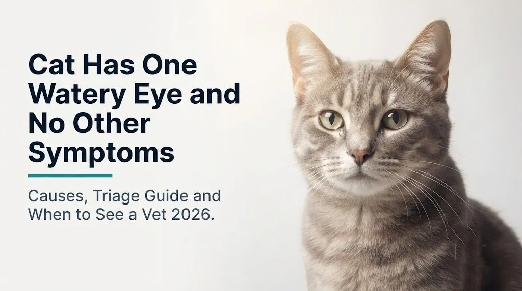
Cat Has One Watery Eye: Causes and When to Act 2026
When a cat has one watery eye and no other symptoms, the most likely explanation is a localized issue affecting just that eye, such as a minor irritant, early feline herpesvirus reactivation, a partially blocked tear duct, or early-stage conjunctivitis, rather than a systemic illness. The fact that only one eye is involved and your cat is otherwise eating, active, and behaving normally actually carries clinical meaning: it narrows the probability considerably toward eye-specific causes rather than upper respiratory or whole-body disease.
According to the Cornell Feline Health Center, eye problems are among the most frequent reasons cats are brought to veterinary attention, with ocular discharge representing one of the most common owner-observed signs. Unilateral (one-sided) discharge in an otherwise-healthy cat is a distinct clinical picture from the bilateral tearing and concurrent sneezing seen in feline upper respiratory infections.
This article covers the specific anatomy behind feline tear production and drainage, the most likely causes ranked by frequency, what discharge color and character tell you, which signs mean you need a vet today versus tomorrow, and which breeds and ages face higher baseline risk. The goal is to help you make a specific, informed decision about your cat’s eye, not leave you with a list of twenty possibilities and no guidance on what to do next.
Cat Has One Watery Eye and No Other Symptoms: What This Means Clinically
A cat with one watery eye and no other symptoms most likely has a localized ocular or adnexal problem affecting only one side, which is the starting point every veterinarian uses to narrow the differential diagnosis.
In clinical feline ophthalmology, the first question is always: one eye or both? Bilateral tearing with concurrent sneezing, nasal discharge, or lethargy points strongly toward systemic infectious causes like feline upper respiratory infection (URI) from feline herpesvirus-1 or feline calicivirus. But one eye watering in a cat that is otherwise eating well, grooming normally, and showing no behavioral change tells a different story. It points toward something affecting that specific eye’s surface, drainage system, or adnexal structures, rather than the whole animal.
The term veterinarians use for excess tearing that overflows the eyelid margin is epiphora. It occurs when tear production exceeds the drainage capacity of the nasolacrimal duct system, or when drainage is obstructed. Either way, tears spill over onto the face rather than draining internally.
What makes the single-eye presentation particularly important is the range of causes it includes. Some are self-resolving within 24 to 48 hours. Others, such as a corneal scratch or early anterior uveitis, require same-day veterinary attention to prevent permanent vision damage.
Things to observe during the first 24 hours at home:
- Is the discharge clear and watery, or thickening to a mucoid or colored consistency?
- Is your cat squinting, blinking rapidly, or holding the eye partially closed?
- Is your cat rubbing or pawing at the affected eye?
- Is the white of the eye (sclera) or the pink tissue (conjunctiva) visibly reddened?
- Is the cornea (the normally crystal-clear front of the eye) showing any cloudiness?
A cat producing clear, watery tears from one eye without squinting, eye-rubbing, or redness, and who is otherwise alert and eating, can be observed for up to 24 to 48 hours. Any of the signs above moves this into same-day veterinary territory.
What Is Epiphora in Cats and Why Does It Happen
Epiphora in cats is the overflow of tears onto the face caused by either excessive tear production, obstruction of the normal drainage pathway, or both simultaneously.
Tears are not just water. Feline tears are a three-layer film: an outer lipid layer secreted by the meibomian glands in the eyelids, a middle aqueous layer produced by the lacrimal gland positioned above the eye, and an inner mucin layer produced by conjunctival goblet cells. This film keeps the cornea hydrated, delivers oxygen to the avascular corneal epithelium, and provides front-line immune protection via secretory IgA and antimicrobial lysozymes.
Normally, this tear film drains through the system continuously. Any disruption to either the production or drainage side of this equation produces visible overflow tearing onto the face. In cats, even mild obstruction of the narrow nasolacrimal duct produces noticeable tearing because the duct diameter is proportionally smaller than in dogs, leaving less reserve drainage capacity.
According to the Merck Veterinary Manual, epiphora in cats is classified as either primary (due to a structural drainage problem) or secondary (due to excess tear production from ocular surface irritation, infection, or inflammation). Both types can present as unilateral tearing in an otherwise-healthy-appearing cat.
Kittens under 8 weeks old may show unilateral or bilateral epiphora from primary FHV-1 infection acquired from their mother, even before other signs of illness appear. In this age group, watery eyes are not a minor finding: a kitten with eye discharge needs veterinary evaluation, not watchful waiting, because the infection can cause permanent corneal scarring if treatment is delayed.
Feline Eye Anatomy: How the Tear Drainage System Works
The feline tear drainage system begins at the ocular surface and ends in the nasal cavity, and understanding this pathway explains why a cat can have a watery eye without having a systemic illness.
Tears produced by the lacrimal gland spread across the eye with each blink. At the inner corner of each eye (the medial canthus), there are two tiny openings called the upper and lower lacrimal puncta. These puncta are the entry points for tear drainage. Tears flow through the puncta into small channels called canaliculi, which merge into the lacrimal sac, and then continue down through the nasolacrimal duct into the nasal cavity. This is why cats sometimes have a moist nose: it is normal tear drainage.
Think of the nasolacrimal duct as a slow drain in a sink. When the sink is producing a normal amount of water and the drain is fully open, everything flows away without visible overflow. If the drain narrows even slightly, or if the faucet suddenly produces a bigger surge of water (from irritation or inflammation), the sink overflows. The cat’s face below the eye is that overflowing sink.
The feline nasolacrimal duct is notably narrow in cross-section compared to canine anatomy. According to the Veterinary Clinics of North America: Small Animal Practice, this anatomical feature makes cats more prone to clinically apparent epiphora from even partial duct obstruction than dogs with equivalent duct pathology.
In brachycephalic breeds, the entire duct pathway is compressed and geometrically distorted by skull shape changes, making full drainage anatomically impossible in some individuals regardless of inflammation or infection. This is why flat-faced cats can have chronic tear staining without any underlying disease process.
What Causes a Cat to Have One Watery Eye
The most common cause of one watery eye in a cat that appears otherwise healthy is localized irritation or early feline herpesvirus-1 reactivation affecting only the ocular surface or adnexal structures of one eye.
Causes ranked by frequency in otherwise-healthy cats:
| Cause | Mechanism | Most Affected Population |
|---|---|---|
| Feline herpesvirus-1 (FHV-1) reactivation | Latent virus reactivates in trigeminal ganglion; infects corneal and conjunctival epithelium unilaterally | Adult cats with prior exposure; stressed or immunocompromised cats |
| Nasolacrimal duct obstruction | Narrowing or blockage of drainage pathway; tears overflow | Brachycephalic breeds; cats with prior URI or facial trauma |
| Foreign body on ocular surface | Grass seed, dust, hair, or debris contacts cornea or conjunctival sac; reflex tearing | Outdoor cats; long-haired breeds |
| Bacterial conjunctivitis (Chlamydia felis, Mycoplasma felis) | Bacterial colonization of conjunctival surface produces inflammatory exudate and reflex tearing | Young adult cats; cats in multi-cat households |
| Corneal scratch or early ulceration | Epithelial disruption triggers reflex tearing via trigeminal nerve ophthalmic branch (V1) | Cats with housemates; outdoor cats |
| Entropion (eyelid rolling inward) | Eyelashes contact corneal surface; chronic irritation produces reflex hyperlacrimation | Persian, Burmese, certain domestic shorthair cats |
| Environmental irritant | Smoke, perfume, cleaning products, or dusty litter irritate conjunctival surface | Any cat; indoor cats in poorly ventilated spaces |
| Early anterior uveitis | Inflammatory cells in anterior chamber produce ocular pain and reflex tearing | Cats with FIV, FeLV, toxoplasmosis; senior cats |
The distinction between these causes based on observation alone is limited. A foreign body in the eye, a corneal scratch, and early uveitis can all look identical from across the room. This is exactly why any persistent or worsening one-eye tearing warrants professional evaluation rather than self-treatment.
Feline Herpesvirus and One Watery Eye in Cats
Feline herpesvirus-1 (FHV-1) is the single most important infectious cause of unilateral ocular disease in otherwise-healthy-appearing adult cats, and its biology explains why a cat can look completely normal except for one watery eye.
FHV-1, like all herpesviruses, establishes lifelong latency after the initial infection. In cats, the virus retreats to and hides within the trigeminal ganglion, the cluster of sensory nerve cell bodies that supply sensation to the face and eyes via the ophthalmic (V1), maxillary (V2), and mandibular (V3) branches of the trigeminal nerve. The virus stays dormant there, undetected by the immune system, until a trigger event disrupts immune surveillance.
Common reactivation triggers include:
- Stressful events: moving home, a new pet, a change in routine, veterinary visits
- Illness from any cause, even a mild gastrointestinal upset
- Concurrent disease in senior cats: feline chronic kidney disease (CKD), hyperthyroidism, or feline immunodeficiency virus (FIV)
- Corticosteroid administration (suppresses immune function)
- Seasonal environmental changes
When FHV-1 reactivates, viral particles travel down the trigeminal nerve axons toward the ocular surface. Crucially, reactivation often involves predominantly one branch of the trigeminal nerve, explaining why a cat can have significant unilateral ocular signs from herpesvirus while appearing completely healthy in every other respect.
According to a study published in the Journal of Feline Medicine and Surgery, the estimated prevalence of FHV-1 seropositivity (evidence of prior infection) in the domestic cat population exceeds 90% in some multi-cat environments, meaning the vast majority of adult cats carry latent virus and are capable of reactivation episodes throughout their lives.
In cats with FHV-1 reactivation, the typical presentation is unilateral clear-to-slightly-mucoid tearing, possible mild conjunctival redness, and occasionally mild blepharospasm (squinting). The cat otherwise eats normally, grooms, and behaves as usual. This presentation pattern is genuinely difficult to distinguish from other causes without veterinary examination.
Blocked Nasolacrimal Duct in Cats: Causes and Signs
A blocked or obstructed nasolacrimal duct is a common structural cause of one watery eye in cats, and it produces chronic, usually non-painful overflow tearing that is often initially overlooked because the cat appears otherwise completely healthy.
The nasolacrimal duct can become obstructed at several points along its path from the lacrimal puncta to the nasal cavity. Obstruction most commonly results from:
- Chronic inflammation from prior upper respiratory infections narrowing the duct lumen through scar tissue formation
- Abnormal facial anatomy in brachycephalic breeds compressing the duct’s natural course
- Grass seeds or debris lodged within the canaliculi or duct
- Dacryocystitis: infection of the lacrimal sac producing swelling and functional duct obstruction
A cat with nasolacrimal duct obstruction typically shows chronic, reddish-brown fur staining below the medial canthus of the affected eye, sometimes called epiphora staining or tear staining. The reddish color comes from porphyrins, iron-containing compounds in cat tears that oxidize on contact with air and light, staining the fur a rust or dark-brown color.
To describe your cat’s nasolacrimal signs accurately to a veterinarian:
- Note which eye is affected and whether the staining has been present for days, weeks, or months
- Observe whether the discharge is clear watery fluid, thicker mucoid material, or has any yellow, green, or blood-tinged color
- Check whether the skin below the eye appears irritated, red, or has any hair loss from chronic moisture contact
- Note whether squinting or eye rubbing accompanies the tearing
- Record any recent illnesses, environmental changes, or new household products that might have triggered or worsened the tearing
The American College of Veterinary Ophthalmologists notes that nasolacrimal duct patency can be confirmed in a clinical setting by a simple flush procedure performed under mild sedation, which is both diagnostic and sometimes temporarily therapeutic.
Key Takeaway: One watery eye with reddish-brown fur staining below the medial canthus, no squinting, and no colored discharge often points to a partial nasolacrimal duct obstruction rather than active infection; this warrants veterinary evaluation but is not an emergency unless squinting or corneal cloudiness develops.
Cat Eye Discharge: What Color and Consistency Tell You
The color and consistency of the discharge from your cat’s watery eye carries direct clinical information about the likely cause and the urgency of the situation.
Clear, watery discharge is the least alarming presentation. It indicates that the discharge is tear fluid itself, not inflammatory exudate, which suggests simple overflow tearing from a drainage problem, irritant exposure, or early FHV-1 reactivation without significant secondary bacterial involvement. A cat producing clear tears from one eye without squinting represents the least urgent presentation on the spectrum.
Discharge that shifts from clear to slightly cloudy or mucoid indicates the presence of mucin from goblet cells or early inflammatory cells from the conjunctival surface. This pattern is consistent with early conjunctivitis or mild FHV-1 keratoconjunctivitis and warrants a veterinary appointment within 24 to 48 hours.
A practical guide to discharge character and clinical urgency:
| Discharge Type | Color/Texture | Likely Cause | Action |
|---|---|---|---|
| Serous | Clear, watery | Drainage obstruction, irritant, early FHV-1 | Monitor 24-48 hours; vet if no improvement |
| Mucoid | Clear, slightly thick | Mild conjunctivitis, FHV-1 reactivation | Vet within 24-48 hours |
| Mucopurulent | Yellow-green, thick | Bacterial secondary infection, Chlamydia felis | Vet same day |
| Purulent | Yellow, green, or blood-tinged; thick | Active bacterial infection, corneal ulcer | Vet same day; urgent |
| Blood-tinged | Pink or red-tinged | Trauma, severe ulceration, uveitis | Emergency vet immediately |
Cats with feline immunodeficiency virus (FIV) or feline leukemia virus (FeLV) often progress to mucopurulent discharge faster than immunocompetent cats because their reduced lymphocyte-mediated immune surveillance allows secondary bacterial colonization of the inflamed conjunctival surface to establish more rapidly. If your cat has a known FIV or FeLV diagnosis and develops unilateral eye discharge of any type, schedule a veterinary appointment the same day rather than monitoring at home.
Cat Squinting and Watery Eye: When It Means More Than Tearing
A cat that is squinting the affected eye alongside producing watery discharge is communicating one clinically important thing: there is likely pain originating from the ocular surface or inside the eye, and this changes the urgency of the situation immediately.
The medical term for involuntary squinting or partial eye closure in response to ocular pain is blepharospasm. In cats, it is produced by activation of the ophthalmic branch (V1) of the trigeminal nerve, which densely innervates the corneal surface. Even microscopic disruptions to the corneal epithelium, such as a tiny scratch from a housemate’s claw or a piece of grass debris, activate corneal nociceptors and trigger the reflex squinting response.
Blepharospasm in cats is never a sign to watch and wait. It indicates that the ocular surface or intraocular structures are painful, and pain-producing eye conditions in cats include corneal ulcers, foreign bodies, acute anterior uveitis, and early glaucoma, all of which worsen rapidly without treatment.
The nictitating membrane (third eyelid) is a unique feline anatomical structure not present in humans. It sits at the medial canthus and sweeps horizontally across the eye when the cat is in pain, systemically ill, or has significant ocular surface disease. Unilateral third-eyelid protrusion in one eye is a strong indicator of localized ocular pain rather than systemic illness. If the third eyelid is elevated in both eyes, systemic causes including dehydration, fever, or parasitism (Dipylidium caninum infection in young cats) become more likely.
Signs that require same-day veterinary evaluation by a licensed veterinarian, not observation at home:
- Any degree of squinting or rapid blinking in the affected eye
- Third eyelid visible in the affected eye and not receding
- Cat rubbing or pawing at the eye
- Any cloudiness, film, or color change visible on the cornea
- Redness of the conjunctiva or sclera that was not present before
Brachycephalic Cats and Chronic Watery Eyes
Brachycephalic cats, specifically Persians, Himalayans, Exotic Shorthairs, Scottish Folds, and Burmese, have a structurally predetermined predisposition to chronic unilateral or bilateral epiphora that is fundamentally different from the pathological causes seen in other cats.
Brachycephalic ocular syndrome in cats refers to a cluster of eye-related anatomical abnormalities produced by selective breeding for flattened facial features. The shortened skull base compresses and distorts the normal course of the nasolacrimal duct, making complete tear drainage physically impossible in many individuals. Simultaneously, the prominent, relatively exposed ocular globes (larger apparent eye size) and shortened eyelid margins mean the corneal surface has greater air and environmental exposure, increasing baseline tear evaporation and reflex compensatory tearing.
Additionally, some brachycephalic cats develop entropion, a condition where the eyelid margin rolls inward, causing eyelashes or facial skin to rub continuously against the corneal surface. This produces constant irritation, reflex tearing, and over time, corneal scarring and vascularization. In Persian cats, the medial lower eyelid is particularly susceptible to this inward rolling pattern.
According to the American College of Veterinary Ophthalmologists, brachycephalic cats showing only tear staining without squinting, corneal changes, or discharge color change can often be managed with regular gentle cleaning of the periocular fur to prevent skin maceration. However, any Persian or Himalayan cat with a sudden change in the character of its chronic tearing, or new squinting, requires veterinary evaluation to rule out a superimposed acute problem on the background of their chronic structural issues.
Reddish-brown tear staining in flat-faced cats is caused by porphyrin oxidation in the tear film and does not itself indicate infection or disease. However, it should not be assumed to be “just how this cat is” without at least one baseline veterinary ophthalmic examination to rule out concurrent corneal disease or entropion.
Key Takeaway: Chronic clear tearing and tear staining in a brachycephalic cat is often structural and not an emergency, but any new squinting, discharge color change, or corneal cloudiness in a Persian or Himalayan cat should be evaluated by a licensed veterinarian the same day.
Senior Cats and Kittens With One Watery Eye: What Changes
Age changes the clinical picture for cats with one watery eye in two opposite directions: kittens face primary infection vulnerability, while senior cats face reactivation risk from immune compromise related to concurrent disease.
In kittens under 12 weeks, the most likely cause of one or both watery eyes is primary infection with FHV-1 or Chlamydia felis, acquired from the mother or from littermates in the first weeks of life. Kittens have immature mucosal immunity, with incompletely developed secretory IgA responses at the conjunctival surface, making them susceptible to primary ocular infections that older cats with prior exposure and established immunity resist. Kittens also lack complete protective responses from maternally derived antibodies by 6 to 8 weeks of age.
Critically, untreated FHV-1 in kittens can produce symblepharon, a permanent adhesion of the conjunctiva to the cornea or to other conjunctival surfaces, causing long-term visual impairment. This means a watery eye in a kitten, even a kitten who seems otherwise playful and eating, warrants veterinary evaluation within 24 hours, not several days of home observation.
In senior cats over 10 years, new-onset unilateral epiphora raises a different set of concerns. The most clinically important is FHV-1 reactivation triggered by systemic disease. Cats with feline chronic kidney disease (CKD), hyperthyroidism, or diabetes mellitus have altered immune surveillance that reduces control over latent viral infections. A new watery eye in a previously unaffected senior cat, particularly one who also shows any appetite change, weight change, or increased drinking or urination, warrants veterinary evaluation that includes a systemic health screen, not just an eye examination.
Senior cats also have a higher baseline prevalence of anterior uveitis from systemic causes including feline infectious peritonitis (FIP), toxoplasmosis, and lymphoma. Uveitis can present initially as a quiet, watery, slightly reddened eye in an otherwise-alert cat before progressing to visible intraocular changes.
Cat One Watery Eye Triage: Monitor at Home or See a Vet Now
Whether to monitor your cat at home or seek veterinary care for one watery eye depends on a specific set of observable signs, not on how long the tearing has been present alone.
The following triage framework is based on clinical presentations observed by veterinary ophthalmologists and general practitioners:
| Finding | At-Home Observation Period | Action |
|---|---|---|
| Clear watery discharge, no squinting, cat eating and alert | Up to 24 to 48 hours | Clean gently, monitor; vet if no improvement |
| Mucoid (slightly thick, clear) discharge, mild eye redness | 24 hours maximum | Call your veterinarian to schedule within 24 hours |
| Any squinting or blepharospasm | Zero hours | Vet same day, do not delay |
| Third eyelid elevated in affected eye | Zero hours | Vet same day |
| Yellow, green, or blood-tinged discharge | Zero hours | Vet same day |
| Cloudy or blue-grey cornea | Zero hours | Emergency vet immediately |
| Cat pawing or rubbing the eye | Zero hours | Vet same day |
| Watery eye in a kitten under 12 weeks | 24 hours maximum | Vet within 24 hours regardless of other signs |
| Watery eye in a senior cat with known CKD, hyperthyroidism | 24 hours maximum | Vet within 24 hours; request systemic health check |
At home, you can gently clean dried discharge from the fur below the eye using a clean, damp cotton ball or gauze pad, wiping from the inner corner outward. Do not use human eye drops, contact lens solution, or any medicated drops in your cat’s eye without veterinary direction. Many human ophthalmic products contain preservatives or active ingredients that are not safe for feline ocular tissue.
The American Veterinary Medical Association emphasizes that self-treating an animal’s eye condition without a confirmed diagnosis carries the risk of worsening underlying disease, particularly if an undiagnosed corneal ulcer is present, since some topical products can impair corneal healing.
How a Veterinarian Diagnoses the Cause of a Cat’s Watery Eye
A veterinarian diagnosing the cause of a cat’s watery eye follows a structured sequence of examinations and tests designed to identify or rule out the most serious causes first.
The standard diagnostic sequence:
- Complete history: duration, onset, character and color of discharge, any recent stress or illness, vaccination status (FHV-1 is included in standard feline vaccines but does not eliminate latency), household pet contacts, and any recent environmental changes
- External examination: assessment of eyelid conformation (entropion, ectropion), medial canthus anatomy, third eyelid position, periocular fur staining pattern, and visible conjunctival redness
- Schirmer tear test: measures aqueous tear production by placing a small strip of filter paper in the conjunctival sac for 60 seconds; rules out keratoconjunctivitis sicca (dry eye), where paradoxically inadequate tears can cause reflex compensatory hypersecretion from the ocular surface
- Fluorescein stain test: a small amount of fluorescent dye is applied to the eye surface; under blue-light illumination, any break in the corneal epithelium glows bright green, confirming or ruling out corneal ulceration
- Tonometry: measurement of intraocular pressure using a handheld device; rules out acute glaucoma, where elevated pressure produces pain and tearing; a normal pressure reading (typically 10 to 25 mmHg in cats) effectively rules out acute glaucoma
- Conjunctival cytology or culture: a gentle swab of the conjunctival surface for microscopic examination or bacterial culture and sensitivity testing, particularly when discharge is colored or mucopurulent
- PCR testing for FHV-1 and Chlamydia felis: polymerase chain reaction testing of conjunctival swabs is the most sensitive method of confirming infectious causes; ordered when the clinical picture is consistent with FHV-1 but confirmation affects treatment decisions
For cases where nasolacrimal duct obstruction is suspected, the veterinarian may perform a nasolacrimal flush: a small catheter is inserted into the upper or lower punctum and saline is gently flushed through to assess patency and clear any debris or inflammatory material.
Corneal Ulcers and Uveitis in Cats: Serious Causes of One Watery Eye
Corneal ulceration and anterior uveitis are the two most serious non-emergency causes of one watery eye in an otherwise-stable cat, and both require prompt veterinary treatment to prevent vision loss.
A corneal ulcer is a breach in the corneal epithelium (the protective outer cell layer of the clear front surface of the eye). In cats, ulcers most commonly result from FHV-1-induced epithelial cell death, a scratch from a housemate’s claw, a foreign body on the ocular surface, or abnormal eyelid hair contact. The corneal epithelium has the highest density of sensory nerve endings of any tissue in the cat’s body, supplied by the ophthalmic branch of the trigeminal nerve (V1). Even a superficial ulcer produces intense pain, explaining why cats with corneal ulcers often show marked squinting, excessive tearing, and strong aversion to light (photophobia).
Think of a corneal ulcer like a raw abrasion on your own skin, except it is covering the surface of your eye, which is one of the most densely innervated tissues in the body. Every blink, every air current, every light source activates those pain fibers. A cat with a corneal ulcer is in significant discomfort and should not be left without treatment.
Anterior uveitis is inflammation of the uveal tract, specifically the iris and ciliary body. In cats, it is caused by systemic conditions including FIV, FeLV, feline infectious peritonitis (FIP), toxoplasmosis, and lymphoma, as well as by direct extension of severe corneal disease. Early uveitis can present as a single mildly watery, slightly reddened eye with the cat appearing otherwise healthy.
According to the Journal of Feline Medicine and Surgery, anterior uveitis in cats is clinically important to detect early because untreated uveitis leads to cataract formation, synechia (adhesions between the iris and lens), secondary glaucoma, and in severe cases, permanent blindness in the affected eye. Any cat whose pupil in the affected eye appears smaller than the other (miosis), or whose iris appears dull or discolored compared to the other eye, needs emergency veterinary evaluation the same day.
Key Takeaway: A cat with one watery eye and any squinting, aversion to light, or one pupil smaller than the other needs a same-day veterinary appointment, not another 24 hours of observation, because corneal ulcers and anterior uveitis worsen rapidly without treatment.
Emergency Signs: When to Take Your Cat to the Vet Immediately
Certain signs accompanying one watery eye in a cat require immediate veterinary evaluation. Do not wait to see if these resolve on their own.
Take your cat to a licensed veterinarian or a veterinary emergency clinic immediately if you observe any of the following:
- A cloudy, blue-grey, or white appearance to the normally clear cornea (indicates corneal edema from ulceration, severe inflammation, or acute glaucoma)
- Visible blood anywhere in or around the eye, including within the normally clear fluid of the anterior chamber (a condition called hyphema)
- The affected pupil is noticeably smaller than the other eye’s pupil (suggests anterior uveitis producing pupillary spasm)
- The affected pupil is noticeably larger and unresponsive to light (suggests acute glaucoma or severe neurological involvement)
- Your cat is in obvious distress: vocalizing, hiding, or reacting aggressively when touched near the face or head
- Any visible protrusion of the eye from its socket (proptosis), even partial
- A visible foreign object embedded in the eye’s surface
- Watery eye in combination with sudden behavior change, loss of appetite, or any systemic signs developing within hours
These signs can indicate corneal perforation, acute glaucoma, severe anterior uveitis, intraocular hemorrhage, or globe proptosis, all of which are sight-threatening and in some cases life-threatening conditions that require emergency surgical or medical intervention.
If you are concerned that your cat has had contact with a household chemical, plant, or substance near the eye, call the ASPCA Animal Poison Control Center at 888-426-4435 immediately and follow their guidance before attempting any home rinsing of the eye. A consultation fee may apply.
These presentations require an emergency veterinary clinic if your regular veterinarian is unavailable, not a next-day appointment.
Home Care and What to Expect at the Vet Appointment
Before your veterinary appointment, there are specific, safe steps you can take to manage your cat’s comfort at home without risking worsening the underlying condition.
Safe at-home management steps before the vet visit:
- Gently clean dried or fresh discharge from the fur below and around the eye using a clean cotton ball or gauze pad dampened with sterile saline or plain warm water. Wipe from the inner corner of the eye outward in a single direction; do not rub back and forth.
- Do not place any drops, ointments, medications, or human eye products in the affected eye before being seen by a veterinarian. Topical steroids, in particular, are absolutely contraindicated in any eye with an active corneal ulcer and can rapidly worsen corneal destruction if used inappropriately.
- Prevent your cat from rubbing or pawing at the eye by keeping them calm and indoors; an Elizabethan collar (cone) from a prior veterinary visit can be used temporarily if rubbing is occurring.
- Note the time of onset, any recent events that may have triggered the issue (new cat in the home, moving, illness, vet visit, change in litter or household products), and whether any other pets in the home show similar signs.
- Photograph the eye in natural light before the appointment; this gives the veterinarian baseline documentation of discharge type, color, and degree of third-eyelid protrusion or conjunctival redness.
At the appointment, your veterinarian will want to know: which eye is affected, how long it has been watering, what the discharge looks like, whether squinting is present, whether your cat has had prior episodes, vaccination status (specifically whether the FVRCP vaccine, which includes FHV-1 coverage, is current), and any recent stressors or systemic health changes.
Expect the examination to include the fluorescein stain and Schirmer tear test at minimum. Bring your cat in a secure carrier to prevent stress-related exacerbation of symptoms en route.
In kittens and senior cats, the veterinarian may recommend bloodwork alongside the eye examination to check for underlying systemic disease, particularly in cats over 10 years with a new-onset eye problem.
Frequently Asked Questions About Cat Has One Watery Eye
Why does my cat have one watery eye but seems fine otherwise?
A cat with one watery eye and no other symptoms most likely has a localized ocular cause, such as feline herpesvirus-1 reactivation, a minor irritant, or a partially blocked nasolacrimal duct, rather than a systemic illness.
The fact that only one eye is affected and your cat is eating, active, and behaving normally narrows the likely cause to something affecting that specific eye rather than the whole cat.
A veterinary examination within 24 to 48 hours is appropriate if the discharge is clear and your cat is not squinting; any squinting, colored discharge, or corneal cloudiness requires same-day evaluation.
Can I wait and see if my cat’s watery eye clears up on its own?
You can monitor a cat with clear, watery discharge from one eye, no squinting, and no colored discharge for up to 24 to 48 hours before seeking veterinary care.
If the discharge thickens, changes color to yellow or green, if squinting develops, or if the eye looks cloudy, do not wait longer; schedule same-day veterinary evaluation.
Kittens under 12 weeks and senior cats with known health conditions should be evaluated within 24 hours regardless of how mild the tearing appears.
What does it mean if my cat’s eye is watering clear liquid?
Clear, watery discharge from one cat eye typically indicates overflow tearing (epiphora) from irritation, early FHV-1 reactivation, or a drainage obstruction rather than active bacterial infection.
Clear discharge is the least urgent presentation on the ocular discharge spectrum, but it does not mean the underlying cause is always minor.
If clear tearing persists beyond 48 hours without improvement, a licensed veterinarian should examine the eye to rule out a corneal scratch, early ulceration, or nasolacrimal duct obstruction.
Is one watery eye in a cat a sign of herpesvirus?
Feline herpesvirus-1 reactivation is one of the most common causes of one watery eye in an adult cat who appears otherwise healthy, according to research published in the Journal of Feline Medicine and Surgery.
FHV-1 establishes lifelong latency in the trigeminal ganglion and can reactivate during stress or illness, producing unilateral ocular signs without any other symptoms of illness.
A definitive FHV-1 diagnosis requires PCR testing of a conjunctival swab, which a licensed veterinarian can arrange if the presentation is consistent with herpesvirus reactivation.
When should I take my cat to the vet for a watery eye?
Take your cat to a licensed veterinarian the same day if the affected eye shows squinting, third eyelid elevation, colored discharge, or any corneal cloudiness.
Take your cat to a veterinary emergency clinic immediately if the cornea appears cloudy or blue-grey, if one pupil is notably different in size from the other, or if your cat is in visible distress near the face.
Clear tearing without squinting in an otherwise-healthy adult cat allows for up to 48 hours of home monitoring, but any worsening during that period should prompt immediate contact with a veterinarian.
Why do Persian and flat-faced cats get watery eyes so often?
Persian and Himalayan cats have brachycephalic skull anatomy that compresses and distorts the nasolacrimal duct, making complete tear drainage structurally difficult or impossible for many individuals.
This anatomical predisposition produces chronic clear tearing and reddish-brown fur staining below the eyes (from porphyrin oxidation) that is structural rather than infection-related in most cases.
A licensed veterinarian or board-certified veterinary ophthalmologist should perform at least one baseline examination to rule out concurrent corneal disease, entropion, or nasolacrimal duct obstruction that requires active treatment.
What to Do With What You Now Know
One watery eye in a cat with no other symptoms is almost never a reason to panic, but it is a reason to pay close attention for the next 24 to 48 hours. Clear, watery discharge in a cat that is eating, alert, and not squinting is the least urgent presentation. Write down when it started, what the discharge looks like, and whether anything changed recently at home.
The two signs that change everything are squinting and corneal cloudiness. Either one means same-day veterinary evaluation, not tomorrow, not the weekend. A board-certified veterinary ophthalmologist is the appropriate specialist for cases that don’t resolve with initial treatment, for suspected FHV-1 with recurrent reactivations, or for brachycephalic cats with structural duct problems requiring surgical consideration.
Your cat can’t tell you the eye is painful. Watching for blepharospasm, third-eyelid protrusion, and discharge color change is the practical way you translate what your cat is experiencing into a timely, appropriate clinical response.