Broken Big Toe Symptoms: What to Watch and When to Act
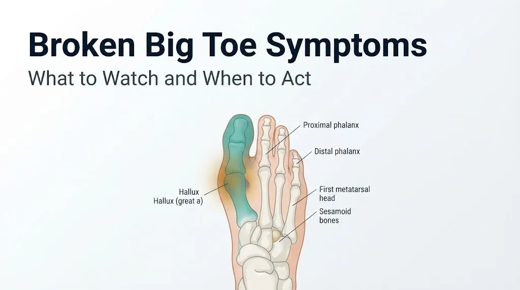
Broken big toe symptoms include immediate, localized pain, swelling that develops within minutes, visible bruising that deepens over 24 to 48 hours, and in many cases an inability to bear weight or bend the toe without sharp pain. The hallux (the medical term for the great toe) is not just another toe: it bears a disproportionate share of body weight and plays a central role in normal walking, which makes a fracture here more functionally impairing than a break in any of the lesser four toes.
The big toe’s mechanical importance is not trivial. According to the American Orthopaedic Foot and Ankle Society, the first ray (the functional unit including the first metatarsal, hallux, and its two sesamoid bones) bears approximately 40 to 60 percent of body weight during the push-off phase of each walking step. Fractures here do not just hurt; they disrupt the most loaded structure in the foot.
This article covers every named symptom of a broken big toe with the anatomical and physiological mechanism behind each one, distinguishes a hallux fracture from its three most common clinical mimics (sprain, turf toe, and sesamoid fracture), addresses which populations face different symptom presentations, and identifies the specific threshold that requires emergency evaluation today rather than a scheduled appointment next week.
Broken Big Toe Symptoms: The Full Clinical Picture
Broken big toe symptoms include sudden severe pain at the site of fracture, rapid onset of swelling, ecchymosis (bruising) visible within 24 to 48 hours, point tenderness directly over the broken bone, and varying degrees of functional impairment depending on which part of the hallux is fractured.
The complete symptom profile of a hallux fracture depends on two key factors: the location of the break within the toe and the mechanism of injury. Fractures of the proximal phalanx (the bone closest to the foot) tend to produce more severe functional impairment than fractures of the distal phalanx (the tip bone), because the proximal phalanx contributes to the first metatarsophalangeal joint and its ligamentous attachments. Distal phalanx fractures often involve the nail bed and produce a subungual hematoma (blood pooling under the toenail) as an additional distinctive feature.
The mechanism of injury predicts the fracture pattern. Direct trauma (dropping a heavy object on the toe, stubbing the toe hard against a fixed surface) most commonly produces transverse or comminuted fractures of the proximal phalanx. Hyperextension injuries at the first MTP joint during athletic activity produce fractures at the base of the proximal phalanx. Repetitive loading in runners and military recruits produces stress fractures of the hallux with an entirely different, insidious symptom progression.
| Fracture Location | Primary Symptom Pattern | Weight-Bearing Status | Key Distinguishing Feature |
|---|---|---|---|
| Proximal phalanx, shaft | Severe dorsal and lateral toe pain, marked swelling | Usually impaired or impossible | First MTP joint tenderness |
| Proximal phalanx, base | First MTP joint pain, pain with toe extension | Impaired | Pain with passive extension of hallux |
| Distal phalanx | Tip pain, subungual hematoma, nail deformity | Often preserved | Blood under toenail within hours |
| Stress fracture of hallux | Insidious activity-related pain over weeks | Initially preserved | No trauma event; progressive course |
Hallux Anatomy: Why the Big Toe Fractures Differently
The hallux fractures differently from the lesser toes because of its unique biomechanical role, its larger bone architecture, and its complex tendon and sesamoid apparatus that collectively make it the primary propulsive structure of the foot.
The proximal phalanx of the hallux is the largest phalanx in the foot, roughly twice the width of any lesser toe’s phalanx. Its base articulates with the first metatarsal head at the first metatarsophalangeal joint, a joint stabilized by the medial and lateral collateral ligaments, the plantar plate, and the flexor hallucis brevis tendons on each side. The joint capsule is reinforced inferiorly by the plantar plate, a dense fibrocartilaginous structure that resists hyperextension during push-off.
Beneath the first metatarsal head sit two small bones embedded within the tendons of the flexor hallucis brevis muscle: the tibial sesamoid (medial) and the fibular sesamoid (lateral). These sesamoids act like pulleys, redirecting the mechanical force of the flexor hallucis brevis and protecting the flexor tendon from direct ground contact during weight-bearing. They bear substantial compressive force with every step: estimates from biomechanical research published in the Journal of Bone and Joint Surgery suggest the sesamoids experience loading forces of up to three times body weight during the push-off phase of running.
Think of the hallux like the ball-and-hinge mechanism of a piston. The proximal phalanx is the piston shaft, the first MTP joint is the hinge, and the sesamoid bones are the ball bearings that keep the mechanism running smoothly. Break the piston shaft and the whole mechanism locks. Damage the ball bearings and the motion becomes painful even if the shaft itself is intact.
This architecture explains why hallux fractures produce more functional impairment than lesser toe fractures: there is no redundant weight-bearing neighbor to compensate, and the first ray cannot be offloaded without altering the entire gait cycle.
Broken Big Toe Pain: Where It Hurts and Why
Broken big toe pain is produced by activation of periosteal nociceptors, specialized pain-sensing nerve endings in the periosteum (the dense connective tissue membrane covering the outer surface of every bone), triggered by the immediate release of prostaglandins E2 and I2 and substance P at the fracture site.
The pain of a hallux fracture is typically immediate and sharp at the moment of injury, rated by most patients at 7 to 9 out of 10 on the Visual Analog Scale in acute complete fractures. This is not coincidence: the periosteum covering the hallux phalanges is richly innervated by branches of the medial dorsal cutaneous nerve (a branch of the superficial peroneal nerve) and the medial and lateral digital nerves of the hallux (branches of the medial plantar nerve). These nerves transmit the pain signal from periosteal nociceptors to the central nervous system within milliseconds of fracture.
Pain location is diagnostically informative. Fractures of the proximal phalanx produce pain that is maximum at the dorsal surface of the base of the big toe and at the first MTP joint line. Fractures of the distal phalanx produce pain at the very tip of the hallux and beneath the toenail. Stress fractures produce a diffuse, deep aching pain along the shaft of the proximal phalanx that is maximum during activity and in the toe’s dorsal midshaft rather than at any joint.
The American Academy of Orthopaedic Surgeons notes that point tenderness directly over the fracture site on physical examination, elicited by pressing firmly with one finger over the specific bone, is the most clinically reliable physical examination finding for confirming a toe fracture before imaging is performed.
People with diabetic peripheral neuropathy experience a significantly altered pain presentation. Nerve damage from chronic hyperglycemia reduces the density and function of peripheral nociceptors, meaning the same fracture that produces 8/10 pain in a healthy adult may produce only 3/10 pain or none at all in a person with advanced neuropathy. This pain reduction is not protective: it delays presentation and allows complications to develop undetected.
Broken Big Toe Swelling and Bruising: Mechanisms and Patterns
Broken big toe swelling begins within minutes of the injury and follows a predictable anatomical pattern that reflects the vascular architecture of the hallux and the tight soft tissue envelope surrounding it.
Swelling develops from two simultaneous processes. First, periosteal and intraosseous blood vessels are torn at the fracture site, producing a periosteal hematoma that fills the space between the bone surface and the overlying periosteum within minutes. Second, the inflammatory mediators released at the fracture site, particularly histamine and bradykinin, increase vascular permeability in the digital arteries of the hallux, allowing plasma proteins and fluid to leak into the surrounding interstitial tissue. Because the skin and soft tissue envelope of the hallux is relatively inelastic compared to larger body regions, this fluid accumulation produces rapid, visible swelling that is tightly constrained to the toe.
Bruising (ecchymosis) follows the fascial planes of the hallux and foot. Blood from the periosteal hematoma tracks along the periosteum and through the loose connective tissue planes of the dorsal foot, producing visible discoloration at the dorsum of the hallux within the first 24 hours. Over the following 24 to 48 hours, this bruising typically spreads toward the web space between the first and second toes and downward to the plantar surface. A bruise confined strictly to one location without this spreading pattern suggests a soft tissue contusion rather than a fracture.
In distal phalanx fractures, blood accumulates beneath the toenail, producing a subungual hematoma that appears as a dark maroon or black discoloration under part or all of the toenail. This is one of the most diagnostically specific features of a distal phalanx fracture. The pressure from the accumulating blood under the nail often produces throbbing pain that is disproportionate to the apparent injury.
Pregnant women experience more pronounced swelling from foot injuries due to baseline physiological peripheral edema and increased total circulating blood volume during pregnancy. Any significant foot or toe injury during pregnancy warrants evaluation by an obstetrician or primary care physician before standard imaging, as radiation protocols need to be considered.
Key Takeaway: Bruising that spreads from the big toe toward the web space and plantar surface within 48 hours is a strong clinical indicator of phalangeal fracture rather than a soft tissue bruise, and a blood blister under the toenail is a characteristic sign of distal phalanx fracture specifically.
Can You Walk on a Broken Big Toe
Whether you can walk on a broken big toe depends on the fracture location, fracture displacement, and which supporting structures of the first ray are intact.
Many people with a non-displaced fracture of the distal phalanx of the hallux can walk, though with significant pain and a limping gait pattern. The distal phalanx does not directly participate in the push-off phase of gait the way the proximal phalanx does, so a fracture here impairs weight-bearing less than a proximal injury. However, walking on any toe fracture without evaluation or appropriate support risks displacing a non-displaced fracture and converting a straightforward injury into one requiring surgical intervention.
Fractures of the proximal phalanx of the hallux impair walking far more severely. The proximal phalanx is the structural foundation of the push-off mechanism. During normal walking, the big toe dorsiflexes at the first MTP joint to approximately 60 to 70 degrees as body weight passes forward over the foot. A proximal phalanx fracture makes this motion acutely painful or mechanically impossible, forcing the person to walk with the foot externally rotated and weight shifted to the outer border, producing a characteristic antalgic gait.
The American Academy of Orthopaedic Surgeons recommends against attempting to assess injury severity by testing how much weight you can bear: preserved weight-bearing does not exclude a fracture, and a person with a non-displaced proximal phalanx fracture who walks on it without support risks creating a displaced fracture that changes the entire management approach.
To assess your functional status before a medical appointment:
- Sit with the foot non-weight-bearing and observe whether the toe rests in a normal or abnormal position at rest
- Note whether the toe is visibly crooked, shortened, or rotated compared to the other foot
- Gently press along the full length of the toe from tip to base, noting the exact location of maximum tenderness
- Attempt gentle range of motion at the tip joint only; stop if pain is severe
- Note whether walking is possible with pain rated at or below 5/10, or whether any weight-bearing produces severe sharp pain
- Bring this assessment to your appointment along with a description of the exact injury mechanism and timing
Broken Big Toe Symptoms vs. Sprained Big Toe
A broken big toe and a sprained big toe produce overlapping symptoms because both injuries involve the same anatomical structures and the same inflammatory response, but specific features of each allow clinical distinction before imaging.
A hallux sprain involves injury to the ligamentous and capsular structures of the first metatarsophalangeal joint without fracture of the phalangeal bones. The medial and lateral collateral ligaments, the plantar plate, or the joint capsule are stretched or partially torn. This produces pain at the joint line, swelling around the first MTP joint, and restricted joint motion, all of which are present in proximal phalanx fractures.
The distinguishing features lie in the pattern of maximum tenderness and the behavior of pain with specific movements:
| Feature | Broken Big Toe (Proximal Phalanx) | Sprained Big Toe |
|---|---|---|
| Maximum point tenderness | Directly over the phalanx shaft or base | At the first MTP joint line (medial or lateral) |
| Pain with axial load test | Severe, along bone shaft | Mild to moderate, at joint |
| Pain with passive toe extension | Present, due to fracture site tension | Present, due to plantar plate or ligament stretch |
| Visible deformity | Possible if displaced | Absent |
| Bruising pattern | Along dorsal toe shaft and web space | Concentrated at first MTP joint |
| X-ray finding | Fracture line visible | Normal bone, possible joint space changes |
| Ability to bear weight | Usually significantly impaired | Often preserved with mild sprain |
Only imaging can definitively differentiate the two. Any injury to the big toe that produces inability to bear weight, visible deformity, or severe point tenderness over the phalangeal shaft should be evaluated with plain radiographs (X-rays) by a primary care physician, urgent care physician, or emergency medicine physician.
Broken Big Toe vs. Turf Toe Symptoms
Turf toe is a specific injury of the plantar plate and surrounding capsular structures of the first metatarsophalangeal joint, produced by forced hyperextension of the hallux, and it produces a symptom pattern that overlaps substantially with a proximal phalanx fracture but has distinct differentiating features.
Turf toe occurs when the hallux is forcibly extended beyond its normal range of approximately 60 to 70 degrees of dorsiflexion. This happens when an athlete’s foot is planted and body weight pushes forward, driving the hallux into extreme extension. The plantar plate, the medial and lateral sesamoid ligaments, and the joint capsule are stretched or torn. The sesamoid bones may also be involved if the sesamoid-phalangeal ligaments are disrupted.
Turf toe symptoms are concentrated on the plantar surface and medial or lateral aspect of the first MTP joint, not on the dorsal toe shaft. Pain is maximum with passive extension of the hallux (reproducing the injury mechanism), whereas in a phalanx fracture, pain is maximum with palpation directly over the bone and is less specifically related to toe extension. Swelling in turf toe concentrates around the first MTP joint rather than along the phalanx shaft.
The American Orthopaedic Foot and Ankle Society classifies turf toe by severity into three grades. Grade 1 involves stretching of the capsuloligamentous complex without disruption: pain is mild, swelling is minimal, and weight-bearing is preserved. Grade 2 involves partial rupture: pain is moderate, swelling is marked, and movement is restricted. Grade 3 involves complete rupture of the plantar plate: pain is severe, weight-bearing is significantly impaired, and the hallux may be unstable at the first MTP joint.
MRI is the definitive imaging tool for confirming turf toe and assessing plantar plate integrity. Plain X-rays may show bony avulsion fragments in severe cases or sesamoid displacement but will not directly visualize plantar plate tearing. A board-certified orthopedic surgeon with foot and ankle specialty or a sports medicine physician should evaluate suspected Grade 2 or Grade 3 turf toe, as mismanagement can lead to chronic hallux instability.
Key Takeaway: Turf toe produces pain primarily on the plantar and medial-lateral surface of the first MTP joint after a forced extension mechanism, while a broken big toe produces maximum tenderness over the phalanx shaft after a direct crush or axial loading mechanism; imaging is required to confirm which injury is present.
Sesamoid Fracture Symptoms Near the Big Toe
A sesamoid fracture produces a symptom pattern that is distinct from a phalangeal fracture of the hallux but is located in the same anatomical region, making it one of the most frequently missed diagnoses in the context of big toe pain.
Sesamoid fractures involve the tibial sesamoid (the medial, larger of the two) or less commonly the fibular sesamoid (the lateral, smaller sesamoid). Because both sesamoids are embedded in the tendons of the flexor hallucis brevis and sit directly beneath the first metatarsal head on the plantar surface of the foot, the pain of a sesamoid fracture is located on the ball of the foot beneath the big toe, not in the toe itself. This plantar location is the single most clinically useful distinguishing feature.
Sesamoid fracture pain is characteristically worsened by direct pressure over the ball of the foot, by toe-off during walking, and by walking barefoot on hard floors. The pain is typically described as a deep, aching throb underneath the first metatarsal head, rather than in the toe itself. Pressing directly over the sesamoid with the thumb from the plantar surface reproduces the exact pain the patient identifies as their primary complaint.
Sesamoid fractures are most common in ballet dancers, runners, athletes who wear high-heeled footwear, and individuals with high-arched feet (cavus foot deformity), because these situations increase loading through the first metatarsal head and sesamoid apparatus. Research published in Foot and Ankle International has documented sesamoid stress fractures in dancers at rates substantially higher than in the general population, attributed to the extreme forefoot loading inherent in classical ballet technique.
Standard foot X-rays may not adequately visualize sesamoid fractures. A dedicated sesamoid axial view (a specific radiographic projection taken with the hallux in maximum dorsiflexion) is required to image the sesamoids on end. If plain radiographs are inconclusive, MRI provides definitive assessment of sesamoid bone integrity, sesamoid-phalangeal ligament status, and associated soft tissue injury.
Broken Big Toe Stress Fracture Symptoms
Broken big toe stress fracture symptoms develop over days to weeks through a fundamentally different biological process than acute phalangeal fractures, and the insidious onset is the defining clinical feature that separates this injury from all acute fracture presentations.
A stress fracture of the hallux occurs when repetitive loading accelerates osteoclastic bone resorption in the trabecular network of the proximal phalanx faster than osteoblastic bone formation can replace it, producing progressive microfracture accumulation and eventual cortical crack propagation. There is no single injury event the person can identify. Instead, the pain begins as a diffuse aching in the big toe during activity, initially appearing late in a run or march and resolving with rest.
As the stress fracture progresses, the pain threshold shifts downward. Pain that once required 45 minutes of running to appear now appears within 10 minutes, then at the start of activity, then during normal walking. The toe may show mild swelling over the proximal phalanx shaft without the acute bruising pattern of a traumatic fracture. Point tenderness directly over the dorsal midshaft of the proximal phalanx is the most reliable clinical finding.
The National Institute of Arthritis and Musculoskeletal and Skin Diseases identifies rapid increases in training intensity, surface changes, and inadequate recovery periods as the primary contributing factors in lower extremity stress fractures, including those affecting the hallux. Runners who increase weekly mileage by more than 10 percent per week, military recruits undergoing intensive basic training, and dancers who increase rehearsal hours rapidly are the populations most commonly affected.
Key features distinguishing stress fracture from acute fracture of the hallux:
- No identifiable single injury event
- Insidious onset over days to weeks
- Pain begins late in activity and progresses to earlier onset over time
- Mild swelling without acute bruising
- Point tenderness over phalanx midshaft, not at joint line
- Normal X-ray in early stages; MRI detects bone marrow edema before cortical crack is visible
Key Takeaway: A stress fracture of the hallux produces progressive activity-related pain with no trauma event, mild swelling without bruising, and a normal initial X-ray, making MRI necessary for diagnosis in any runner or military recruit with persistent big toe pain that worsens with activity over multiple weeks.
Broken Big Toe Symptoms in Children and Adolescents
Broken big toe symptoms in children and adolescents carry an additional clinical consideration not present in adults: the risk of growth plate injury at the base of the proximal phalanx of the hallux.
The physis (growth plate) of the proximal phalanx sits at its base, the point where the bone attaches to the first metatarsal head at the first MTP joint. In children and adolescents whose bones have not yet reached skeletal maturity (typically before age 16 in girls and 18 in boys), this growth plate is the mechanically weakest point in the entire first ray. A force that would produce a ligament sprain in an adult (whose bones are stronger than the ligaments) produces a Salter-Harris physeal fracture in an adolescent, because the growth plate fails before the ligaments do.
Salter-Harris classification of growth plate fractures ranges from Type I (a pure physeal separation, often visible only on MRI because plain X-rays appear normal) through Type V (crush injury of the growth plate, the most serious type). Type I and Type II fractures of the hallux proximal phalanx are the most common in clinical practice. Type I fractures produce pain and swelling at the base of the big toe that is clinically indistinguishable from a ligament sprain without imaging, but failure to recognize a growth plate injury risks disrupting the growth mechanism and producing a permanent deformity.
Any child or adolescent with pain, swelling, and point tenderness at the base of the big toe after an injury should be evaluated by a pediatric emergency medicine physician or primary care physician the same day, with plain X-rays as a minimum. If X-rays are normal but clinical suspicion remains high, an MRI or stress views (supervised gentle stressing of the joint under fluoroscopic imaging) are the next appropriate step. Parents should specifically inform the physician of the child’s age and whether they have had a growth evaluation recently.
Broken Big Toe Symptoms With Diabetes and Poor Circulation
Broken big toe symptoms in people with diabetes mellitus present a fundamentally different clinical picture that demands same-day medical evaluation regardless of how mild the symptoms appear.
Diabetes produces three independent processes that alter how a big toe fracture presents, heals, and complications develop. First, diabetic peripheral neuropathy damages the small fiber nerves that transmit pain signals from the periosteal nociceptors of the hallux, reducing or eliminating the acute pain response to fracture. A person with severe neuropathy may sustain a complete phalangeal fracture with only mild discomfort or none at all. Second, peripheral arterial disease (narrowing of the tibial, peroneal, and digital arteries from atherosclerosis accelerated by chronic hyperglycemia) reduces blood flow to the fractured bone and surrounding tissue, impairing the vascular-dependent processes of bone healing and infection response. Third, immune dysfunction from chronic hyperglycemia impairs neutrophil function and reduces the body’s ability to contain bacterial infection, dramatically increasing the risk of osteomyelitis (bone infection) if the skin overlying a fracture is broken, abraded, or macerated.
The combination of these three processes creates the risk of Charcot neuroarthropathy, a progressive destruction of bone and joint architecture that occurs in the insensate diabetic foot when repeated unrecognized trauma accumulates without protective modification of activity. An unrecognized or inadequately treated hallux fracture in a person with diabetic neuropathy can initiate or accelerate Charcot changes at the first MTP joint.
Any person with diabetes who suspects a big toe injury, regardless of pain severity, should be evaluated by their primary care physician or a podiatrist with expertise in diabetic foot care the same day. They should bring a description of the injury mechanism, their current diabetes management regimen, any history of prior foot ulcers or infections, and their most recent HbA1c level to that appointment. Do not wait for swelling or bruising to develop: in diabetic neuropathy, these signs may be absent or delayed even in the presence of a fracture.
Broken Big Toe Symptoms in Older Adults With Osteoporosis
Older adults with osteoporosis sustain hallux fractures from lower-energy mechanisms than younger adults, and the initial symptom severity may be lower, leading to delayed presentation and underestimated injury.
Osteoporosis reduces the trabecular bone density of the phalanges, meaning the structural lattice inside the proximal and distal phalanges of the hallux is less dense and less capable of withstanding normal load levels. The National Institutes of Health reports that osteoporosis affects approximately 10 million Americans, with fracture risk increasing substantially with each decade after age 50. In the foot, this translates to hallux fractures occurring from mechanisms that would produce only bruising in younger adults: stepping off a curb awkwardly, catching the toe on a rug edge, or a low-energy stumble.
The initial pain level in older adults with hallux fractures may be lower than expected for the fracture severity, for several reasons. First, reduced pain nociceptor density with aging is documented in the clinical literature. Second, pre-existing baseline toe pain from hallux valgus (bunion deformity), degenerative joint changes at the first MTP joint, or prior injuries may mask the new fracture pain. Third, some older adults attribute the new pain to their existing arthritis or gout and delay evaluation.
Older adults taking long-term oral corticosteroids (for conditions including rheumatoid arthritis, chronic obstructive pulmonary disease, or inflammatory bowel disease) face an additional risk layer. Corticosteroids suppress osteoblastic bone formation, further reducing bone mineral density and increasing fracture susceptibility even when baseline osteoporosis has not been formally diagnosed.
Any older adult who has had a fall, a direct impact to the toe, or a new onset of toe pain after any injury mechanism should have plain radiographs of the foot performed by their primary care physician or an emergency medicine physician the same day, with the explicit clinical question of whether the hallux shows a fracture. Age-appropriate pain reduction and protected weight-bearing should be initiated based on imaging findings, not based on pain severity alone.
Key Takeaway: Older adults with osteoporosis can sustain a complete hallux fracture from a low-energy mechanism with pain levels lower than expected for the fracture severity, making imaging after any big toe injury mandatory rather than discretionary in this age group.
Broken Big Toe Diagnosis: X-Ray and Clinical Evaluation
Diagnosing a broken big toe accurately requires a combination of clinical examination findings and plain radiographic imaging, with the imaging sequence tailored to the suspected fracture type and location.
For an acute hallux fracture, the standard imaging protocol consists of three plain radiographic views of the foot: anteroposterior (AP), oblique, and lateral views. These three views together visualize the full extent of the proximal and distal phalanges, the first MTP joint, and the first metatarsal head. A fracture line, cortical disruption, displacement, or angulation is visible on plain X-rays in the majority of complete acute phalangeal fractures. The degree of displacement, the presence of intra-articular extension (fracture entering the first MTP joint), and the angular alignment of fracture fragments are the key imaging features that determine whether surgical or non-surgical management is appropriate.
For suspected sesamoid fractures, the standard three-view foot series is insufficient. A dedicated sesamoid axial view radiograph is required, taken with the hallux in maximum passive dorsiflexion to project the sesamoids in isolation. Even with this dedicated view, distinguishing a sesamoid fracture from a bipartite sesamoid (a normal anatomical variant in which the sesamoid develops from two ossification centers and appears as two fragments on X-ray in up to 30 percent of the population) can be difficult. MRI is the preferred next step when sesamoid pathology is suspected and X-rays are equivocal.
For stress fractures of the hallux, plain X-rays are frequently normal in the first 2 to 4 weeks of injury. MRI is the gold standard for early stress fracture detection because it identifies bone marrow edema (the earliest pathological change in the trabecular bone) before any cortical crack is radiographically visible. A CT scan provides superior cortical detail when a stress fracture has progressed to a visible crack and surgical planning is under consideration.
The healthcare provider appropriate for initial evaluation is a primary care physician, urgent care physician, or emergency medicine physician depending on severity. Definitive management of displaced fractures, intra-articular fractures, or complex injuries involving the sesamoid apparatus should involve a board-certified orthopedic surgeon with foot and ankle fellowship training or a podiatrist with surgical training in forefoot reconstruction.
| Imaging Modality | What It Shows | Best Used For | Limitation |
|---|---|---|---|
| Plain X-ray (3 views) | Fracture line, displacement, joint involvement | Acute complete fractures | Misses early stress fractures, poor sesamoid visualization |
| Sesamoid axial view | Sesamoid fracture or bipartite sesamoid | Suspected sesamoid injury | Cannot differentiate fracture from bipartite variant alone |
| MRI | Bone marrow edema, soft tissue, plantar plate | Stress fractures, turf toe, equivocal X-rays | Cost, availability |
| CT scan | Cortical detail, fracture displacement | Surgical planning for complex fractures | Radiation, poor soft tissue visualization |
| Bone scan (technetium) | Increased metabolic activity at fracture | Stress fractures when MRI unavailable | Low specificity |
Broken Big Toe Complications to Watch For
Broken big toe complications develop in a predictable range that spans from cosmetic deformity from malunion to serious neurovascular emergencies requiring surgical intervention.
Malunion is the most common complication of hallux fractures that are inadequately immobilized or that receive delayed treatment. Malunion occurs when the fracture heals in an angulated or rotated position rather than anatomically correct alignment. In the hallux, malunion of the proximal phalanx can produce a permanent toe deformity, altered gait mechanics, shoe-fitting difficulties, and post-traumatic arthritis at the first MTP joint over months to years. The risk of malunion is highest in displaced fractures that are managed without adequate reduction and fixation.
Post-traumatic arthritis of the first MTP joint develops when a fracture extends into the joint surface (intra-articular fracture) and the articular cartilage is damaged or heals with an irregular surface. Cartilage has limited capacity for self-repair, and even a small step-off in the joint surface can produce progressive cartilage wear over months to years, eventually producing joint stiffness, chronic pain with activity, and reduced hallux range of motion. This complication is most common in Sanders-type intra-articular fracture patterns and in fractures treated without anatomic reduction.
Osteomyelitis (bone infection) is a rare but serious complication occurring most commonly when a hallux fracture is an open fracture (bone or bone fragments penetrate the skin) or when a diabetic or immunocompromised patient sustains a fracture with any overlying skin injury. The hallux is particularly vulnerable because its blood supply can be compromised by both the fracture itself and by the tight skin envelope limiting soft tissue swelling accommodation.
Complications to watch for after initial injury and during recovery:
- Progressive worsening pain rather than gradual improvement after the first week (suggests malunion, infection, or developing arthritis)
- Fever, increasing redness, warmth, or purulent discharge from any skin break near the fracture (suggests infection or osteomyelitis)
- Permanent toe deviation or angulation developing after initial swelling resolves (suggests malunion)
- Chronic stiffness and pain at the first MTP joint months after healing (suggests post-traumatic arthritis)
- Numbness or tingling in the hallux that develops or worsens after injury (suggests digital nerve compression from swelling or hematoma)
When Broken Big Toe Symptoms Require Emergency Care
Certain symptoms associated with a broken big toe require immediate emergency evaluation. Do not wait to see if these resolve on their own.
Call 911 or go to the nearest emergency room immediately if you experience:
- A wound or skin break over or near the toe with visible bone or bone fragments: this is an open fracture requiring emergency surgical debridement within hours to prevent bone infection
- Progressive, severe foot and toe pain that worsens disproportionately to the apparent injury and is not relieved by elevation: this is the cardinal warning sign of acute compartment syndrome of the foot, a surgical emergency requiring fasciotomy within hours
- Numbness, complete loss of sensation, or persistent tingling in the toes that develops after a toe or foot injury: indicates compression or injury to the digital nerves from edema within the tight compartments of the foot
- A toe that has turned cold, pale, blue, or white after injury: indicates compromise of the medial or lateral digital arteries of the hallux, a vascular emergency requiring immediate evaluation
- Visible deformity with the toe pointing in an abnormal direction that is accompanied by severe pain and swelling: indicates significant fracture displacement or dislocation of the first MTP joint requiring emergency reduction
These presentations can indicate open fracture, vascular injury, nerve compression, or acute compartment syndrome, all of which require emergency surgical evaluation, not an urgent care appointment scheduled for the next morning.
For individuals with diabetes who sustain any big toe injury: evaluation by a primary care physician or podiatrist is required the same day regardless of pain severity. Peripheral neuropathy may mask the warning symptoms listed above, making self-assessment insufficient in this population.
Frequently Asked Questions About Broken Big Toe Symptoms
How do I know if my big toe is broken or just badly bruised?
A broken big toe typically produces point tenderness directly over the phalangeal bone that is reproducible on pressing, while a bruise produces surface-level tenderness without a firm bony focus.
Bruising that spreads from the toe toward the web space and plantar arch within 48 hours, visible toe deformity, and inability to bear weight without sharp pain are all features that favor a fracture over a simple bruise.
Only plain X-rays can definitively distinguish a phalanx fracture from a soft tissue injury, and any big toe injury with these features warrants same-day imaging at an urgent care center, emergency department, or primary care physician’s office.
Can you walk on a broken big toe?
Walking may be possible with a non-displaced distal phalanx fracture of the hallux, but it is not clinically safe without evaluation and appropriate protection.
A fracture of the proximal phalanx typically impairs walking substantially due to the hallux’s role in the push-off phase of gait, producing sharp pain at the first metatarsophalangeal joint with each step.
Attempting to walk through a suspected hallux fracture without imaging risks displacing a non-displaced fracture and converting a non-surgical injury into one requiring surgical fixation.
What are the signs that a broken big toe needs emergency care?
A broken big toe requires emergency evaluation if the skin over the fracture is broken with visible bone, if foot pain is worsening progressively and out of proportion to the apparent injury, if the toe is cold, pale, or has lost sensation, or if visible deformity is present with severe swelling.
These signs indicate open fracture, compartment syndrome, vascular injury, or significant fracture displacement, all of which require surgical evaluation within hours.
Call 911 or go to the nearest emergency room immediately if any of these features are present; do not drive yourself if the pain prevents safe operation of a vehicle.
How long does a broken big toe take to heal?
Non-displaced fractures of the distal phalanx of the hallux typically heal in 4 to 6 weeks with buddy taping and a stiff-soled shoe.
Fractures of the proximal phalanx of the hallux, particularly those involving the first MTP joint, may require 6 to 8 weeks or longer with protected weight-bearing, and displaced fractures requiring surgical fixation may need 10 to 12 weeks before return to full activity.
Stress fractures of the hallux typically require 6 to 10 weeks of activity modification, and return to running or impact activity should be guided by a primary care physician or orthopedic surgeon rather than pain tolerance alone.
What is the difference between a broken big toe and turf toe?
A broken big toe produces maximum tenderness directly over the phalanx shaft and is caused by direct trauma or axial loading, while turf toe produces maximum tenderness at the plantar surface of the first MTP joint and is caused by forced hyperextension of the hallux.
Turf toe does not involve a phalangeal bone fracture; it is an injury to the plantar plate and capsuloligamentous complex of the first MTP joint.
Plain X-rays typically show normal bone in turf toe, while MRI is needed to visualize plantar plate disruption; phalangeal fractures are visible on X-ray in most acute cases.
Should someone with diabetes see a doctor for a broken big toe right away?
Yes, any person with diabetes who suspects a big toe injury requires same-day evaluation by a primary care physician or a podiatrist with expertise in diabetic foot care, regardless of pain severity.
Diabetic peripheral neuropathy reduces pain perception, meaning the injury may feel mild even when the fracture is severe or when early infection is developing.
Peripheral arterial disease and immune dysfunction in diabetes dramatically increase the risk of impaired bone healing and osteomyelitis following any foot fracture, making prompt evaluation and monitoring non-optional in this population.
Closing
The symptom pattern of a broken big toe tells a specific anatomical story. Pain located directly over the phalangeal shaft, bruising that spreads toward the web space within two days, inability to perform the push-off phase of walking without sharp pain, and point tenderness reproducible on direct pressure are the features that distinguish a hallux fracture from a sprain, a bruise, or a turf toe injury. None of these features alone confirms a fracture, but their combination in the context of a clear injury mechanism should prompt same-day imaging.
Your next step depends on your specific situation. Any deformity, open wound, cold or pale toe, or severe worsening pain means the emergency room now, without delay. Any suspected fracture without those emergency features means plain X-rays at an urgent care center or primary care physician’s office the same day or the next morning. Any person with diabetes skips the wait entirely and goes to a physician today regardless of pain level.
What you’ve read here gives you the anatomical language and the clinical framework to describe your injury accurately and ask the right questions. Bring the injury mechanism, the exact location of maximum tenderness, and a description of your functional limitations to that appointment, and you’ll give your provider everything they need to make the right call.