Eyelash Mites Symptoms: What Every Patient Needs in 2026
Eyelash mites symptoms include persistent eyelid itching that worsens at night, waxy or crusty debris at the base of the eyelashes, burning or dry eyes, and in advanced infestations, thinning or loss of eyelashes. These symptoms occur because two microscopic species of the Demodex genus colonize the hair follicles and glands along the eyelid margin, triggering an inflammatory response that disrupts normal eyelid and tear film function.
The prevalence of Demodex colonization increases sharply with age. According to research published in Optometry and Vision Science, Demodex infestation is detectable in approximately 84 percent of adults over age 60 and approaches universal prevalence in adults over 80. Many infestations remain asymptomatic at low mite densities, but when mite populations exceed a physiological threshold, the immune suppression they rely on fails and inflammatory symptoms emerge.
This article covers every clinically recognized symptom of eyelash mite infestation with the specific anatomical and physiological reason behind each one. It distinguishes the two Demodex species, explains which populations face the highest risk, identifies the red flags that require same-day ophthalmology evaluation, and clarifies what a proper clinical diagnosis involves.
Eyelash Mites Symptoms: What They Are and Why They Occur
Eyelash mites symptoms occur when the population density of Demodex folliculorum or Demodex brevis exceeds the threshold at which their surface proteins can suppress the eyelid’s innate immune response, triggering cytokine-driven inflammation along the eyelid margin.
At low colonization densities, which is the normal state for most adults, these mites live without producing symptoms. They use a specific immune evasion strategy: their outer cuticle proteins downregulate toll-like receptor 2 (TLR2) signaling on the eyelid epithelial cells. TLR2 is a key pattern recognition receptor of the innate immune system. When its signaling is suppressed, the host mounts no meaningful inflammatory response to the mites’ presence.
When mite populations grow, this suppression fails. The eyelid’s immune cells recognize mite waste products and shed cuticle fragments as foreign antigens. The result is a local inflammatory cascade involving interleukin-1beta, interleukin-6, and tumor necrosis factor-alpha (TNF-alpha) released from eyelid epithelial cells and recruited inflammatory cells. This cascade produces the redness, itching, swelling, and glandular obstruction that characterize symptomatic Demodex infestation.
Recognized eyelash mites symptoms in clinical order of frequency:
- Persistent eyelid itching, most intense at the eyelid margin
- Burning, stinging, or foreign body sensation in one or both eyes
- Crusty, sticky debris at the base of eyelashes, particularly on waking
- Cylindrical, waxy collarettes (cylindrical dandruff) encircling lash bases
- Redness and swelling of the eyelid margin
- Thinning, falling, or misdirected eyelashes
- Dry, gritty eye symptoms that do not respond to standard eye drops
- Blurred vision that clears temporarily with blinking (from tear film instability)
Immunocompromised individuals, including those receiving chemotherapy or living with untreated HIV disease, can develop severe symptomatic infestations at much lower mite population densities because their TLR2-mediated immune control is globally impaired.
What Are Eyelash Mites and How Do They Colonize the Eyelid?
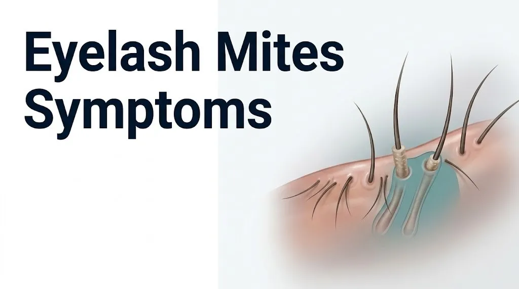
Eyelash mites are microscopic arachnids of the genus Demodex that colonize the pilosebaceous units of the eyelid margin, completing their entire life cycle within or immediately adjacent to the eyelash hair follicles and associated sebaceous and meibomian glands.
Two species colonize the human eyelid. Demodex folliculorum measures approximately 0.3 to 0.4 mm in length and lives within the eyelash hair follicle shaft itself, typically lodging head-first in groups of two to six mites per follicle. Demodex brevis is shorter (approximately 0.2 mm) and colonizes deeper structures: the sebaceous glands of Zeis and the meibomian glands (the lipid-secreting glands embedded in the tarsal plate of the eyelid).
Think of the eyelid follicle and gland system like a tightly packed apartment building. Demodex folliculorum occupies the main corridors (the follicle shafts), while Demodex brevis moves into the smaller utility rooms (the oil-secreting glands deeper in the eyelid). Both tenants cause different types of disruption depending on where they set up residence.
The mite life cycle takes approximately 14 to 18 days, passing through five stages: egg, larva, protonymph, deutonymph, and adult. Mites feed on sebaceous gland secretions (sebum) and eyelid epithelial cells. They are nocturnal migrants, emerging from the follicle at night to mate on the skin surface and re-entering follicles before daylight. This nocturnal activity pattern explains why eyelid itching and discomfort are often reported as being worse at night.
According to the American Academy of Ophthalmology, Demodex is the most prevalent ectoparasite in humans, with colonization rates rising progressively with age.
Early Signs of Eyelash Mites You Should Not Ignore
The earliest signs of eyelash mite infestation are mild but persistent eyelid margin itching, a sensation of slight gritty discomfort in the eyes, and barely visible waxy debris at the base of one or more eyelashes that does not wash away completely with routine face washing.
At this early stage, the mite population is growing but has not yet exceeded the TLR2 suppression threshold. The symptoms are subtle and easy to attribute to seasonal allergies, digital eye strain, or dry air. The key clinical distinction is persistence: early Demodex-related itching does not fully resolve with antihistamines or lubricating eye drops the way allergic or environmental irritation typically does.
A practical self-assessment at this stage involves looking closely at the lash bases in a well-lit mirror. Early collarette formation appears as a slight roughness or slight white scale at the very base of individual lashes, distinct from ordinary sleep discharge (which appears as a soft, removable crust over the entire lid margin and inner corner rather than as a tight wrap around individual lash shafts).
Quick Tip:
- Look for debris that appears wrapped tightly around the base of individual lashes rather than sitting loosely along the lid margin. That pattern is specific to Demodex folliculorum.
- Track whether the itching is distinctly worse at night than during the day. Nocturnal worsening aligns with Demodex mites’ documented nocturnal migration behavior.
- Older adults over 60 who notice new, unexplained eyelid irritation should mention it at their next comprehensive eye examination with an optometrist or ophthalmologist, as Demodex infestation in this age group is far more prevalent than other causes of new-onset eyelid itching.
Key Takeaway: Early eyelash mite infestation produces mild, persistent eyelid itching and gritty eye discomfort that does not respond to antihistamines or lubricating eye drops, and the itching is distinctly worse at night due to the mites’ documented nocturnal activity pattern.
Itching and Irritation as Eyelash Mite Symptoms
Eyelid itching from eyelash mites is produced by direct mechanical stimulation of the follicle epithelium by mite movement, by the inflammatory cytokine response to mite waste products accumulating in the follicle, and in some individuals by IgE-mediated allergic sensitization to Demodex cuticle proteins.
The itch is anatomically specific. It concentrates at the eyelid margin itself, the narrow strip of tissue between the line of lashes and the inner wet conjunctival surface, rather than spreading across the eyelid skin or into the eye. This location reflects the precise anatomical distribution of the Demodex colonies within the follicle orifices at the lid margin.
The inflammatory component involves mast cell degranulation stimulated by local cytokine release, releasing histamine into the eyelid tissue. This histamine-driven component explains why some patients find partial, temporary relief from antihistamine eye drops. However, the mechanical and cytokine-driven components persist regardless of histamine blockade, which is why the itch typically returns fully once antihistamine effect wanes.
The Cornea journal has published multiple studies linking high Demodex density to measurable increases in eyelid inflammatory markers, with itch severity correlating with mite count per eyelash examined. Research from the same journal found that patients with more than 5 mites per epilated eyelash had substantially higher symptom scores on validated ocular discomfort scales compared to patients with lower counts.
Contact lens wearers may experience amplified eyelid irritation from Demodex infestation because the lens edge interacts with the inflamed lid margin on every blink, creating friction at the exact site of maximum mite-driven inflammation. This population should specifically mention contact lens wear when being evaluated by an optometrist for eyelid symptoms.
Cylindrical Dandruff and Collarettes: The Most Specific Sign of Eyelash Mites
Cylindrical dandruff, also called collarettes, are tight, sleeve-like accumulations of waxy, keratinized material wrapping around the base of an eyelash shaft, and they are the most pathognomonic clinical sign of Demodex folliculorum infestation.
Collarettes form because the follicle is physically occupied by a cluster of mites lodged head-first within the follicle canal. Their bodies block normal desquamation (shedding of dead follicle epithelial cells) and sebum flow from the follicle orifice. The blocked material, a mixture of dead epithelial cells, mite waste products, and retained sebum, accumulates and hardens into the distinctive cylindrical collar that surrounds the lash shaft. Unlike ordinary sleep discharge or dried tear debris, this material does not wipe away easily and tends to recur at the same lash bases day after day.
The American Academy of Ophthalmology identifies collarettes at the lash bases as the clinical hallmark of Demodex blepharitis, distinguishable from the scaling of seborrheic blepharitis (which produces flaky, dandruff-like scales along the lid margin rather than tight cylindrical sleeves around individual lashes) and from the hard, crusted exudate of staphylococcal blepharitis (which occurs over the entire lid margin rather than specifically encircling lash bases).
To report this finding accurately to an eye care professional:
- Examine your eyelashes in a magnifying mirror under bright light.
- Look for visible debris that appears wrapped around the individual lash shaft, starting at the point where the lash exits the eyelid skin.
- Try to gently slide the debris along the lash with a dampened cotton swab. Collarettes from Demodex tend to be adherent and resist easy removal.
- Note how many lashes are affected and whether upper or lower lashes predominate.
- Photograph the finding if possible before your appointment to help the clinician correlate what you observed at home with their slit-lamp examination findings.
Women who use waterproof mascara may have difficulty distinguishing cosmetic residue from true collarettes. A dermatologist or ophthalmologist can differentiate these during a slit-lamp examination.
Eyelash Loss and Follicle Damage From Eyelash Mite Infestation
Eyelash loss, called madarosis, occurs as a consequence of long-standing, high-density Demodex folliculorum infestation when the sustained inflammatory process within the follicle damages the hair follicle architecture and disrupts the normal eyelash growth cycle.
The eyelash follicle, like all hair follicles, cycles through growth (anagen), regression (catagen), and rest (telogen) phases. The follicle wall normally provides structural and nutritional support for the growing lash. When a follicle is densely occupied by Demodex mites and simultaneously inflamed by the cytokine response their presence triggers, the follicle epithelium degenerates. The lash may grow back in an altered direction, a condition called trichiasis (inward-growing eyelashes that contact the corneal surface), or may fail to regrow at all.
Trichiasis is more than a cosmetic problem. An eyelash growing inward and contacting the corneal epithelium with every blink abrades the corneal surface repeatedly. This can produce persistent corneal erosions, corneal scarring, and, if superinfected, bacterial keratitis with risk of permanent vision loss.
According to research in the Ocular Surface journal, patients with moderate to severe Demodex blepharitis have measurably higher rates of eyelash irregularities, including lash thinning, misdirection, and loss, compared to control populations without Demodex infestation.
Older adults with long-standing, untreated Demodex blepharitis may present with noticeable eyelash thinning that they attribute to aging rather than to a treatable infestation. Any adult who notices progressive eyelash thinning or loss without a history of thyroid disease, chemotherapy, or other known cause should be evaluated by an ophthalmologist to assess for Demodex as a contributing factor, with a specific request for eyelash microscopy at the appointment.
Key Takeaway: Cylindrical collarettes wrapping tightly around individual lash bases are the single most specific sign of Demodex folliculorum infestation and distinguish it from other forms of blepharitis. Eyelash loss in the absence of a known systemic cause warrants ophthalmology evaluation to rule out Demodex follicle damage.
Dry Eye Symptoms Caused by Demodex Brevis and Meibomian Gland Dysfunction
Dry eye symptoms in eyelash mite infestation are caused specifically by Demodex brevis colonizing the meibomian glands, obstructing their orifices, and degrading the quality and quantity of the lipid layer of the tear film, which normally prevents rapid tear evaporation.
The meibomian glands are elongated sebaceous glands embedded vertically in the tarsal plate of both the upper and lower eyelids. In healthy eyes, they secrete meibum, a complex lipid mixture that forms the outermost layer of the tear film. This lipid layer acts like a seal on the tear surface, dramatically slowing the rate at which the aqueous (watery) layer of tears evaporates between blinks.
When Demodex brevis colonizes the meibomian gland ducts, the mite bodies, along with accumulated debris from their feeding and reproduction, obstruct the gland orifice. Meibum cannot flow freely onto the tear surface. The lipid layer thins or becomes irregular, and the aqueous layer of the tear film evaporates much faster than normal between blinks. This evaporative dry eye pattern produces tear film hyperosmolarity, a state in which the remaining tear film becomes concentrated, irritating the corneal epithelial cells and conjunctival surface cells through osmotic stress.
The result is the classic dry eye triad: burning, stinging, and a persistent feeling of something in the eye (foreign body sensation). These symptoms are often reported as worse in environments with airflow (fans, air conditioning, outdoor wind) because evaporation is accelerated in those conditions.
The National Eye Institute identifies meibomian gland dysfunction as the most frequent underlying cause of dry eye disease in adults. Demodex brevis is one of the recognized contributors to MGD that is often overlooked when patients are evaluated only for environmental and hormonal causes of dry eye.
Postmenopausal women are at elevated risk for both meibomian gland dysfunction and Demodex brevis infestation simultaneously, as estrogen decline reduces meibum secretion quality and increases sebaceous gland vulnerability to Demodex colonization. Any postmenopausal woman with persistent dry eye symptoms not responding to standard lubricating drops should ask their ophthalmologist specifically about Demodex evaluation.
Demodex Folliculorum vs. Demodex Brevis: Different Symptoms Explained
Demodex folliculorum primarily causes anterior blepharitis symptoms including eyelid margin itching, collarettes, and lash loss, while Demodex brevis primarily causes posterior blepharitis and meibomian gland dysfunction symptoms including dry eye, foreign body sensation, and frothy tear film.
This distinction matters because a patient presenting predominantly with anterior eyelid itching and collarette formation has a different predominant species driving their symptoms than a patient presenting predominantly with burning dry eyes and abnormal meibum secretion. While both species often coexist, the predominant symptoms point toward the anatomically relevant species and guide treatment focus.
| Feature | Demodex Folliculorum | Demodex Brevis |
|---|---|---|
| Size | 0.3 to 0.4 mm (longer) | 0.2 mm (shorter) |
| Primary anatomical location | Eyelash hair follicle shaft | Meibomian glands and sebaceous glands of Zeis |
| Type of blepharitis produced | Anterior blepharitis | Posterior blepharitis |
| Primary symptoms | Eyelid margin itching, collarettes, lash loss | Dry eye, burning, foreign body sensation, frothy tear film |
| Characteristic finding | Cylindrical dandruff encircling lash bases | Frothy, foamy discharge at inner eyelid corner; thickened meibum on lid margin expression |
| Tear film effect | Indirect (lid margin inflammation disrupts tear spreading) | Direct (meibomian gland obstruction reduces lipid layer) |
| Worse timing | Nocturnal (mite surface migration at night) | Throughout the day with prolonged screen use or dry environments |
The frothy, foam-like discharge sometimes noticed at the inner corner of the eye by patients with Demodex brevis infestation is produced by the abnormal meibum mixing with the aqueous tear layer. Normal meibum is a smooth oil that stays at the tear surface. Demodex-degraded meibum has altered surface tension properties that cause it to emulsify and foam when mixed with aqueous tears, producing the visible foam that some patients describe as “soap bubbles” at the inner eye corner.
Eyelash Mite Symptoms in Older Adults and Age-Related Risk
Eyelash mite symptoms in adults over 60 tend to be more pronounced and longer-standing than in younger adults because the sebaceous gland output that Demodex feeds on increases with age-related skin changes, and because the immune surveillance capacity of older eyelid tissue is reduced, allowing larger mite populations to establish before immune responses mount.
The age-related increase in Demodex prevalence is well-documented in the ophthalmological literature. Research published in Optometry and Vision Science found that Demodex infestation is detectable in approximately 48 percent of adults in their 30s, rising to approximately 84 percent in adults over 60, and approaching universal prevalence in those over 80 years of age. High-density, symptomatic infestation follows a similar age-related pattern.
Aging skin produces a relative increase in the proportion of oleic acid in sebum composition and a thickening of the keratinized follicle walls. These changes create a more favorable habitat for Demodex mites. Additionally, the age-related decline in T-cell function and natural killer cell activity reduces the immune pressure that normally keeps mite populations in check.
Symptoms in older adults may be attributed incorrectly to dry eye disease from menopause (in women), to the normal aging of the ocular surface, or to the side effects of systemic medications. Several classes of medications commonly used by older adults, including antihistamines, antidepressants, and diuretics, reduce tear production and produce dry eye symptoms that can mask or overlap with Demodex-driven ocular surface disease.
Any older adult with new or worsening eyelid itching, eye burning, or unexplained eyelash thinning should ask their optometrist or ophthalmologist for a Demodex-specific examination, including slit-lamp inspection of the eyelash bases for collarettes, at their next scheduled comprehensive eye examination.
Eyelash Mites and Rosacea: Overlapping Symptoms and Shared Risk
Patients with rosacea have dramatically higher rates of Demodex infestation than the general population, with studies in the British Journal of Dermatology finding Demodex counts up to four times higher in rosacea-affected skin than in control skin, and their eyelash mite symptoms are frequently more severe and more resistant to standard lid hygiene measures.
The physiological link is sebaceous gland hyperactivity. Rosacea involves dysregulation of the innate immune response in sebaceous gland-rich facial skin, producing chronic inflammation, vasodilation, and increased sebaceous secretion. Demodex mites feed on sebum produced by the sebaceous glands of Zeis associated with eyelash follicles. The elevated sebum production in rosacea skin creates an abundant food supply that supports a larger, denser mite population.
Ocular rosacea, the ophthalmic manifestation of systemic rosacea, produces eyelid margin inflammation, meibomian gland dysfunction, and recurrent chalazia (blocked meibomian gland cysts) that overlap substantially with Demodex blepharitis symptoms. In fact, Demodex infestation may be a contributing driver of ocular rosacea severity in some patients, rather than a separate coexisting condition. Research published in the Cornea journal has found an association between higher Demodex density and more severe ocular rosacea symptoms.
Patients with rosacea who are receiving dermatological treatment for their skin disease should specifically ask their treating dermatologist whether their eyelid symptoms might represent Demodex blepharitis, and request a referral to an ophthalmologist for eyelid-specific evaluation if they have ongoing eyelid itching, crusting, or ocular burning that is not resolving with their current rosacea management.
Key Takeaway: Rosacea patients have four times higher Demodex mite counts than the general population, making them at high risk for severe eyelash mite symptoms that may not respond to standard lid hygiene alone, and they should be evaluated for Demodex blepharitis specifically rather than generic blepharitis management.
Eyelash Mite Symptoms in Immunocompromised Individuals
Immunocompromised individuals can develop severe, high-density Demodex infestation with dramatically more pronounced eyelash mite symptoms at younger ages and lower colonization thresholds than immunocompetent adults, because the T-cell-mediated and innate immune mechanisms that normally limit mite population growth are impaired.
The categories of immunosuppression most associated with severe Demodex infestation include HIV disease (particularly with CD4 counts below 200 cells per microliter), organ transplant recipients receiving calcineurin inhibitors or other anti-rejection drugs, patients receiving systemic corticosteroids for autoimmune conditions, and patients receiving chemotherapy for hematological or solid tumor malignancies.
In these populations, Demodex folliculorum can produce a distinctive and more severe pattern of eyelid disease called Demodex folliculitis, in which the follicle becomes overtly inflamed with visible erythema (redness), pustule formation at follicle orifices, and accelerated eyelash loss. The eyelid skin surrounding the follicles may also show scaling and crusting that extends beyond the lid margin onto the eyelid skin itself.
Topical or systemic corticosteroids deserve particular mention. While corticosteroids are often prescribed for eyelid inflammatory conditions, using them in a patient with undiagnosed Demodex blepharitis can worsen the infestation by suppressing the immune response that limits mite population growth, paradoxically intensifying symptoms after an initial apparent improvement.
Any immunocompromised individual with eyelid symptoms should be evaluated by an ophthalmologist rather than managing symptoms at home, because the risk of rapid progression to corneal involvement (Demodex-associated keratitis) is substantially higher in these patients than in immunocompetent adults. Clinicians should ask specifically about immunosuppressive medications before treating any blepharitis presentation without a confirmed etiology.
Eye Mites Symptoms vs. Allergic Conjunctivitis and Blepharitis From Other Causes
Eye mites symptoms differ from allergic conjunctivitis primarily by their location (eyelid margin vs. the conjunctival surface), by the presence of collarettes at lash bases (specific to Demodex), and by the pattern of persistence (chronic, year-round, and nocturnal versus seasonal or allergen-dependent).
Allergic conjunctivitis produces prominent redness and swelling of the conjunctiva (the pink membrane covering the white of the eye and lining the eyelid inner surface), intense watery discharge, and bilateral itching that tracks with allergen exposure. Eyelash mite infestation produces eyelid margin symptoms rather than conjunctival symptoms as the primary presentation, with collarettes as a pathognomonic finding not seen in allergic conjunctivitis.
| Feature | Eyelash Mites (Demodex) | Allergic Conjunctivitis | Staphylococcal Blepharitis | Seborrheic Blepharitis |
|---|---|---|---|---|
| Collarettes at lash bases | Yes, pathognomonic | No | No | No |
| Eyelid scaling type | Cylindrical around lash shaft | Absent | Hard crusts along lid margin | Greasy, flaky scales on lid margin |
| Conjunctival injection | Mild, if present | Prominent, bilateral | Mild | Mild |
| Discharge type | Waxy debris at lash bases | Watery, bilateral | Sticky, purulent | Greasy, minimal |
| Itching location | Eyelid margin, worse at night | Conjunctiva, bilateral, worse with allergen | Eyelid margin | Eyelid margin |
| Response to antihistamines | Partial and temporary | Good response | No response | No response |
| Age group most affected | Adults over 40, increasing with age | Any age, often childhood onset | Any age | Adults with seborrheic dermatitis |
The differentiation from staphylococcal blepharitis is important because staphylococcal blepharitis may require different treatment, including topical antibiotic therapy, while Demodex blepharitis requires anti-mite treatment. Both can coexist: Demodex mites carry Bacillus oleronius, a bacterial symbiont found in the mite’s gut, which can serve as a secondary source of inflammatory antigens at the eyelid.
A primary care physician can provide an initial evaluation and referral, but definitive differentiation between these blepharitis types requires slit-lamp examination by an optometrist or ophthalmologist.
When Eyelash Mite Symptoms Indicate a Serious Eye Condition
Eyelash mite symptoms indicate a potentially serious eye condition when they are accompanied by changes in vision, photophobia, eye pain, visible corneal opacity, or rapidly worsening redness that does not improve with eyelid hygiene, as these signs suggest progression to Demodex-associated keratitis (corneal inflammation) with risk of permanent vision impairment.
Demodex-associated keratitis develops when the corneal epithelium is directly invaded or severely irritated by Demodex organisms or their associated inflammatory products. The cornea is exquisitely sensitive: it is the most densely innervated tissue in the body per unit area. Even mild corneal epithelial disruption produces intense pain, severe photophobia, and tearing. Vision may blur if the corneal surface irregularity interferes with the normal refractive surface.
Corneal involvement is more likely when Demodex infestation is untreated for a prolonged period, when the patient is immunocompromised, when trichiasis (inward-growing lashes abrading the cornea) has gone unrecognized, or when the patient has been using topical corticosteroid preparations that suppress corneal immune defenses while allowing mite populations to expand.
Go to an ophthalmologist or emergency eye care clinic the same day if you notice any of the following alongside eyelid symptoms:
- Sudden or progressive vision blurring not explained by tears or discharge
- Eye pain that is new, severe, or worsening
- Sensitivity to light (photophobia) that makes opening the eye outdoors difficult
- A visible white or gray spot on the colored part of the eye (cornea)
- Significant eye redness that is worsening rather than stable over 48 hours
- Eyelashes growing inward and visibly contacting the eye surface
These presentations can indicate Demodex keratitis, corneal ulceration, or secondary bacterial keratitis and require emergency ophthalmological assessment, not an urgent care appointment or home management.
How Eyelash Mites Are Diagnosed by an Eye Care Professional
Eyelash mites are definitively diagnosed by epilating (removing) two to three eyelashes from the affected eyelid margin, placing them on a glass slide, and examining them under a microscope at 4x to 16x magnification to visualize adult mites, eggs, or larval forms attached to the lash shaft.
This simple, non-painful procedure can be performed in an optometrist’s or ophthalmologist’s office during a standard examination. The American Academy of Ophthalmology identifies epilated lash microscopy as the clinical standard for Demodex confirmation. The finding of cylindrical dandruff on the lash sample at lower magnification, combined with visualization of mite forms at higher magnification, confirms the diagnosis.
A slit-lamp biomicroscope examination of the eyelid margin before lash epilation allows the clinician to assess the extent of collarette formation, the degree of anterior lid margin inflammation, the appearance of the meibomian gland orifices, and the quality of expressed meibum. All of these findings together provide a complete clinical picture of whether both Demodex folliculorum and Demodex brevis are likely contributors.
More advanced diagnostic technology includes in vivo confocal microscopy (IVCM), which allows real-time imaging of Demodex mites within the follicles and meibomian glands without lash removal, providing mite count data per follicle. This technology is available at academic medical centers and some tertiary referral ophthalmology practices.
To prepare effectively for your appointment with an ophthalmologist or optometrist:
- Do not remove existing eyelash debris or clean the lid margin immediately before the appointment, as the collarettes and debris are part of the diagnostic finding.
- Bring a photograph taken at home of the lash base debris, if you were able to capture it.
- List all eye drops, lid hygiene products, and cosmetics currently in use, as some products can partially reduce visible collarettes.
- Report how long symptoms have been present, whether they are worse at night, and whether standard dry eye treatments have provided any relief.
- Ask specifically about being evaluated for Demodex, as this is not always part of a routine blepharitis workup unless the clinician specifically examines for collarettes.
Eyelash Mite Symptoms and Treatment Options in 2026
Eyelash mite infestation producing symptomatic Demodex blepharitis has, as of 2023, a specifically FDA-approved treatment: lotilaner ophthalmic solution 0.25%, marketed as Xdemvy, the first and only anti-Demodex ophthalmic medication approved by the U.S. Food and Drug Administration for this indication, representing a defined advance in the management of this condition beyond previously available off-label options.
Before the 2023 FDA approval of lotilaner ophthalmic solution, treatment relied primarily on lid hygiene measures using products containing terpinen-4-ol (the active antiparasitic component of tea tree oil), physical cleaning of the eyelid margins to mechanically remove mites and collarettes, and off-label use of agents including topical ivermectin and metronidazole gel. These approaches required prolonged use and did not always achieve complete resolution.
Lotilaner works by blocking inhibitory glutamate-gated chloride channels in the Demodex nervous system, producing mite paralysis and death. Clinical trials submitted to the FDA demonstrated statistically meaningful reductions in collarette scores and mite counts over a six-week treatment course. A board-certified ophthalmologist can assess whether this treatment is appropriate based on symptom severity, mite count, and patient health history.
Lid hygiene measures remain an evidence-supported adjunct regardless of primary treatment approach. These include daily warm compress application to the closed eyelids for 5 to 10 minutes to soften meibomian gland secretions, followed by gentle mechanical eyelid massage, and eyelid margin cleaning with diluted baby shampoo or commercially available tea tree oil-based eyelid cleanser products.
People with rosacea undergoing systemic treatment with oral doxycycline for their skin condition may have some partial benefit for their eyelid Demodex symptoms as a secondary effect, as doxycycline has anti-inflammatory properties relevant to meibomian gland dysfunction. However, this is not a primary anti-mite treatment and should not substitute for ophthalmology evaluation and targeted therapy.
Frequently Asked Questions About Eyelash Mites Symptoms
How do I know if I have eyelash mites?
The most specific indicator of eyelash mites is cylindrical, waxy debris encircling the base of individual eyelash shafts (collarettes), combined with persistent eyelid margin itching that is worse at night.
These findings are distinct from standard blepharitis because collarettes wrap tightly around individual lashes rather than appearing as general lid margin scaling or crusting.
Confirmation requires a microscopic examination of epilated eyelashes by an optometrist or ophthalmologist; self-assessment can suggest the diagnosis but cannot confirm it.
Can you feel eyelash mites moving on your eyelashes?
Most people cannot directly feel individual mites moving because Demodex are 0.2 to 0.4 mm in length and move slowly within and between follicles.
The nocturnal itching and crawling sensation that some patients report is not individual mite movement but rather the eyelid inflammatory response and the physical friction of mites emerging from follicles to mate on the skin surface at night.
If you are experiencing a distinctly crawling or creeping sensation on the eyelids at night, report this specifically to your ophthalmologist, as it is a recognized patient description of high-density Demodex infestation.
Do eyelash mites cause eyelashes to fall out?
Yes: high-density Demodex folliculorum infestation can cause eyelash loss (madarosis) by damaging the hair follicle architecture through sustained inflammation and physical obstruction of the follicle canal.
Long-standing infestation may also cause eyelashes to regrow in an inward direction (trichiasis), where they contact and abrade the corneal surface with every blink.
Any unexplained eyelash thinning or loss in an adult without a known systemic cause (thyroid disease, chemotherapy, alopecia areata) warrants evaluation by an ophthalmologist, who should include eyelash microscopy as part of the workup.
Are eyelash mites contagious?
Demodex mites are transmitted through close personal contact, sharing of eye cosmetics, pillowcases, or towels, and possibly through face-to-face proximity, though the precise transmission route in humans is not fully established by controlled research.
According to the American Academy of Ophthalmology, they are not classified as a contagious disease in the public health sense, and normal social contact does not typically result in clinically significant transmission between adults.
Household members of patients with symptomatic Demodex blepharitis may benefit from individual assessment by an eye care provider, particularly if they share bedding or cosmetics with the affected individual.
Can eyelash mites affect your vision?
Eyelash mites can affect vision indirectly through tear film instability from meibomian gland dysfunction, which produces transient blurring that clears with blinking, and directly through corneal involvement (Demodex keratitis) in advanced or immunocompromised cases.
Tear film-related blurring from Demodex brevis infestation is temporary and fluctuating, typically worst during prolonged reading or screen use and temporarily improved by blinking or lubricating eye drops.
Persistent vision blurring, eye pain, or photophobia in the context of eyelid symptoms should be evaluated by an ophthalmologist the same day, as corneal keratitis from Demodex can cause permanent corneal scarring if not treated promptly.
What is the white stuff at the base of my eyelashes?
The white, waxy material encircling the base of individual eyelashes is called cylindrical dandruff or collarettes, and it is the most pathognomonic finding of Demodex folliculorum infestation.
It forms because the mites occupying the hair follicle canal block normal shedding of follicle epithelial cells and sebum flow, causing accumulated dead skin cells, mite debris, and retained secretions to compact into a tight cylindrical sleeve around the lash shaft.
If you can see this material and it recurs at the same lash bases after washing, schedule an appointment with an optometrist or ophthalmologist and ask specifically for a Demodex evaluation, as this is not a finding produced by any other common eyelid condition.
Eyelash mite infestation is far more prevalent than most people realize, and far more treatable than most patients with unexplained blepharitis symptoms are ever told. The symptoms you have been attributing to allergies, dry eyes, or aging eyelids may have a specific, identifiable cause at the base of your lashes.
The single most practical action is this: look at your lash bases in a well-lit mirror for tight, cylindrical debris wrapped around individual shafts. If you see it, that finding alone is worth an appointment with an optometrist or ophthalmologist. Ask them specifically to evaluate for Demodex at the slit lamp, and ask whether epilated lash microscopy is indicated.
Persistent eyelid itching, burning, and lash changes that have not responded to standard lubricating drops or basic eyelid hygiene deserve a proper diagnosis. You now have the clinical framework to describe your symptoms precisely and ask the right questions.