Pneumonia Symptoms: Full 2026 Clinical Guide for Adults
Pneumonia symptoms include fever, a productive or dry cough, sharp chest pain that worsens with breathing, and shortness of breath that may develop gradually or arrive suddenly, depending on the type and severity of the lung infection. These symptoms occur because pathogens reach the alveoli (the tiny air sacs deep in the lungs) and trigger an inflammatory cascade that fills those air sacs with fluid and immune cells, directly impairing the lung’s ability to transfer oxygen into the bloodstream.
According to the Centers for Disease Control and Prevention, pneumonia is responsible for more than 40,000 deaths annually in the United States and accounts for approximately 1.5 million emergency room visits each year. It remains the leading infectious cause of hospitalization in adults and children nationwide, which is why recognizing its full symptom range, including the atypical presentations that affect older adults and immunocompromised individuals, carries real clinical weight.
This article covers every recognized symptom of pneumonia with the physiological reason behind each one. It distinguishes bacterial from viral and atypical presentations, explains how symptoms change by age and health status, identifies the specific thresholds that require emergency care, and prepares you to communicate your clinical picture accurately to the provider who evaluates you.
Pneumonia Symptoms: What They Are and Why They Occur
Pneumonia produces its symptoms through two parallel processes: localized lung inflammation that impairs gas exchange, and a systemic immune response that generates fever, fatigue, and body aches throughout the body.
When a pathogen reaches the alveolar space, resident alveolar macrophages detect it through pattern recognition receptors (specifically toll-like receptors) and immediately release pro-inflammatory cytokines. This cytokine release is the starting point of virtually every symptom that follows.
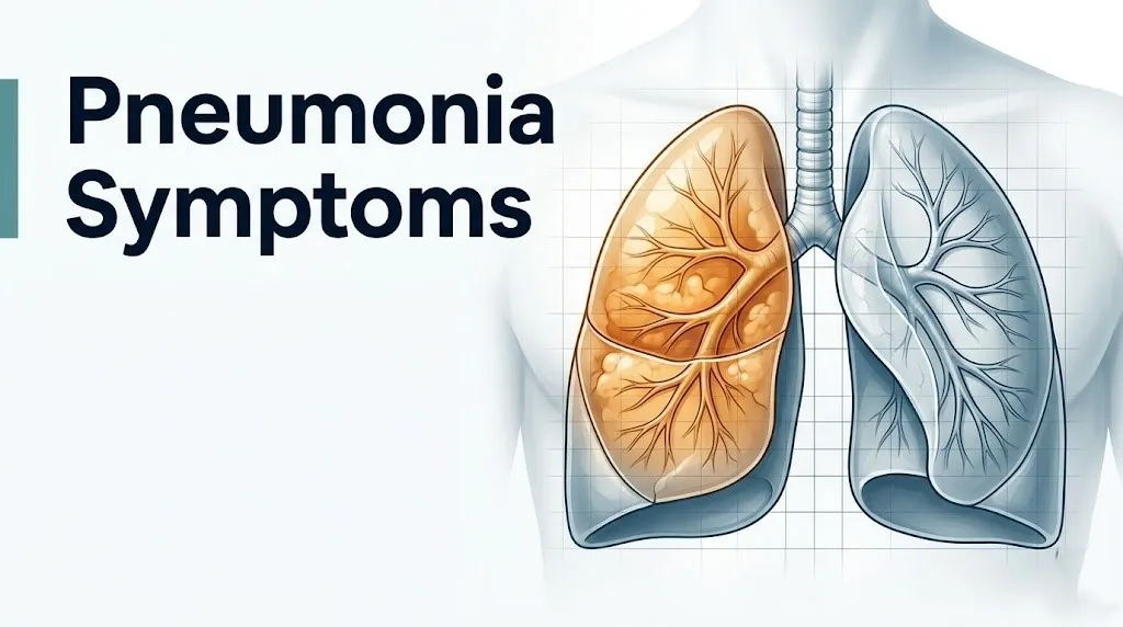
Think of the alveoli like millions of tiny inflatable bubbles lining the inside of each lung. Each bubble transfers oxygen into the blood and removes carbon dioxide from it. When infection fills those bubbles with fluid, mucus, and inflammatory debris, the exchange stops. The lung essentially develops waterlogged pockets that cannot breathe, and the body’s oxygen supply drops accordingly.
The localized consequences (cough, chest pain, difficulty breathing) come from the physical disruption of the lung architecture. The systemic consequences (fever, chills, fatigue, muscle aches, headache) come from cytokines released into the bloodstream, traveling to the brain and distant tissues.
Primary pneumonia symptoms in order of clinical frequency:
- Cough, productive or dry depending on the causative organism
- Fever (temperature at or above 38°C / 100.4°F), often with chills and sweating
- Shortness of breath, ranging from mild on exertion to severe at rest
- Chest pain that sharpens with each breath or with coughing
- Fatigue that is often disproportionate to other symptoms
- Rapid breathing (respiratory rate above 20 breaths per minute)
- Rapid heart rate (tachycardia above 100 beats per minute)
- Confusion or altered mental status, particularly in adults over 65
Immunocompromised individuals, including people receiving chemotherapy or living with HIV/AIDS, may present with fewer and milder symptoms despite having extensive lung consolidation on imaging, because the blunted immune response produces less cytokine signaling.
Early Signs of Pneumonia to Recognize Immediately
The earliest signs of pneumonia are a worsening cough that develops in the days following an upper respiratory illness, a low-grade fever that rises progressively over 24 to 48 hours, and fatigue that feels disproportionately severe compared to a typical cold.
Many pneumonia cases begin as a viral upper respiratory infection that appears to improve and then suddenly worsens. This pattern, sometimes called the “two-hit” course, occurs because the initial viral infection damages the mucociliary escalator, the layer of ciliated cells lining the airways that normally sweeps pathogens upward and out of the lung. With that defense compromised, a secondary bacterial infection can reach the alveoli more easily.
At the very earliest stage, the symptoms are frustratingly similar to influenza or a severe cold: muscle aches, fatigue, low fever, and a cough that begins dry and then turns productive. The distinguishing early feature is chest involvement. A cough that produces pain when you take a deep breath, or a sensation of heaviness in the chest that worsens rather than improves after three to five days of illness, points toward lower respiratory tract involvement rather than a simple upper airway infection.
Quick Tip:
- Track whether your fever is rising or falling over 24-hour periods. A fever that peaks and then climbs again after a brief improvement is a clinical flag for bacterial superinfection.
- Note whether coughing produces colored sputum. Rust-colored or brownish sputum is associated with Streptococcus pneumoniae infection. Yellow-green sputum is consistent with purulent bacterial infection generally.
- Children under 5 and adults over 65 should have a lower threshold for evaluation when any of these early signs appear, because both age groups can deteriorate quickly before classic pneumonia symptoms fully declare themselves.
Fever and Chills in Pneumonia: The Cytokine Mechanism Explained
Fever in pneumonia is produced when interleukin-1beta (IL-1beta), interleukin-6 (IL-6), and tumor necrosis factor-alpha (TNF-alpha), released by activated alveolar macrophages in response to the pathogen, reach the brain and stimulate the hypothalamic thermoregulatory center to raise the body’s temperature set point via prostaglandin E2 synthesis.
This is not the body simply overheating. It is a precisely orchestrated immune signal. The hypothalamus receives the cytokine message, interprets it as evidence of a serious threat, and deliberately raises the temperature set point to make the environment hostile to pathogens. The shaking chills that often accompany a high fever in pneumonia are the body’s muscle-shivering mechanism generating heat to reach the new, elevated set point quickly.
The magnitude of the fever corresponds roughly to the intensity of the inflammatory response. Bacterial pneumonia, particularly lobar pneumonia from Streptococcus pneumoniae, often produces high fevers in the range of 39 to 40°C (102 to 104°F) with dramatic chills. Atypical pneumonia from Mycoplasma pneumoniae tends to produce lower-grade fevers because the organism triggers a different and slower cytokine profile.
According to the National Heart Lung and Blood Institute (NHLBI), fever is among the most consistent presenting features of bacterial pneumonia in adults, present in approximately 80 percent of hospitalized pneumonia cases.
Older adults over 65 are an important exception. Immunosenescence, the age-related decline in immune cell function, reduces the magnitude of cytokine release from aging macrophages. As a result, elderly patients with confirmed pneumonia on chest X-ray often have temperatures below 38°C, and some present with no fever at all. Absence of fever in an older adult does not rule out pneumonia and should not reassure caregivers when other symptoms are present.
Key Takeaway: Fever in pneumonia is a deliberate cytokine-driven immune response, not random heat generation. Elderly patients may not develop fever even with confirmed pneumonia due to age-related reduction in macrophage cytokine output, making other symptom patterns more important in this age group.
Cough in Pneumonia: What It Sounds Like and What It Means
The cough of pneumonia is produced when inflammatory fluid and debris in the infected alveoli and adjacent bronchioles stimulate cough receptors in the airway epithelium, driving a forced expiratory effort to clear material from the lower respiratory tract.
In bacterial pneumonia, the cough is typically productive early in the illness, producing sputum that may be yellow, green, rust-colored (a finding particularly associated with Streptococcus pneumoniae, where red blood cells from the alveolar exudate produce the characteristic color), or blood-tinged. The sputum is the actual inflammatory exudate being expelled from the infected lung tissue.
Atypical pneumonia from Mycoplasma pneumoniae or Chlamydophila pneumoniae tends to produce a prominent, persistent dry cough because these organisms damage the respiratory epithelium through direct cell toxicity rather than producing the thick exudate of bacterial lobar pneumonia. The cough can be severe enough to cause chest wall muscle soreness and disturb sleep for weeks.
Viral pneumonia from influenza or SARS-CoV-2 initially produces a dry cough that may become productive if a secondary bacterial infection develops.
The American Thoracic Society notes that a new cough producing purulent (pus-containing) sputum is one of the three key clinical criteria, alongside fever and radiographic consolidation, used to confirm community-acquired pneumonia diagnosis.
A productive cough with blood (hemoptysis) is a red flag that requires same-day evaluation by a primary care physician or emergency medicine physician. While minor blood streaking can occur from airway irritation, substantial hemoptysis suggests either significant lung tissue destruction, a necrotizing pneumonia process, or a complicating condition such as a pulmonary embolism.
Chest Pain With Pneumonia and Why It Happens
Chest pain in pneumonia is pleuritic chest pain: a sharp, stabbing discomfort that worsens with each breath, deep inhalation, or cough, produced by inflammation of the parietal pleura (the outer lining of the lung cavity) where it is richly supplied by intercostal nerve branches.
The lungs are surrounded by two membrane layers: the visceral pleura (adhering to the lung surface) and the parietal pleura (lining the chest wall). Normally, these layers slide frictionlessly against each other during breathing. When pneumonia involves the peripheral lung tissue adjacent to the pleural surface, the inflammatory process spreads to the visceral pleura and then across the pleural space to the parietal pleura. The parietal pleura is the layer with sensory innervation from the intercostal nerves. When inflamed, every expansion of the chest wall during inhalation stretches these inflamed nerves, producing the characteristically sharp, breath-dependent pain.
This type of pain is clinically different from cardiac chest pain. Cardiac pain typically does not change with breathing or body position. Pleuritic pain is sharply worse with inhalation and often eases when you hold your breath or press on the chest wall.
The location of pleuritic chest pain corresponds to the location of the affected lung. Left lower lobe pneumonia produces left lower chest pain. Right lower lobe pneumonia produces right lower chest pain that can radiate toward the right shoulder, occasionally mimicking gallbladder or liver pathology.
People with underlying rib fractures (older adults with osteoporosis, for example) may experience more severe pain with the same degree of pleural inflammation because rib fractures amplify intercostal nerve irritation with every breath. These individuals may underventilate (take shallow breaths to avoid pain) and develop respiratory complications more rapidly.
Shortness of Breath in Pneumonia: How Alveolar Consolidation Impairs Breathing
Shortness of breath in pneumonia is caused by ventilation-perfusion (V/Q) mismatch: the infected, consolidated areas of the lung continue to receive blood flow from the pulmonary circulation, but those areas can no longer transfer oxygen into that blood because the alveolar air spaces are filled with inflammatory fluid instead of air.
The consequence is that oxygen-poor blood leaves the consolidated lung segments and mixes with oxygenated blood from healthy lung segments, lowering the overall oxygen concentration of the blood leaving the heart. This is called intrapulmonary shunting. The brain detects the falling blood oxygen level (measured by chemoreceptors in the carotid and aortic bodies) and drives increased respiratory rate and effort to compensate.
The degree of shortness of breath directly correlates with how much of the lung is consolidated. A small area of pneumonia in one lobe may produce only mild dyspnea on exertion, noticed when climbing stairs. Bilateral pneumonia involving multiple lobes can produce shortness of breath at rest, with oxygen saturation dropping rapidly during even minimal movement.
The Infectious Diseases Society of America (IDSA) and American Thoracic Society joint clinical guidelines identify a respiratory rate above 30 breaths per minute as one of the key severity thresholds in the CURB-65 score, indicating a need for hospital-level evaluation.
Measuring breathing rate is something anyone can do at home. Count how many times your chest rises in 60 seconds while sitting quietly. A resting rate above 20 is above normal. Above 25 warrants urgent evaluation by a primary care physician or urgent care provider. Above 30 warrants going to an emergency room the same day.
Pregnant women are at elevated risk for severe respiratory compromise from pneumonia because the growing uterus elevates the diaphragm and reduces total lung capacity by approximately 4 to 5 percent, reducing the available respiratory reserve when consolidation further limits gas exchange.
Key Takeaway: Shortness of breath in pneumonia is not just “feeling winded.” It reflects measurable oxygen deprivation from alveolar consolidation, and a resting respiratory rate above 30 breaths per minute is a validated emergency threshold that warrants hospital evaluation the same day.
Fatigue, Headache, and Body Aches as Pneumonia Symptoms
Fatigue, headache, and widespread muscle aches (myalgia) in pneumonia are systemic symptoms driven by the same cytokine cascade that produces fever: TNF-alpha suppresses appetite and promotes generalized fatigue through direct action on the hypothalamus, while prostaglandins distributed systemically through the bloodstream sensitize pain receptors in muscles and connective tissue.
These symptoms are not incidental. They are the body’s deliberate response to redirect energy toward immune function. TNF-alpha specifically suppresses muscle protein synthesis and promotes skeletal muscle catabolism to liberate amino acids for immune cell production and acute phase protein synthesis in the liver. The result is a profound sense of physical weakness that is often disproportionate to the apparent respiratory symptoms, particularly in the first 24 to 48 hours of illness.
Headache is present in up to 50 percent of Mycoplasma pneumoniae cases, according to clinical case series reviewed in the American Journal of Respiratory and Critical Care Medicine, and is more prominent in atypical pneumonia than in typical bacterial pneumonia. The mechanism involves direct CNS penetration of cytokines across the blood-brain barrier, stimulating prostaglandin production within cerebral tissues.
Loss of appetite (anorexia) accompanies these systemic symptoms in most moderate to severe pneumonia cases, driven by IL-1beta and TNF-alpha acting on appetite-regulating hypothalamic circuits. This is clinically relevant because dehydration and inadequate caloric intake during a pneumonia illness can accelerate deterioration, particularly in older adults and those with diabetes who are managing blood glucose through oral intake.
Adults with chronic kidney disease are particularly vulnerable to the systemic consequences of the cytokine response, as impaired renal function reduces clearance of inflammatory mediators and may amplify the magnitude and duration of systemic symptoms.
Walking Pneumonia Symptoms and How They Differ From Typical Pneumonia
Walking pneumonia is an informal term for atypical pneumonia, most commonly caused by Mycoplasma pneumoniae, characterized by a gradual onset of dry cough, low-grade fever, headache, and fatigue that is mild enough that many people continue to function and move around rather than feeling acutely ill.
The name comes from the observation that patients can literally keep walking around. Unlike typical lobar bacterial pneumonia, which often strikes with dramatic, acute-onset high fever, rigors, and chest pain severe enough to force bed rest, Mycoplasma pneumonia creeps in over one to three weeks with symptoms that initially resemble a persistent cold.
The reason for this milder presentation lies in the organism’s mechanism of injury. Mycoplasma pneumoniae lacks a cell wall and cannot invade lung tissue directly. Instead, it adheres to the ciliated respiratory epithelium via a surface protein called the P1 adhesin, disrupting ciliary function and releasing hydrogen peroxide that damages the epithelium over time. This damages the airway lining gradually rather than triggering the massive neutrophilic exudate that consolidates a lobe rapidly in Streptococcal pneumonia.
| Feature | Typical Pneumonia (Streptococcal) | Walking Pneumonia (Mycoplasma) |
|---|---|---|
| Onset | Abrupt, within 24 to 48 hours | Gradual, over 1 to 3 weeks |
| Fever | High (39 to 40°C), with rigors | Low-grade (37.5 to 38.5°C) |
| Cough | Productive, purulent, rust-colored | Dry, persistent, prominent |
| Chest pain | Sharp, pleuritic, breath-dependent | Mild chest discomfort or absent |
| Headache | Less prominent | Often prominent |
| Ability to function | Usually significantly impaired | Often partially maintained |
| Age group most affected | All ages, highest in elderly | School-age children, young adults |
Walking pneumonia is common in school-age children and young adults, and outbreaks occur in closed settings like schools and college dormitories. A board-certified primary care physician can distinguish these presentations with chest X-ray and specific testing, including Mycoplasma IgM serology or a respiratory PCR panel.
Bacterial vs. Viral Pneumonia Symptoms
Bacterial pneumonia typically produces an abrupt onset with high fever, rigors, productive cough, and pleuritic chest pain, while viral pneumonia tends to develop more gradually with prominent dry cough, milder fever, and more prominent myalgia and fatigue.
The difference in symptom pattern reflects the different inflammatory pathways these organisms trigger. Bacterial pathogens (particularly Streptococcus pneumoniae and Haemophilus influenzae) stimulate a massive neutrophilic inflammatory response in the alveoli, producing dense, lobar consolidation rapidly. The large volume of exudate irritates the pleura quickly and produces the dramatic pleuritic pain and high fever characteristic of typical bacterial pneumonia.
Viral pneumonia, caused by influenza virus, RSV, or SARS-CoV-2, produces a different pattern. Viruses replicate intracellularly within respiratory epithelial cells. The immune response is driven by interferon signaling and natural killer cell activity rather than neutrophilic exudate, producing diffuse interstitial inflammation rather than lobar consolidation. This interstitial pattern causes widespread gas exchange impairment with less focal chest pain but more prominent shortness of breath and oxygen desaturation.
According to clinical data reviewed in The Lancet Respiratory Medicine, the COVID-19 pneumonia pattern is notable for bilateral interstitial infiltrates on CT imaging with prominent hypoxemia that may be disproportionate to the degree of subjective breathlessness, a phenomenon called “silent hypoxia” or “happy hypoxia” in early publications. This makes pulse oximetry monitoring particularly important in suspected COVID-related pneumonia.
Staphylococcus aureus pneumonia deserves specific mention. This organism can produce necrotizing pneumonia, characterized by rapid tissue destruction, cavitation, hemoptysis, and severe sepsis. It occurs most commonly as a post-influenza complication. Any patient who had influenza and develops dramatically worsening chest symptoms with bloody sputum warrants emergency room evaluation the same day, as this presentation has high mortality without rapid antibiotic treatment.
Key Takeaway: Bacterial pneumonia typically hits fast with high fever and productive cough. Viral pneumonia tends to arrive more gradually with prominent fatigue, dry cough, and sometimes severe oxygen desaturation that appears before the person feels as sick as their blood oxygen level suggests.
Pneumonia Symptoms in Children: What Parents Need to Know
Pneumonia symptoms in children under 5 years old include fast breathing (the most reliable early sign), noisy or labored breathing, persistent fever, reduced feeding or drinking, and unusual irritability or lethargy, and warrant same-day evaluation by a pediatrician or pediatric emergency medicine physician when present.
Children’s respiratory systems differ from adults’ in ways that change the symptom picture. Their airways are proportionally smaller, so even moderate swelling and secretions from a lower respiratory infection produce more resistance and work of breathing than the same degree of inflammation would in an adult. A child breathing fast is working much harder per breath than it might appear from the outside.
The American Academy of Pediatrics defines tachypnea (fast breathing) in young children as a respiratory rate above 50 breaths per minute in infants under 2 months, above 40 in children aged 2 months to 12 months, and above 30 in children aged 1 to 5 years. Any child meeting these thresholds with fever or cough warrants evaluation.
Signs of breathing difficulty in children that require urgent evaluation:
- Breathing so fast the belly visibly moves with each breath (abdominal breathing)
- Skin pulling inward between the ribs or at the throat notch with each breath (retractions)
- Nostril flaring with every breath
- Grunting sound at the end of each exhalation
- Blue or gray color around the lips (central cyanosis)
- Child too tired to feed, drink, or respond normally
- High-pitched stridor on inhalation
Respiratory syncytial virus (RSV) is the leading cause of viral pneumonia and bronchiolitis in infants under 12 months. In premature infants and those with congenital heart disease, RSV pneumonia can produce severe respiratory failure rapidly and requires immediate evaluation.
Pneumonia Symptoms in Adults Over 65: Atypical Presentation Explained
Pneumonia symptoms in adults over 65 frequently include confusion, sudden falls, reduced activity tolerance, and loss of appetite rather than the classic triad of cough, fever, and chest pain, because age-related immune changes blunt the inflammatory signals that produce typical pneumonia symptoms in younger adults.
The specific mechanism is immunosenescence: the progressive decline in immune cell number and function with age. Aging alveolar macrophages produce lower quantities of IL-1beta, IL-6, and TNF-alpha in response to the same pathogen load that would trigger robust cytokine release in a younger immune system. Without a strong cytokine signal, the hypothalamus does not raise the temperature set point as high, resulting in absent or minimal fever.
Simultaneously, the aging brain is more sensitive to the neurological effects of cytokines and hypoxia. Even modest drops in blood oxygen concentration, which younger adults may tolerate without obvious cognitive change, can produce acute confusion and disorientation in older adults with pre-existing cerebrovascular disease or reduced cognitive reserve.
A 2019 study published in JAMA Internal Medicine examining pneumonia presentation in elderly patients found that approximately 30 percent of adults over 75 with radiographically confirmed pneumonia did not meet the classic symptom criteria of fever plus cough plus shortness of breath. Their presenting complaint was most often functional decline, confusion, or a fall.
If an older adult in your care develops sudden confusion without an obvious cause, stops eating or drinking, becomes uncharacteristically drowsy, or begins falling without their usual gait, request same-day evaluation by their primary care physician and specifically ask about a chest X-ray to rule out pneumonia.
Adults over 65 who smoke or have COPD are at elevated risk for pneumonia caused by Haemophilus influenzae in addition to Streptococcus pneumoniae, and their blunted respiratory reserve means consolidation that a healthy adult would tolerate can produce respiratory failure much faster in this population.
Pneumonia Symptoms Without Fever and Silent Pneumonia
Pneumonia without fever is a recognized clinical presentation that occurs in specific populations: elderly adults, immunocompromised individuals, and those taking corticosteroids or other immune-suppressing medications, all of whom have a blunted cytokine response that fails to trigger the hypothalamic fever mechanism even with significant lung consolidation.
The term “silent pneumonia” refers loosely to pneumonia that produces minimal or atypical symptoms, sometimes discovered incidentally on a chest X-ray or CT scan performed for another reason. This is distinct from the classic presentation and creates a genuine diagnostic challenge: the patient does not feel sick enough to seek care, yet their lungs may be substantially compromised.
In people taking oral corticosteroids for conditions like rheumatoid arthritis, asthma, or inflammatory bowel disease, corticosteroid-mediated suppression of macrophage and neutrophil function can dramatically reduce the inflammatory response to a pulmonary infection. Fever may be absent, cough may be mild, and the chest X-ray finding can appear ahead of significant symptoms.
People with HIV/AIDS present an important specific case. Pneumocystis jirovecii pneumonia (PCP), the most common opportunistic lung infection in people with advanced HIV disease and CD4 counts below 200 cells per microliter, typically produces progressive shortness of breath over days to weeks with minimal productive cough and little or no fever. The breathlessness worsens with exertion and progresses to rest dyspnea. Without treatment, PCP is life-threatening. Any immunocompromised person with progressive unexplained breathlessness should be evaluated by an infectious disease specialist without delay.
The practical implication is that the absence of fever does not rule out pneumonia, particularly in older adults, those on immunosuppressive therapy, or those with HIV. Any progressive unexplained dyspnea or cough lasting more than one week in these populations warrants a chest X-ray ordered by a primary care physician.
Key Takeaway: Fever-free pneumonia is a documented pattern in elderly adults, corticosteroid users, and immunocompromised individuals. In these groups, progressive breathlessness, confusion, or functional decline should prompt a chest X-ray even without a typical fever or cough.
Signs That Pneumonia Is Getting Worse
Signs that pneumonia is getting worse include worsening shortness of breath at rest (not just with activity), rising or spiking fever after a period of improvement, confusion or disorientation that is new or worsening, a respiratory rate that is climbing rather than falling, and oxygen saturation dropping below 94% on a pulse oximeter.
A well-known clinical pattern is apparent initial improvement followed by sudden deterioration, particularly when a viral pneumonia is complicated by bacterial superinfection. The bacteria exploit the disrupted mucociliary defenses from the viral illness to colonize the alveolar space, producing a second, often more severe inflammatory wave. This pattern was documented extensively in post-influenza Staphylococcus aureus pneumonia and in bacterial superinfections following COVID-19.
Measuring oxygen saturation at home with a pulse oximeter provides actionable clinical information. According to the IDSA and ATS joint pneumonia guidelines, an oxygen saturation below 94% on room air in a previously healthy adult is a threshold that warrants evaluation in an emergency setting, not a phone call or a next-available appointment.
| Symptom Change | Home Monitoring Appropriate | Urgent Care Same Day | Emergency Room Now |
|---|---|---|---|
| Mild improvement each day | Yes | No | No |
| Plateau with no improvement after 48 hours on treatment | No | Yes | No |
| Fever rising after initial improvement | No | Yes | No |
| New confusion or disorientation | No | No | Yes |
| Oxygen saturation below 94% | No | No | Yes |
| Respiratory rate above 30/min | No | No | Yes |
| Blue lips or fingertips | No | No | Yes |
| Inability to speak a full sentence | No | No | Yes |
People with COPD or asthma require closer monitoring because their baseline lung function is already reduced. A decline that might be manageable at home in a healthy adult may represent a critical event in someone with 50% predicted FEV1 at baseline.
Pneumonia Symptoms vs. Bronchitis, Flu, and COVID-19
Pneumonia is distinguished from acute bronchitis by the presence of lung consolidation visible on chest X-ray and by systemic symptoms (high fever, oxygen desaturation, pleuritic chest pain) that are not characteristic of airway-limited bronchitis.
Acute bronchitis inflames the bronchial airways but does not reach the alveoli. The cough produces clear or yellow mucus, fever is mild or absent, and breathing difficulty is minimal in otherwise healthy adults. There is no alveolar consolidation, no gas exchange impairment, and no pleuritic pain. Pneumonia, by contrast, involves the alveoli directly and produces the systemic inflammatory response and gas exchange impairment that acute bronchitis does not.
| Feature | Pneumonia | Acute Bronchitis | Influenza | COVID-19 Pneumonia |
|---|---|---|---|---|
| Alveolar involvement | Yes | No | Sometimes (viral pneumonia) | Yes (bilateral interstitial) |
| Fever | High, often with rigors | Low-grade or absent | High, abrupt onset | Variable; may be absent |
| Cough | Productive or dry, with chest pain | Productive, no chest pain | Dry initially, then productive | Dry, prominent |
| Chest pain | Pleuritic, breath-related | Sore sternum from coughing | Myalgic chest wall pain | Variable |
| Shortness of breath | Present, often at rest in severe cases | Minimal | Mild to moderate | Can be severe with silent hypoxia |
| Oxygen desaturation | Common in moderate to severe cases | Absent | Present in severe cases | Common, may precede symptoms |
| Duration | Weeks with treatment | 1 to 3 weeks | 1 to 2 weeks | Variable |
The clinical difficulty is that influenza, COVID-19, and bacterial pneumonia all produce fever, cough, and fatigue, and they can coexist: a person can have influenza and develop bacterial pneumonia as a secondary complication within the same illness course. Clinical evaluation with chest X-ray and specific laboratory testing is the only reliable way to distinguish these conditions.
Any person whose influenza-like illness was improving and then sharply worsens on day 5 to 7 with rising fever and new chest pain warrants same-day evaluation by a primary care physician or emergency medicine physician to rule out bacterial superinfection.
Emergency Symptoms: When to Call 911 or Go to the ER for Pneumonia
Certain symptoms associated with pneumonia require immediate emergency evaluation. Do not wait to see if these resolve on their own.
Call 911 or go to the nearest emergency room immediately if you or someone you are caring for experiences:
- Oxygen saturation below 90% on a pulse oximeter (indicating severe hypoxemia and impending respiratory failure)
- Blue or gray discoloration of the lips, tongue, or fingertips (cyanosis, indicating critical oxygen deprivation)
- Inability to complete a sentence without stopping to breathe
- Respiratory rate above 30 breaths per minute at rest, confirmed over 60 seconds
- Sudden confusion, agitation, or inability to recognize familiar people or surroundings (indicating hypoxic encephalopathy or sepsis)
- Systolic blood pressure below 90 mmHg with dizziness or near-fainting (hypotension suggesting septic shock)
- High fever above 40°C (104°F) that does not respond to fever-reducing measures and is accompanied by rigors and confusion
- Bloody cough (hemoptysis) producing more than a streak of blood
- Chest pain so severe that breathing becomes extremely shallow (risk of respiratory failure from inadequate tidal volume)
- Any pneumonia symptoms in a newborn, infant under 3 months, or immunocompromised individual that include any degree of breathing difficulty
These presentations can indicate respiratory failure, septic shock, or hypoxic brain injury. They require emergency medical assessment with oxygen supplementation, intravenous antibiotics, and potentially mechanical ventilatory support. An urgent care appointment or a phone call is not the appropriate response to any item on this list.
How Pneumonia Is Diagnosed and What to Tell Your Doctor
Pneumonia is diagnosed through a combination of clinical history, physical examination findings, chest imaging, and laboratory tests, with a chest X-ray showing pulmonary infiltrates (areas of consolidation or interstitial changes) serving as the standard confirmatory test in most outpatient and emergency settings.
A primary care physician, urgent care physician, or emergency medicine physician will typically evaluate breathing rate, oxygen saturation by pulse oximetry, heart rate, blood pressure, and temperature before examining the chest with a stethoscope. The hallmark physical examination finding in bacterial pneumonia is bronchial breath sounds and dullness to percussion over the consolidated lung segment, produced because dense consolidated tissue transmits sound differently than air-filled lung tissue.
To describe your symptoms accurately to your provider:
- State when symptoms started, whether they began suddenly or gradually, and whether they followed a prior illness.
- Describe your cough: dry or productive, sputum color, presence of blood.
- Report your temperature, both the highest reading and when it occurred.
- State whether your shortness of breath is present only with exertion, with light activity, or at rest.
- Describe the chest pain: whether it is sharp or dull, whether it worsens with breathing, whether it is localized or diffuse.
- Report your current oxygen saturation if you have a home pulse oximeter.
- List all medications including corticosteroids, immunosuppressants, and any antivirals already taken.
The IDSA/ATS joint guidelines recommend a chest X-ray for all patients with suspected community-acquired pneumonia. If the X-ray is negative but clinical suspicion remains high (particularly in immunocompromised patients or those with an atypical presentation), a CT scan of the chest provides higher sensitivity for early or atypical infiltrates.
Blood tests including a complete blood count (CBC) with differential, C-reactive protein (CRP), and procalcitonin help distinguish bacterial from viral pneumonia and gauge severity. Blood cultures are recommended for hospitalized patients to identify the causative organism and guide antibiotic selection. A urinary antigen test for Legionella and Streptococcus pneumoniae antigens can confirm specific pathogens within hours without waiting for culture results.
Frequently Asked Questions About Pneumonia Symptoms
How do I know if I have pneumonia or just a bad cold?
Pneumonia produces lower respiratory tract involvement with fever, shortness of breath, and chest pain, while a cold stays in the upper airway with nasal congestion, sore throat, and minimal fever.
The key distinguishing features are pleuritic chest pain (sharp, breath-related), oxygen saturation below 94%, and a respiratory rate above 20 breaths per minute, none of which occur with a typical cold.
Any cough illness lasting more than one week with worsening fever or new shortness of breath warrants evaluation by a primary care physician to rule out pneumonia with a chest X-ray.
Can you have pneumonia without a fever?
Pneumonia without fever is documented in elderly adults, immunocompromised individuals, and people taking corticosteroids, because these populations have a blunted cytokine response that does not reliably trigger the hypothalamic fever mechanism.
According to clinical data reviewed in JAMA Internal Medicine, approximately 30 percent of adults over 75 with confirmed pneumonia did not present with fever as a primary symptom.
Absence of fever should not reassure caregivers when an older adult develops new confusion, reduced appetite, or functional decline, because these can be the presenting signs of pneumonia in this age group.
What does pneumonia chest pain feel like?
Pneumonia chest pain is pleuritic: sharp, stabbing, and directly worsened by taking a deep breath, coughing, or sneezing, localized to the side of the chest corresponding to the affected lung.
It is produced by inflammation of the parietal pleura (the chest cavity lining), which is innervated by intercostal nerves that are stretched and irritated with each breath.
Pneumonia chest pain is distinct from cardiac chest pain because it changes sharply with breathing and body position, whereas cardiac pain is typically constant and unaffected by respiratory effort.
How long do pneumonia symptoms last?
Most symptoms of mild to moderate community-acquired pneumonia improve within 3 to 5 days of starting appropriate antibiotic or antiviral treatment, with fatigue and residual cough persisting for 2 to 4 weeks after the acute illness.
The Infectious Diseases Society of America notes that radiographic clearing (resolution of the chest X-ray abnormality) typically lags behind clinical improvement by several weeks and should not be used as the sole criterion for return to full activity.
Older adults and immunocompromised patients often have longer recovery timelines, with fatigue and reduced exercise tolerance persisting for 4 to 8 weeks, and require follow-up chest X-ray at 6 to 8 weeks to confirm resolution.
What oxygen level is dangerous with pneumonia?
An oxygen saturation below 94% on room air in an adult with pneumonia warrants urgent hospital-level evaluation, according to the IDSA and ATS joint clinical practice guidelines for community-acquired pneumonia management.
An oxygen saturation below 90% represents severe hypoxemia and is a clinical emergency requiring immediate supplemental oxygen and emergency room assessment.
Home pulse oximeters provide useful monitoring during pneumonia recovery, but a reading in the normal range does not rule out early respiratory deterioration, particularly in patients with COVID-19 pneumonia who can have silent hypoxia with normal subjective sensation of breathing.
What are the first signs of pneumonia in an elderly person?
The first signs of pneumonia in an elderly person are often confusion, reduced appetite, sudden falls, unusual drowsiness, or a noticeable decrease in their normal activity level, rather than the classic cough and fever seen in younger adults.
This atypical presentation occurs because immunosenescence reduces cytokine output from aging alveolar macrophages, blunting fever, while even modest drops in blood oxygen from alveolar consolidation produce cognitive changes in older adults with reduced cerebrovascular reserve.
Any elderly person who develops sudden unexplained confusion or functional decline should be evaluated by their primary care physician the same day, with a chest X-ray requested specifically to rule out pneumonia.
Pneumonia is one of those conditions where the difference between “I’ll see how I feel tomorrow” and “I need to go to the ER now” is a measurable number: your breathing rate and your oxygen saturation. Count your breaths at rest for 60 seconds. Check your oxygen with a pulse oximeter if you have one. A resting rate above 30 or an oxygen reading below 94% are the two numbers that change the decision from urgent care to emergency room.
For elderly family members, the symptom to watch for is not a dramatic cough or a high fever. It is confusion, a fall, or an unusual quietness. Get a chest X-ray ordered by their primary care physician before assuming it is something else.
You now have the physiological framework to understand what is happening in your lungs and the specific clinical thresholds that separate monitoring at home from seeking care immediately. Use that information to act on the right timeline.