Dislocated Hip Symptoms: Clinical Signs and Emergencies
Dislocated hip symptoms include sudden, severe hip pain, a visible or palpable change in limb position, complete inability to bear weight, and in many cases numbness or weakness in the leg or foot from associated nerve injury. A hip dislocation is a medical emergency, not a condition to monitor at home, because the blood supply to the femoral head begins to fail within minutes of the dislocation and avascular necrosis risk rises sharply if reduction is delayed beyond six hours.
According to the American Academy of Orthopaedic Surgeons, traumatic hip dislocation accounts for a relatively small but clinically serious fraction of major joint injuries, with posterior dislocation representing approximately 85 to 90 percent of all cases. The injury typically results from high-energy trauma, but a subset of patients including those with total hip replacements, connective tissue hypermobility, or developmental hip dysplasia can dislocate with surprisingly low-force mechanisms.
This guide covers every recognized symptom of a dislocated hip with the anatomical and physiological reason behind each one. It addresses the different presentations of posterior, anterior, and partial dislocations, how age and clinical context change the picture, the critical sciatic nerve and vascular complications, and exactly when this injury requires a 911 call rather than a drive to urgent care.
Dislocated Hip Symptoms: What They Are and Why They Happen
Dislocated hip symptoms are produced by forced displacement of the femoral head out of the acetabulum, tearing the hip joint capsule and its supporting ligaments, disrupting the blood supply to the femoral head, and potentially compressing or stretching adjacent nerves.
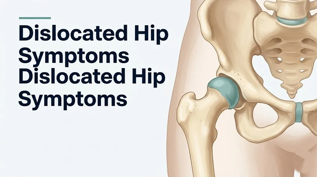
The hip is a ball-and-socket joint. The femoral head, the spherical upper end of the femur, normally sits deeply within the acetabulum, the cup-shaped socket formed by the pelvis. Three ligaments reinforce the hip capsule: the iliofemoral ligament (the strongest ligament in the body, anteriorly), the ischiofemoral ligament (posteriorly), and the pubofemoral ligament (inferiorly). When dislocation forces overcome these structures, the femoral head exits through the path of least resistance determined by the direction of applied force.
The immediate symptoms arise from three simultaneous events. First, tearing of the capsular ligaments and acetabular labrum activates dense nociceptive nerve fibers in those structures, releasing substance P and bradykinin and generating the severe, immediate pain. Second, the displaced femoral head compresses or stretches whatever nerve structure lies in its path (the sciatic nerve posteriorly, the femoral nerve anteriorly). Third, the medial circumflex femoral artery (MCFA) and its retinacular branches, which supply the majority of blood to the femoral head in adults, are disrupted by the displacement, beginning the ischemic clock.
Complete symptom inventory with mechanisms and clinical significance:
| Symptom | Mechanism | Clinical Significance |
|---|---|---|
| Sudden severe hip pain | Capsular tear activating substance P and bradykinin in ligamentous nociceptors | Most consistent symptom; intensity reflects energy of dislocation |
| Visible limb deformity (rotation, shortening) | Femoral head displaced from socket; muscle pull maintains displaced position | Limb position differs by dislocation type (key diagnostic clue) |
| Complete inability to bear weight | Disrupted joint mechanics; pain-mediated inhibition of weight-bearing musculature | Universal in complete dislocation |
| Numbness or weakness in leg or foot | Sciatic nerve stretch (posterior) or femoral nerve compression (anterior) | Present in 10 to 20 percent of posterior dislocations |
| Swelling and bruising around hip | Capsular vessel disruption, fracture hematoma, inflammatory edema | Develops over hours; may be limited in acute phase |
| Groin pain | Periarticular muscle spasm from capsular disruption and joint instability | More prominent in anterior dislocation |
Patients taking chronic corticosteroids for conditions including rheumatoid arthritis or systemic lupus erythematosus have weakened joint capsules and may experience dislocation with a lower-energy mechanism than otherwise healthy adults.
What Does a Dislocated Hip Feel Like
A dislocated hip typically feels like a sudden, catastrophic, deep pain in the hip region at the moment of injury, often described as a “pop,” a “snap,” or a sensation of the leg “giving way” violently, followed immediately by inability to move the limb and severe, unremitting pain.
The pain of a hip dislocation is not like muscle soreness or joint aching. It is deep, severe, and constant from the moment of injury. It does not ease with positional change in the way a muscle cramp or ligament sprain might briefly improve. The hip joint capsule is richly innervated with afferent pain fibers. When it tears under the force of dislocation, those fibers fire intensely and continuously.
Many people describe a sense that something has moved to a place it fundamentally should not be. The leg may look visibly shorter or rotated. Touching the hip or attempting any movement of the leg produces an immediate, sharp escalation in pain. The sense of helplessness is a clinical feature in itself: most patients with complete hip dislocation describe being completely unable to lift or reposition the leg without external assistance.
The sensation differs depending on dislocation direction. Posterior dislocation tends to produce pain concentrated in the buttock and posterior hip, sometimes radiating down the back of the thigh if the sciatic nerve is involved. Anterior dislocation often produces groin pain and a sense of the hip being “stuck” in an externally rotated position.
The American Academy of Orthopaedic Surgeons states that traumatic hip dislocation almost always produces immediate, severe pain and functional loss at the moment of injury, distinguishing it from the more gradual presentations of hip osteoarthritis flares or trochanteric bursitis.
Children, whose hip capsule is more elastic and whose ligamentum teres foveal artery contributes a greater proportion of femoral head blood supply than in adults, may describe the injury sensation somewhat differently, with pain less immediately catastrophic than in adults, though the injury is equally serious and requires the same emergency evaluation.
Signs of a Dislocated Hip
The recognizable signs of a dislocated hip include visible limb malpositioning, inability to move or bear weight on the affected leg, swelling and tenderness around the hip joint, and, in posterior dislocations, the characteristic posture of the limb in internal rotation, adduction (turned inward and crossed toward the midline), and mild flexion.
The limb position sign is one of the most diagnostically informative physical findings for hip dislocation. A clinician or trained observer looking at a patient with a posterior dislocation will see a leg that appears shortened, internally rotated, and adducted. The knee and foot of the affected leg point inward and may cross over or toward the uninjured leg. This position is produced by the posterior displacement of the femoral head and the pull of the attached hip musculature, particularly the iliopsoas and piriformis muscles, which maintain the limb in that position.
The greater trochanter, the bony prominence on the outer side of the upper thigh, may feel more prominent or palpably displaced compared to the uninjured side when the examiner places hands on both hips simultaneously. This asymmetry is a useful clinical sign when the limb position is ambiguous.
Key clinical signs of a dislocated hip by examination finding:
- Leg appearing shorter on the affected side compared to the uninjured leg (limb length discrepancy from proximal femoral head displacement)
- Internal rotation, adduction, and flexion of the affected leg at rest in posterior dislocation
- External rotation, abduction, and extension of the affected leg in anterior dislocation
- Severe tenderness on deep palpation of the groin or buttock, depending on dislocation direction
- Inability to actively move the hip in any direction without severe pain
- Possible skin bruising or swelling developing over the lateral hip and buttock within one to four hours
- Numbness or tingling in the posterior thigh, lateral leg, or foot (sciatic nerve involvement in posterior dislocation)
People with Ehlers-Danlos syndrome or generalized ligamentous hypermobility may present with less dramatic limb deformity because their capsular laxity allows greater femoral head displacement without the characteristic “locked” muscle spasm that maintains the typical abnormal limb position in normal-capsule patients.
Key Takeaway: The leg appearing shorter, internally rotated, and pulled toward the midline after a hip injury is the single most recognizable physical sign of a posterior hip dislocation and requires immediate emergency evaluation, not watchful waiting.
Hip Out of Place Symptoms: Limb Position and Deformity
The symptom of a “hip out of place” is most visibly expressed through an abnormal limb position that the patient cannot self-correct, produced by the mechanical displacement of the femoral head and the reflex muscle guarding that locks the limb in the position of least capsular tension.
Think of the hip joint like a ball pressed firmly into a suction cup. Normally, the ball sits centered and the suction holds. When the ball is forced out, it does not float freely. The surrounding muscles, which attach to the femur, immediately contract in protective spasm to stabilize whatever position the bone has landed in. That spasm is what makes the limb appear “locked” in an abnormal rotation and keeps the patient from being able to self-correct the position.
The specific “out of place” appearance depends entirely on which direction the femoral head displaced. Posterior displacement produces internal rotation and adduction. Anterior displacement produces external rotation and abduction. These are opposite-appearing deformities, and distinguishing them on visual examination has direct clinical relevance because they carry different neurological risk profiles and require different reduction techniques.
The leg may also appear functionally shortened. In posterior dislocation, the femoral head sits above and behind the acetabulum, effectively shortening the mechanical length of the lower limb. Laying the patient flat and comparing both leg lengths reveals a discrepancy of one to several centimeters on the dislocated side.
According to the Mayo Clinic, any visible deformity of the hip region following trauma, particularly in combination with inability to bear weight and severe pain, should be treated as a potential hip dislocation until imaging confirms the diagnosis.
Pregnant women in the third trimester present a specific diagnostic challenge. The pelvic relaxin-mediated ligamentous laxity of late pregnancy can both increase dislocation risk and partially reduce the visible muscle spasm component of the limb position sign, making the deformity less obviously abnormal than in non-pregnant patients.
Posterior Hip Dislocation Symptoms
Posterior hip dislocation symptoms include the classic triad of severe hip and buttock pain, a limb held in flexion, adduction, and internal rotation, and a shortened-appearing leg, often accompanied by sciatic nerve symptoms including numbness, tingling, or weakness in the foot.
Posterior dislocation is by far the most frequently occurring type, accounting for approximately 85 to 90 percent of traumatic hip dislocations according to clinical epidemiological data cited by the American Academy of Orthopaedic Surgeons. The typical mechanism is a high-energy axial force transmitted through a flexed knee into a flexed, adducted hip, classically described as the “dashboard injury” in motor vehicle collisions, where the knee strikes the vehicle dashboard and the force drives the femoral head posteriorly.
The characteristic limb position of posterior dislocation is immediately recognizable: the hip is flexed (bent forward), adducted (the knee pointing toward or crossing the midline), and internally rotated (the foot pointing inward). The patient cannot actively change this position because the displaced femoral head has exited through the posterior capsule, and the surrounding musculature locks it there.
The posterior capsular tear places the sciatic nerve at direct risk. The sciatic nerve trunk, formed by the L4, L5, S1, S2, and S3 nerve roots, exits the pelvis through the greater sciatic foramen and passes posterior to the hip joint, directly adjacent to the posterior capsule. When the femoral head is forced posteriorly, the sciatic nerve may be stretched, compressed, or trapped between the displaced femoral head and the posterior acetabular rim.
Symptom profile of posterior hip dislocation:
| Symptom | Specificity | Frequency |
|---|---|---|
| Severe hip and buttock pain | High | Nearly universal |
| Flexion, adduction, internal rotation posture | High | Nearly universal in complete dislocation |
| Shortened-appearing limb | Moderate-high | Majority of complete dislocations |
| Sciatic nerve symptoms (numbness, foot weakness) | High when present | Approximately 10 to 20 percent of posterior dislocations |
| Foot drop (inability to dorsiflex foot) | High when present | Subset with severe sciatic nerve injury |
| Swelling and ecchymosis of buttock | Low acutely | Develops over hours to days |
Older adults sustaining posterior dislocation in a motor vehicle accident face higher rates of associated acetabular posterior wall fracture because age-related bone density loss reduces the acetabular rim’s resistance to impact, turning what might be a pure dislocation in a younger patient into a fracture-dislocation requiring surgical management.
Anterior Hip Dislocation Symptoms
Anterior hip dislocation symptoms differ distinctly from the posterior pattern, producing a limb held in external rotation, abduction, and extension, with pain concentrated in the groin rather than the buttock, and placing the femoral nerve and femoral vessels at greater risk than the sciatic nerve.
Anterior dislocation is the less frequent type, accounting for approximately 10 to 15 percent of traumatic hip dislocations. The mechanism typically involves forced abduction and external rotation of the hip, sometimes with hyperextension, as in a fall from a horse, a twisting sports injury, or a specific vehicle impact pattern. The femoral head exits anteriorly through the anterior capsule and pubofemoral ligament.
The visual appearance is opposite to posterior dislocation. The affected leg lies in external rotation (the foot points outward), abduction (the leg is pushed away from the midline), and extension (the knee is relatively straight). The leg may appear slightly longer than the uninjured side, in contrast to the shortening seen in posterior dislocation, because the femoral head has displaced inferiorly and anteriorly rather than superiorly and posteriorly.
Pain in anterior dislocation is typically felt in the groin and anterior hip, directly over the displaced femoral head, which now sits anterior to the hip joint. A fullness or firm mass may be palpable in the groin as the femoral head bulges anteriorly beneath the skin.
The femoral nerve, which passes from the lumbar plexus (L2-L4 nerve roots) through the femoral triangle anterior to the hip, is at risk in anterior dislocation. Femoral nerve compression by the displaced femoral head can produce weakness of the quadriceps femoris muscle (making knee extension difficult) and numbness over the anterior thigh in the L2-L4 dermatomal distribution.
Women have a wider pelvis and greater hip capsule laxity due to estrogen receptor-mediated effects on capsular collagen, producing relatively greater hip range of motion. This anatomical difference may make anterior dislocation slightly more likely in women than in men under equivalent force vectors, though the overall dislocation frequency still favors males due to occupational and recreational exposure.
Partial Hip Dislocation Symptoms (Subluxation)
Partial hip dislocation (subluxation) symptoms are more subtle than complete dislocation, often producing intermittent hip pain, a sense of instability or “giving way,” reduced range of motion, and a vague sense that the hip “is not right,” without the dramatic limb deformity of complete dislocation.
Hip subluxation means the femoral head has partially exited the acetabulum but has not completely displaced out of the joint. The femoral head remains in partial contact with the acetabular cartilage surface. This partial contact preserves some joint function and may allow the patient to bear some weight, making subluxation a significantly more difficult diagnosis than complete dislocation.
The symptom pattern of subluxation can mimic a bad hip sprain, a labral tear, or a hip flexor strain. The absence of dramatic limb deformity is the feature most likely to lead to missed or delayed diagnosis. A patient with subluxation may walk into an emergency department with an abnormal gait rather than being carried in, which instinctively signals less severity to receiving staff.
Key clinical features that should raise suspicion for subluxation rather than a simple soft tissue injury include: pain that is clearly centered at the hip joint rather than diffusely muscular, a sense of clicking, catching, or instability with specific hip movements (particularly at the end-range of hip flexion and internal rotation), and a mechanism of injury consistent with a lateral force or torsional load on the hip.
Clinical Orthopaedics and Related Research has published case series documenting significant delays in diagnosing hip subluxation in patients who presented ambulatory after athletic injuries, with several cases progressing to avascular necrosis from undertreated persistent subluxation because the initial assessment categorized the injury as a soft tissue sprain.
Symptoms distinguishing hip subluxation from complete dislocation:
- Limb position: near-normal or mildly abnormal rather than dramatically rotated and fixed
- Weight-bearing: partial weight-bearing may be possible versus complete inability in full dislocation
- Pain intensity: severe but potentially slightly less catastrophic than complete dislocation
- Deformity: absent or subtle versus obvious in complete dislocation
- Instability: a distinct sense of the hip “shifting” or “wanting to go out” with movement
Athletes with underlying acetabular dysplasia (a shallow acetabulum) are disproportionately at risk for recurrent subluxation because the shallow socket provides less containment of the femoral head against lateral and torsional forces.
Key Takeaway: Hip subluxation can occur without obvious deformity and may allow partial weight-bearing, making it easy to mistake for a bad sprain; a sense of hip instability or “giving way” after a hip injury warrants imaging evaluation even if the limb position looks relatively normal.
Can You Walk With a Dislocated Hip
Most people cannot walk with a complete hip dislocation because the disrupted joint mechanics and severe pain from the torn capsular ligaments make any weight-bearing on the affected leg functionally impossible.
The inability to bear weight is one of the most reliable clinical indicators distinguishing a complete hip dislocation from less severe hip injuries. When the femoral head is fully displaced from the acetabulum, the hip joint has no structural articulation to transfer ground reaction forces from the foot up through the pelvis. Any attempt to load the limb drives the displaced femoral head against surrounding soft tissue rather than through the normal joint mechanism, producing immediate, severe pain that prevents weight-bearing reflexively.
There is a critical exception to this rule: hip subluxation. As discussed in the previous section, partial dislocation may allow some degree of weight-bearing, typically with a clearly antalgic (pain-avoidant) gait in which the patient leans heavily toward the injured side, takes very short steps, and bears minimal load on the affected limb. This limited ambulatory capacity in subluxation is the main reason partial dislocation gets missed.
A person who was in a traumatic event, reports sudden hip pain, and is walking with a markedly abnormal gait should not be reassured that their hip is “fine” simply because they are ambulatory. The clinical rule from the American College of Emergency Physicians is that any patient with a mechanism of injury consistent with hip dislocation and hip pain or abnormal gait requires imaging, regardless of ambulation status.
Practical guidance if you or someone with you may have a dislocated hip:
- Do not attempt to walk or bear weight on the affected leg
- Do not attempt to straighten, rotate, or reposition the injured limb; maintain the position found
- Call 911 if the person cannot be safely transported; transport by ambulance is preferable for hip dislocation to prevent movement of the displaced femoral head
- If transport by private vehicle is unavoidable, support the injured leg in the position found without forcing any change; do not allow the leg to hang unsupported
- Do not give food or drink in case procedural sedation or anesthesia is needed for reduction at the emergency department
Children who continue walking after a hip injury, even with a limp, must not be assumed to have a minor injury. Pediatric hip subluxation after athletic injury can present with surprisingly preserved ambulatory function.
Dislocated Hip in Elderly Patients
Dislocated hip symptoms in elderly patients (those over age 65) differ from younger adults because the combination of reduced bone density, weaker capsular ligaments from age-related collagen crosslinking changes, and higher prevalence of hip osteoarthritis means dislocation can occur with lower-energy trauma and is more likely to involve an associated acetabular or femoral neck fracture.
The osteoporotic skeleton of an older adult provides less structural resistance to the forces that cause hip dislocation. A fall from standing height, which would typically produce only bruising in a younger healthy adult, can produce a hip fracture-dislocation in an older adult whose trabecular bone has thinned due to age-related estrogen deficiency (in postmenopausal women) or age-related testosterone decline and reduced osteoblast activity (in older men).
Hip osteoarthritis, which has a prevalence in adults over age 65 estimated by the National Institute of Arthritis and Musculoskeletal and Skin Diseases at approximately 10 to 25 percent, alters the articular surface geometry and joint space dimensions. Arthritic changes can reduce the natural containment of the femoral head within the acetabulum, lowering the force threshold required for dislocation.
A clinically important nuance for elderly patients: the limb position sign of classic posterior dislocation (flexion, adduction, internal rotation) may be less dramatically evident in older patients because reduced muscle mass and baseline hip stiffness from osteoarthritis may limit the degree of muscle spasm that locks the limb in the characteristic position. This can make the dislocation appear less obvious to an untrained observer.
Risk factors for hip dislocation in older adults:
- Osteoporosis (T-score below negative 2.5 on DEXA scan), which weakens the acetabular rim and increases fracture-dislocation risk
- Hip osteoarthritis, which reduces articular containment
- History of prior hip surgery other than total hip replacement (e.g., prior ORIF for femoral neck fracture)
- Balance impairment, neuropathy, or muscle weakness increasing fall frequency
- Chronic corticosteroid use producing steroid-induced osteoporosis
- Parkinson’s disease or other neurological conditions affecting protective reflexes
Any older adult who reports sudden hip pain, change in leg position, or inability to bear weight after a fall should be taken to an emergency department for imaging, even if they can verbalize their pain and seem alert. Cognitive impairment from dementia can cause older adults to underreport pain severity, masking the urgency of the injury.
Dislocated Hip After Hip Replacement
Dislocation of a total hip arthroplasty (THA) is the most frequent early complication of hip replacement surgery, occurring in approximately 2 to 3 percent of primary THA cases and up to 5 to 10 percent of revision THA cases, according to registry data published in the Journal of Arthroplasty.
The prosthetic hip is dramatically more vulnerable to dislocation than a native hip for several anatomical reasons. During hip replacement surgery, the native hip joint capsule is partially or completely resected to allow component insertion. The capsular ligaments that normally stabilize the femoral head in the acetabulum are no longer intact. The prosthetic femoral head and acetabular cup must rely on component positioning, soft tissue repair, and patient movement restriction to maintain stability. Even small deviations from the correct component angle or the prohibited movement pattern can cause the prosthetic ball to pop out of the artificial cup.
The symptom presentation of prosthetic hip dislocation is similar to traumatic native hip dislocation: sudden severe hip pain, abnormal limb position, and inability to bear weight. One key difference is that prosthetic dislocation can occur with much lower-energy provocations. Bending too far forward to pick something off the floor, crossing the legs, rotating the hip internally beyond the restricted range, or sitting in a low chair can all dislocate a prosthetic hip in the early post-surgical healing period.
Positions that typically trigger prosthetic hip dislocation in posterior approach THA:
- Hip flexion beyond 90 degrees (bending at the waist past a right angle)
- Internal rotation of the hip beyond neutral
- Adduction past the midline (crossing legs)
- Combination of flexion, adduction, and internal rotation simultaneously
Patients with prior hip dislocation before THA, prior spinal fusion altering pelvic tilt mechanics, abductor muscle weakness, or obesity face elevated prosthetic dislocation risk. Any patient with a hip replacement who experiences sudden hip pain, a “pop” sensation, or abnormal limb position should go to the emergency room immediately and inform staff that they have a hip replacement, because reduction technique differs from native hip dislocation management.
Key Takeaway: A patient with a hip replacement who feels a “pop” and sudden hip pain with an abnormal leg position needs emergency evaluation immediately, because prosthetic hip dislocation can occur from ordinary daily movements and requires urgent closed reduction.
Hip Dislocation in Athletes
Hip dislocation in athletes typically occurs through sport-specific mechanisms that apply the dislocation force vector to the hip at positions of athletic loading, and can occur with lower-energy trauma than civilian hip dislocations when the athlete has underlying acetabular dysplasia, labral pathology, or connective tissue hypermobility.
The most commonly implicated sports include football (particularly when a tackled athlete is hit while the hip is flexed), rugby, skiing, basketball, and martial arts. Athletic hip dislocation tends to occur in younger individuals with denser, stronger bone than the elderly population, so associated acetabular fractures are less common than in older adults. Pure dislocation without fracture is therefore more typical in athletic presentations.
Athletes with acetabular dysplasia, a condition in which the acetabular socket is shallower than normal, face a substantially elevated dislocation risk because the reduced depth of the socket provides less mechanical containment of the femoral head. Dysplasia is more prevalent in women, occurring at approximately four to five times the rate in female athletes compared to male athletes, according to clinical observation data cited in Orthopedic Clinics of North America.
The symptom presentation in athletes mirrors the general posterior dislocation pattern (the most frequently occurring type in sports), but athletes may initially underreport pain severity due to adrenaline, training-induced pain tolerance, and the social pressure to minimize injury on the field. A coaching staff member, athletic trainer, or teammate who observes an athlete suddenly unable to bear weight with an internally rotated leg position after a collision should initiate emergency protocols immediately, regardless of the athlete’s verbal report.
Return to sport after traumatic hip dislocation requires a minimum of several months of rehabilitation under the supervision of a board-certified orthopedic surgeon and a sports physical therapist, with sport-specific assessment of hip stability before clearance.
Quick Tip:
- Athletes with a history of hip “clicking,” “popping,” or a prior sense of hip instability during sport should be evaluated for acetabular dysplasia or labral pathology before a dislocation event occurs, because early identification allows prophylactic surgical stabilization in appropriate cases
- Any athlete who sustains a hip dislocation and then has a recurrence at lower force thresholds should be evaluated for acetabular dysplasia by a board-certified hip preservation orthopedic specialist
- Female athletes face higher dysplasia prevalence and should receive hip stability screening as part of pre-participation evaluation in high-risk contact sports
Hip Dislocation Sciatic Nerve Symptoms
Sciatic nerve injury from posterior hip dislocation produces numbness, tingling, weakness, or complete loss of motor function in the territory supplied by the sciatic nerve trunk, which exits the pelvis through the greater sciatic foramen and passes directly posterior to the hip capsule, placing it at direct risk when the femoral head is displaced posteriorly.
The sciatic nerve is the largest nerve in the human body, formed by the L4, L5, S1, S2, and S3 anterior primary rami from the lumbar and sacral plexuses. When the femoral head is forced posteriorly in a hip dislocation, the nerve may be compressed between the displaced femoral head and the posterior acetabular rim, stretched by the traction force of displacement, or in severe cases, directly contused against the bone.
Sciatic nerve injury occurs in approximately 10 to 20 percent of posterior hip dislocations, according to clinical series published in the Journal of Bone and Joint Surgery. The peroneal division of the sciatic nerve (which becomes the common fibular nerve below the knee) is more susceptible to injury than the tibial division because the peroneal fiber bundles are positioned more laterally within the nerve trunk and are therefore more directly exposed to the compressive force of the displaced femoral head.
Sciatic nerve injury symptom patterns by division affected:
| Division Affected | Sensory Symptoms | Motor Symptoms | Clinical Sign |
|---|---|---|---|
| Common peroneal (fibular) division | Numbness over lateral leg and dorsum of foot | Inability to dorsiflex foot (foot drop), weakness of toe extension | High-stepping gait, foot drop |
| Tibial division | Numbness over sole of foot and posterior calf | Weakness of plantarflexion and toe flexion | Difficulty rising on tiptoe |
| Full sciatic trunk | Numbness over posterior thigh, lateral leg, and entire foot | Combined foot drop and plantarflexion weakness | Severe functional deficit |
The single most important clinical action when sciatic nerve symptoms are present after a hip dislocation is urgent reduction. Research in the Journal of Bone and Joint Surgery has found that sciatic nerve injuries from hip dislocation have a higher rate of functional recovery when reduction is achieved within six hours of injury. Prolonged neural compression or traction dramatically worsens prognosis for nerve recovery.
Patients with pre-existing peripheral neuropathy from diabetes or chronic alcohol use have baseline reduced nerve function and may experience more severe sciatic deficit from the same degree of nerve compression than neurologically intact patients, and face a higher risk of incomplete recovery even after timely reduction.
Hip Dislocation Avascular Necrosis Risk
Avascular necrosis of the femoral head after hip dislocation is produced by disruption of the medial circumflex femoral artery (MCFA) and its superior and inferior retinacular branches, which are the dominant blood supply to the femoral head in adults, initiating ischemia of the femoral head bone tissue that progresses to cellular death and structural collapse if perfusion is not restored.
Think of the femoral head’s blood supply like a garden sprinkler system buried under a grass lawn. The sprinkler lines (the MCFA retinacular branches) run along the femoral neck to reach the head. When the femoral head is forcibly displaced, those lines are kinked, stretched, or torn. The grass (the femoral head bone cells) begins to die within hours. Replanting the sprinkler head in its original position (reduction) reopens the lines and allows perfusion to resume, but only if done quickly enough.
The time-critical nature of this mechanism is what makes hip dislocation a vascular emergency rather than a simple mechanical injury. The National Institute of Arthritis and Musculoskeletal and Skin Diseases states that avascular necrosis of the femoral head is a recognized complication of hip dislocation, with risk increasing substantially with duration of dislocation before reduction.
Multiple cohort studies published in the Journal of Bone and Joint Surgery have found that hip dislocations reduced within six hours of injury carry substantially lower rates of avascular necrosis than those reduced after six hours, with some studies reporting rates as low as 4 to 5 percent with early reduction versus rates exceeding 40 percent with reduction delayed beyond 24 hours.
Avascular necrosis does not produce symptoms immediately after the dislocation event. It is a progressive process that may not produce clinical symptoms (groin pain, worsening hip stiffness, reduced range of motion) until weeks to months after the original injury. This delayed symptom onset means patients who have had a hip dislocation reduced must continue orthopedic follow-up with serial imaging, even if they feel “recovered,” to detect early avascular necrosis before femoral head structural collapse occurs.
Patients who smoke have significantly elevated avascular necrosis risk after femoral head ischemia because nicotine-mediated vasoconstriction further compromises the already-impaired retinacular blood flow after dislocation.
Hip Dislocation vs Hip Fracture Symptoms
Distinguishing hip dislocation from hip fracture symptoms is clinically important because both cause severe hip pain and inability to bear weight after trauma, but the limb position, mechanism, patient population, and treatment urgency differ in specific ways that guide emergency management.
Hip fracture (fracture of the femoral neck or intertrochanteric region) and hip dislocation share their two most obvious symptoms: immediate severe hip pain and inability to bear weight. Both injuries are orthopedic emergencies. The key distinguishing features are found in the limb position and the patient population.
In a typical femoral neck fracture, the affected leg lies in external rotation and shortening but without the marked internal rotation and adduction seen in posterior hip dislocation. The reason is mechanical: a femoral neck fracture allows the distal fragment (the leg) to fall into external rotation under gravity because the neck is no longer maintaining the femoral head in its forward-facing orientation. There is no displacement of the femoral head out of the acetabulum, so the leg position is externally rotated rather than internally rotated as in posterior dislocation.
The patient population also differs. Hip fractures occur predominantly in older adults with osteoporosis, often after a relatively minor fall. Traumatic hip dislocation occurs predominantly in younger adults after high-energy trauma (motor vehicle accidents, sports), though prosthetic hip dislocation in older adults with hip replacements blurs this demographic distinction.
Hip dislocation vs. hip fracture: key differentiating features:
| Feature | Posterior Hip Dislocation | Femoral Neck Fracture |
|---|---|---|
| Limb position | Internally rotated, adducted, flexed | Externally rotated, shortened |
| Typical patient | Younger adult, high-energy trauma | Older adult, low-energy fall |
| Neurovascular risk | Sciatic nerve, MCFA retinacular branches | Medial circumflex femoral artery (similar) |
| Deformity character | “Locked” in abnormal rotation | Leg falls passively into external rotation |
| Associated injury | Acetabular fracture possible | Intertrochanteric or subcapital fracture |
| Diagnostic imaging | AP pelvis X-ray, CT for fracture-dislocation | AP pelvis and hip X-ray |
The honest clinical reality, stated clearly by the American College of Emergency Physicians, is that clinical examination alone cannot reliably distinguish dislocation from fracture in the emergency setting. Imaging is mandatory for both.
Patients with osteoporosis severe enough to produce femoral neck fracture from low-energy falls may also have an acetabular bone density low enough to sustain an acetabular fracture-dislocation simultaneously, making the combined injury more likely in the osteoporotic elderly than in younger adults.
Key Takeaway: A leg lying externally rotated after a fall in an older adult suggests hip fracture; a leg locked in internal rotation and adduction after high-energy trauma suggests hip dislocation; both are emergencies requiring the same immediate action: go to the emergency room.
How Is a Dislocated Hip Diagnosed
A dislocated hip is diagnosed through clinical examination of limb position and neurovascular status combined with plain film radiography of the pelvis and hip, followed by computed tomography (CT) of the pelvis when fracture, intra-articular fragments, or complex dislocation patterns are suspected.
The diagnostic process begins at the moment of emergency department arrival. The evaluating emergency medicine physician performs a rapid physical examination assessing limb position, skin perfusion, distal pulses, and neurovascular status of the leg and foot. The characteristic limb position of posterior dislocation, flexion, adduction, and internal rotation, is often visible before the patient is moved to the imaging suite.
Standard imaging protocol begins with an anteroposterior (AP) pelvis radiograph, which allows simultaneous comparison of both hip joints and identifies the displaced femoral head position, associated femoral neck fracture, and acetabular fracture. In posterior dislocation, the femoral head appears smaller than the contralateral side on AP X-ray because it has moved away from the X-ray beam (posteriorly), creating an apparent size discrepancy.
CT scanning of the pelvis is performed after initial reduction in the majority of cases to identify any intra-articular bone fragments that could block complete reduction or damage the articular cartilage, and to characterize the acetabular fracture pattern in fracture-dislocations. Research published in Clinical Orthopaedics and Related Research has confirmed that CT scanning after reduction identifies clinically relevant intra-articular fragments in a meaningful proportion of apparently successfully reduced hip dislocations, making post-reduction CT a standard of care in most trauma centers.
Diagnostic process for suspected hip dislocation:
- Emergency department arrival: rapid clinical assessment of mechanism, limb position, pain level, neurovascular status (pedal pulses, capillary refill, sensory testing of foot)
- AP pelvis radiograph: confirms dislocation, identifies direction of displacement, screens for associated fractures
- Emergent orthopedic surgery consultation: initiated based on radiographic confirmation of dislocation
- Closed reduction under sedation or anesthesia: performed urgently, ideally within six hours of injury
- Post-reduction AP pelvis and lateral hip X-ray: confirms concentric reduction (femoral head properly seated in acetabulum)
- Post-reduction CT pelvis: identifies intra-articular fragments, characterizes acetabular injury
- MRI of hip (at follow-up, typically within days to weeks): assesses femoral head viability, labral injury, and capsular disruption
Patients with prior hip replacement presenting with suspected prosthetic dislocation require specific radiographic protocol using views appropriate for prosthetic component assessment, and a board-certified orthopedic hip reconstruction specialist should be involved in the evaluation.
Dislocated Hip Treatment and Recovery
Dislocated hip treatment centers on urgent closed reduction under sedation or general anesthesia, followed by a structured rehabilitation program, with surgical intervention reserved for fracture-dislocations, irreducible dislocations, and prosthetic dislocations requiring component revision.
Closed reduction is performed in the emergency department under procedural sedation or in the operating room under general anesthesia. The two most commonly used techniques are the Allis technique, in which the patient lies supine while the physician applies upward traction on the flexed hip, and the Stimson gravity technique, in which the patient lies prone with the injured leg hanging off the table edge while gentle downward pressure is applied to restore the femoral head to the acetabulum.
Once reduction is confirmed by X-ray with the femoral head concentrically seated in the acetabulum, the patient is typically admitted for observation, pain management, and neurovascular monitoring. The injured limb is kept in a neutral position with the hip abducted (legs apart) to reduce the risk of re-dislocation in the early post-reduction phase, particularly for posterior dislocations.
The recovery process is staged. Immediate post-reduction (first one to two weeks): bed rest with limited weight-bearing; neurovascular checks to monitor sciatic nerve status. Early rehabilitation (weeks two to eight): protected weight-bearing with crutches; passive and active range-of-motion exercises under physiotherapy supervision. Later rehabilitation (months two to six): progressive strengthening of the hip abductors, external rotators, and iliopsoas; gait normalization.
Recovery timeline by injury pattern:
| Injury Type | Time to Weight-Bearing | Return to Work (Sedentary) | Return to Heavy Labor or Sport |
|---|---|---|---|
| Simple dislocation, no fracture | Two to four weeks with crutches | Six to twelve weeks | Three to six months minimum |
| Dislocation with small acetabular fracture (managed non-operatively) | Four to six weeks restricted | Twelve to sixteen weeks | Six to twelve months |
| Fracture-dislocation requiring ORIF | Six to twelve weeks post-surgery | Twelve to twenty-four weeks | Twelve months or longer |
| Prosthetic hip dislocation (recurrence) | Dependent on stability; specialist-guided | Variable | Dependent on revision surgery outcome |
A board-certified orthopedic surgeon should manage all follow-up imaging after hip dislocation reduction, with MRI typically performed at six weeks to three months post-injury to assess early avascular necrosis, and repeat imaging at six months, twelve months, and two years in high-risk cases, because avascular necrosis may not be radiographically apparent for months after the initial injury.
Emergency Symptoms: When to Call 911 or Go to the ER
Certain symptoms associated with a dislocated hip require immediate emergency evaluation. Do not wait to see if these resolve on their own.
Call 911 or go to the nearest emergency room immediately if you or someone with you experiences:
- Sudden severe hip pain with an abnormal leg position after any traumatic event: hip dislocation is a vascular emergency with a six-hour reduction window before avascular necrosis risk escalates
- Complete inability to bear weight on the affected leg after a fall, impact, or sports injury: this indicates a high-probability joint or bone injury requiring same-day imaging
- Leg lying in internal rotation, adduction, and flexion after a trauma: the characteristic position of posterior hip dislocation requiring emergency closed reduction
- Leg lying in external rotation, abduction, and extension after a trauma with groin pain: the characteristic position of anterior hip dislocation
- Numbness, tingling, or inability to move the foot or ankle after a hip injury: possible sciatic nerve injury from posterior dislocation requiring urgent reduction
- Foot drop (inability to lift the front of the foot when walking) after a hip injury: indicates peroneal division sciatic nerve injury
- A “pop” sound or sensation followed by severe hip pain and positional change in a patient who has had hip replacement surgery: prosthetic hip dislocation requiring immediate emergency evaluation
- Severe hip pain with a cold, pale, or pulseless foot after trauma: possible vascular injury requiring immediate vascular surgery consultation
These presentations can indicate complete hip dislocation with time-critical vascular compromise of the femoral head, sciatic nerve injury, or prosthetic hip dislocation, all of which require emergency department evaluation and immediate orthopedic surgery consultation, not an urgent care appointment or phone-based triage.
Frequently Asked Questions About Dislocated Hip Symptoms
What are the first signs of a dislocated hip?
The first signs of a dislocated hip are sudden, severe hip pain at the moment of injury combined with immediate inability to move or bear weight on the affected leg.
Most patients with posterior dislocation will notice the affected leg lying in an internally rotated, adducted position that they cannot self-correct.
Any combination of sudden severe hip pain, abnormal limb position, and inability to bear weight after trauma requires emergency evaluation regardless of pain tolerance or perceived severity.
Can a hip be dislocated without severe pain?
A complete hip dislocation almost always produces severe pain because the capsular tear and nerve stretch at the moment of dislocation activate dense nociceptive fibers in the hip joint capsule and surrounding soft tissue.
Partial dislocation (subluxation) can sometimes produce only moderate or intermittent pain, which is why it is more frequently misdiagnosed as a sprain.
Athletes with high pain tolerance may underreport pain severity immediately after dislocation due to adrenaline, but functional loss, particularly the inability to bear normal weight or maintain limb position, will still be present.
How long does a dislocated hip take to heal?
Simple hip dislocation without associated fracture typically requires six to twelve weeks before return to sedentary activities, and three to six months for return to physically demanding work or sport.
Recovery from fracture-dislocation requiring surgical fixation is longer, typically twelve months or more before full functional restoration.
Avascular necrosis, if it develops, can require hip replacement surgery months to years after the original dislocation, extending the overall recovery process significantly.
What happens if a dislocated hip is not treated quickly?
Delayed reduction of a hip dislocation substantially increases the risk of avascular necrosis of the femoral head, because the medial circumflex femoral artery retinacular branches that supply the femoral head are disrupted by the dislocation and perfusion is only restored when the femoral head is returned to the acetabulum.
Cohort studies published in the Journal of Bone and Joint Surgery have found avascular necrosis rates exceeding 40 percent when reduction is delayed beyond 24 hours, compared to rates below 5 percent with reduction within six hours.
Delayed treatment also allows sciatic nerve injury to progress, reducing the chance of full nerve recovery.
Can you dislocate a hip by falling?
A fall can cause a hip dislocation, although the required mechanism and force typically depend on the patient’s age, bone health, and underlying hip anatomy.
In younger adults with healthy bone, hip dislocation from a fall usually requires a high-energy impact at a specific joint angle, such as landing on a flexed, adducted hip.
In older adults with total hip replacements, bending during a fall in a specific direction can dislocate the prosthetic joint with relatively low force, making fall-related prosthetic dislocation common in the early post-surgical period.
How do doctors tell the difference between a broken hip and a dislocated hip?
Doctors distinguish a broken hip from a dislocated hip primarily through plain film radiography of the pelvis, which shows the position of the femoral head relative to the acetabulum and identifies any fracture lines.
Clinically, posterior hip dislocation produces a leg in internal rotation and adduction, while femoral neck fracture typically produces a leg in external rotation with limb shortening.
The American College of Emergency Physicians states that imaging is mandatory in all cases because clinical examination alone has insufficient accuracy to distinguish dislocation from fracture, and because the two conditions can coexist simultaneously as a fracture-dislocation.
The Six-Hour Window
The single most important clinical fact about a dislocated hip is the time urgency. The blood supply to the femoral head begins failing at the moment of dislocation. Every hour of delay increases the probability of permanent femoral head damage from avascular necrosis.
If you or someone near you has sudden severe hip pain with an abnormal leg position after trauma, the appropriate action is not to wait and see. It is not to drive to urgent care and sit in a waiting room. It is to go directly to an emergency department, tell the intake team that a hip dislocation is suspected, and specifically state how long ago the injury occurred. That time stamp drives the urgency of the orthopedic team’s response.
You now have the clinical picture, the specific symptom patterns by dislocation type, the vascular mechanism that makes this injury time-critical, and the exact emergency threshold to apply.