Tilted Pelvis Symptoms: Signs, Causes & When to Act
Tilted pelvis symptoms range from dull lower back ache and uneven hip height to sharp nerve pain and leg length differences that alter your entire gait. The specific symptoms you experience depend on which direction your pelvis has rotated and which muscles, joints, and nerve structures are bearing the abnormal load.
The National Institute of Arthritis and Musculoskeletal and Skin Diseases (NIAMS) estimates that lower back pain, which pelvic malalignment directly contributes to, affects approximately 80% of adults at some point in their lives, making disorders of spinal and pelvic alignment among the most clinically significant musculoskeletal concerns in primary care. Research published in Clinical Biomechanics has consistently shown that pelvic position is one of the primary determinants of lumbar spinal load distribution, meaning a tilted pelvis doesn’t just affect the pelvis itself — it changes how every structure above and below it functions.
This article covers the complete clinical picture of pelvic tilt: the three main types, the specific anatomical reason each type produces each symptom, how symptoms differ between men and women, what changes during pregnancy and athletic training, which symptoms point to a serious underlying problem, and exactly what kind of provider to see and when.
Tilted Pelvis Symptoms: What Is Actually Happening in Your Body
A tilted pelvis produces symptoms because the pelvis is the structural base of the entire axial skeleton and lower extremity kinetic chain, and any deviation from neutral alignment shifts mechanical load to structures that weren’t designed to handle it.
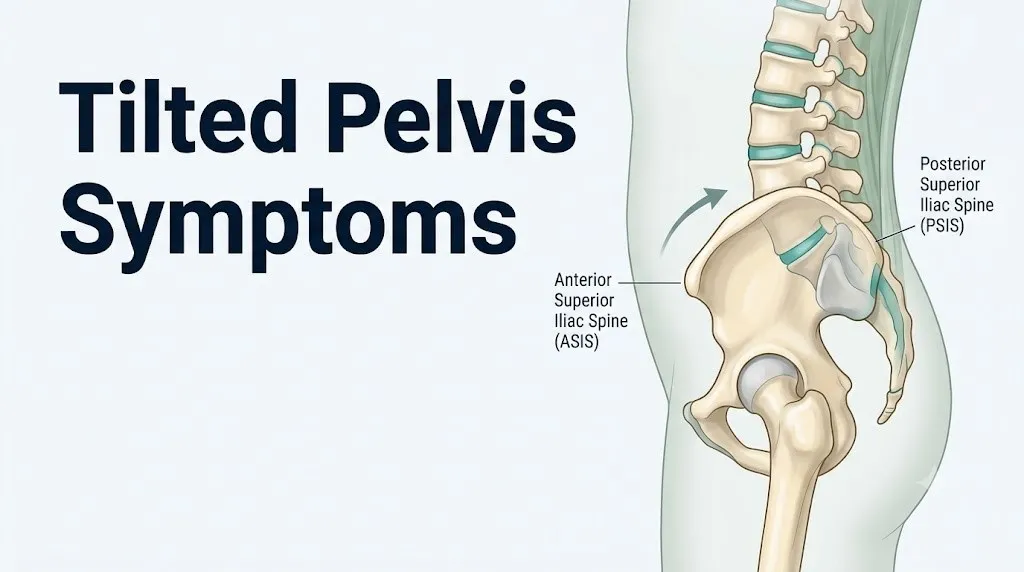
Your pelvic ring consists of the two iliac bones, the sacrum, and the pubic symphysis. In a neutral position, the anterior superior iliac spine (ASIS) and the posterior superior iliac spine (PSIS) sit at approximately the same horizontal level when viewed from the side, and both iliac crests sit at equal height when viewed from behind. When this neutral relationship is disrupted, the cascade of effects moves up the lumbar spine and down into the hip joints, knees, and ankles.
Think of the pelvis like the foundation of a house. If one corner sinks or tilts, the walls above develop cracks, the doors stop closing properly, and the floors start to slope. The “cracks” in pelvic malalignment show up as muscle spasm, joint compression, nerve irritation, and postural compensation patterns.
The three recognized directional subtypes are anterior tilt (pelvis tips forward), posterior tilt (pelvis tips backward), and lateral tilt, also called pelvic obliquity (one side sits higher than the other). Each produces a distinct but overlapping symptom pattern, which is why the same complaint of “low back pain” can look completely different clinically depending on which type a person has.
Key clinical symptoms across all tilt types include:
- Lower back pain, often dull and persistent, worsening with prolonged standing or sitting
- Hip pain or stiffness, typically at the front, back, or outer hip depending on tilt direction
- Visible postural asymmetry (one hip appearing higher, uneven shoulder level, forward head position)
- Fatigue or tension in the paraspinal muscles of the lower lumbar region
- Discomfort or pain that worsens after physical activity and improves briefly with movement before returning
Pelvic Misalignment Symptoms: The Full Clinical Picture
Pelvic misalignment symptoms affect multiple body regions simultaneously because the pelvis connects the axial skeleton to the appendicular skeleton, making it a load-transfer junction for nearly every movement the body performs.
The sacroiliac joints (the joints between the sacrum and the iliac bones on each side) transmit force from the lumbar spine to the femoral heads with every step. When pelvic alignment is off, these joints absorb asymmetric loads, which produces inflammation within the sacroiliac joint capsule and surrounding ligaments. Sacroiliac joint pain typically presents as a deep, aching pain at the dimples of the lower back, occasionally radiating into the buttock, and is notoriously difficult to distinguish from lumbar disc pain without a targeted clinical examination.
Muscle imbalance is the other major driver of symptom spread. A 2019 study published in the Journal of Orthopaedic and Sports Physical Therapy found that pelvic malalignment is consistently associated with asymmetric activation patterns in the gluteus medius and erector spinae, meaning some muscles become overworked and develop trigger points while their antagonists become inhibited and weakened. This imbalance explains why the symptoms of pelvic misalignment don’t stay local — they travel upward to the thoracic spine and downward to the knee.
Postural compensation is visible to a trained observer. Uneven iliac crest height, a visible lateral shift of the lumbar spine, one shoulder sitting higher than the other, and the head translating forward relative to the center of gravity are all observable signs of significant pelvic obliquity or tilt.
| Symptom Region | Typical Clinical Description | Anatomical Source |
|---|---|---|
| Lower back | Dull ache, worse with prolonged standing | Facet joint compression, paraspinal overload |
| Sacroiliac area | Deep ache at lower back dimples | SI joint capsule inflammation |
| Hip | Stiffness, aching, reduced range of motion | Altered femoral head loading |
| Buttock | Aching, occasional referral into thigh | Piriformis tension, SI joint referral |
| Thigh or leg | Radiating pain or numbness | Nerve root irritation (L4, L5, S1) |
| Knee | Aching, patellofemoral tracking problems | Altered Q angle, IT band tension |
What Causes a Tilted Pelvis
A tilted pelvis develops when a persistent muscle length or strength imbalance pulls the pelvis out of its neutral position, or when structural factors like leg length inequality, prior injury, or degenerative joint disease force compensatory pelvic shifts.
Muscle imbalance is the most common cause. The pelvis is held in position by the coordinated tension of the hip flexors (iliopsoas, rectus femoris), hip extensors (gluteus maximus, hamstrings), hip abductors (gluteus medius), hip adductors, and lumbar extensors (erector spinae, multifidus). When one group becomes chronically tight or overactive and the opposing group becomes weak or inhibited, the pelvis rotates in the direction the dominant group pulls it.
Prolonged sitting, which shortens the iliopsoas and rectus femoris while allowing the gluteus maximus and hamstrings to lengthen, is the single most common lifestyle driver of anterior pelvic tilt in adults.
Less common but clinically important causes include:
- True structural leg length discrepancy from prior long bone fracture, joint replacement, or developmental dysplasia
- Scoliosis, where lateral spinal curvature forces the pelvis into a compensatory oblique position
- Hip osteoarthritis, which limits hip joint range of motion and causes the pelvis to compensate
- Prior lumbar spine surgery that altered normal spinal biomechanics and secondary pelvic positioning
- Inflammatory arthropathies such as ankylosing spondylitis, which directly affect sacroiliac joint mobility and alignment
- Habitual postural asymmetry (consistently shifting weight to one leg, carrying loads on one side)
Older adults are at elevated risk because age-related reductions in paraspinal and hip muscle mass (sarcopenia) reduce the stability margin needed to maintain neutral pelvic positioning, particularly in the presence of concurrent hip osteoarthritis.
Key Takeaway: Muscle imbalance from prolonged sitting is the most common cause of a tilted pelvis, but structural causes like leg length discrepancy and hip osteoarthritis require clinical evaluation to identify and manage properly.
Anterior Pelvic Tilt Symptoms
Anterior pelvic tilt produces a distinct symptom pattern driven by the forward rotation of the ASIS below and ahead of the PSIS, which increases lumbar lordosis and compresses the posterior elements of the lumbar spine.
When the ASIS tips forward, the lumbar spine responds by increasing its inward curve (hyperlordosis). This compresses the posterior facet joints at L4-L5 and L5-S1, the two motion segments that bear the greatest mechanical load in the lower lumbar spine. Sustained facet joint compression triggers the release of substance P and prostaglandin E2 from the joint capsule, sensitizing local nociceptors and producing the characteristic deep, aching lower back pain that worsens with prolonged standing and extension-based movements.
The iliopsoas and rectus femoris are chronically shortened in anterior tilt. This creates a pulling sensation in the front of the hip and groin, and limits full hip extension during walking, which forces the lumbar spine to compensate and increases compressive load at L5-S1 with every stride.
According to the American Physical Therapy Association (APTA), anterior pelvic tilt is the most frequently observed postural malalignment pattern in sedentary populations, and it is characterized by a visible “Donald Duck” posture: prominent buttocks, a protruding belly that is not related to body weight, and an exaggerated lumbar curve.
Symptoms specific to anterior pelvic tilt:
- Exaggerated lumbar inward curve visible from the side
- Chronic lower back pain that worsens with standing and arching backward
- Tightness or pulling in the front of the hip and groin
- Weakness or “disconnection” feeling in the glutes during standing or walking
- Occasional anterior thigh tightness from rectus femoris shortening
- Mild abdominal protrusion without weight-related cause
Adolescents experiencing rapid growth spurts are at elevated risk for anterior tilt because bone length temporarily outpaces musculotendinous growth, leaving the hip flexors in a relatively shortened state relative to the elongated femur.
Posterior Pelvic Tilt Symptoms
Posterior pelvic tilt occurs when the ASIS rotates upward and backward relative to the PSIS, flattening the normal lumbar lordosis into a relative kyphosis, and producing a “flat back” posture.
The flat back posture shifts the lumbar spine’s center of gravity anterior to the posterior elements, loading the intervertebral discs at L4-L5 and L5-S1 with increased anterior compressive force rather than the facet joint posterior compression seen in anterior tilt. This altered disc loading pattern increases intradiscal pressure during sitting and forward bending, and research published in Spine has documented that posterior pelvic tilt during seated posture increases intradiscal pressure at L4-L5 by approximately 40% compared to a neutral lumbar curve.
The hamstring group (biceps femoris, semimembranosus, semitendinosus) and the gluteus maximus become chronically shortened in posterior tilt, limiting hip flexion and producing a stiff, shuffling gait pattern. The hip flexors (iliopsoas and rectus femoris) lengthen and weaken.
The sacrotuberous ligament and sacrospinous ligament, which connect the sacrum to the ischial tuberosity and ischial spine respectively, are placed under persistent tension in posterior tilt, contributing to a diffuse aching pain at the sit bones (ischial tuberosities) and across the base of the sacrum.
Symptoms specific to posterior pelvic tilt:
- Flattened lower back curve visible from the side (no inward arch)
- Deep aching at the base of the spine and sacrum
- Pain or tenderness at the sit bones (ischial tuberosities)
- Hamstring tightness and limited ability to flex at the hip during walking
- Discomfort during prolonged sitting, particularly on hard surfaces
- Occasional thoracic kyphosis (rounding of the upper back) as a compensatory upward postural change
Older adults are disproportionately affected by posterior tilt because age-related hip extensor weakness and the increased tendency to stand with slightly flexed hips and knees both drive the pelvis into posterior rotation.
Key Takeaway: Anterior tilt compresses the posterior lumbar facet joints and produces aching back pain that worsens with standing and extension; posterior tilt loads the intervertebral discs and sacral ligaments, producing pain that worsens with sitting and forward bending.
Lateral Pelvic Tilt Symptoms
Lateral pelvic tilt, also called pelvic obliquity, occurs when one iliac crest sits measurably higher than the other when the person stands with both feet flat on the ground, creating a visible and functional asymmetry across the entire pelvis.
The higher side typically reflects overactivation or shortening of the quadratus lumborum on that side, a deep lumbar muscle that runs from the 12th rib to the posterior iliac crest and has direct mechanical attachment to the iliac crest. When the quadratus lumborum shortens and pulls the iliac crest upward, the sacroiliac joint on that same side is compressed while the contralateral sacroiliac joint is distracted. Both produce pain, but in different locations and with different qualities.
Lateral tilt also creates a functional leg length discrepancy, meaning the two legs appear unequal in length not because of a bone length difference but because the pelvis sits unevenly over them. A functional discrepancy of as little as 5 millimeters has been associated with measurable gait asymmetry in research published in Clinical Biomechanics, and discrepancies exceeding 15 millimeters are associated with meaningful increases in lumbar paraspinal muscle activity on the side of the apparent shorter leg.
Symptoms specific to lateral pelvic tilt:
- One hip appearing visibly higher than the other
- Uneven shoulder height as the spine curves laterally to compensate
- Pain concentrated on one side of the lower back or buttock
- A feeling that one leg is shorter than the other when walking or standing
- Uneven wear patterns on shoe soles (inside or outside heel wear on one foot more than the other)
- Gait asymmetry, such as dipping to one side with each step (positive Trendelenburg pattern)
Women are statistically more likely to present with lateral tilt secondary to unilateral hip abductor weakness, a pattern that the American Academy of Orthopaedic Surgeons (AAOS) identifies as a common contributor to hip and pelvis pain in physically active women.
Tilted Pelvis Back Pain: Why It Happens and Where You Feel It
Tilted pelvis back pain is produced by the mechanical overloading of specific lumbar spinal structures that would not be stressed in a neutrally aligned pelvis, and the location and character of the pain directly reflects which structure is under strain.
Anterior tilt directs compressive force into the posterior facet joints at L4-L5 and L5-S1. These joints have synovial capsules that are richly innervated by the medial branch of the dorsal rami. When the capsule is compressed or inflamed, it generates a deep, bilateral aching pain localized to the lower lumbar region, typically two to three finger-widths lateral to the midline spinous processes. This pain often has a characteristic “belt of ache” distribution across the lower back.
Posterior tilt shifts load onto the anterior disc annulus. The outer third of the intervertebral disc annulus fibrosus is innervated by the sinuvertebral nerve (recurrent meningeal nerve). Increased intradiscal pressure from posterior tilt can sensitize this nerve, producing a central lower back ache that is often diffuse and harder to localize than facet pain.
The quadratus lumborum, which spans from the 12th rib to the posterior iliac crest on each side, is one of the primary active stabilizers of the pelvis against lateral tilt. In lateral pelvic obliquity, the quadratus lumborum on the elevated iliac crest side contracts continuously, developing myofascial trigger points that produce a sharp, sometimes debilitating lateral lower back pain that can be mistaken for kidney pain because of its location.
To describe tilted pelvis back pain accurately to a healthcare provider:
- Note whether the pain is central (midline), bilateral, or concentrated on one side.
- Record whether it worsens with standing, walking, sitting, or backward arching.
- Observe whether it improves briefly when you first start walking and then worsens with continued activity.
- Check whether forward bending relieves or worsens the pain (extension worsening suggests facet/anterior tilt; flexion worsening suggests disc loading/posterior tilt).
- Note any pain radiation pattern below the buttock or into the thigh.
Tilted Pelvis Hip Pain and Leg Length Discrepancy
Tilted pelvis hip pain arises because any departure from pelvic neutral alignment changes the angle at which the femoral head sits within the acetabulum, altering contact pressures within the hip joint and the mechanical advantage of the surrounding muscles.
In anterior tilt, the pelvis tips forward, which shifts the femoral head slightly anterior within the acetabular cup. This increases anterior acetabular rim loading and can produce a pinching sensation at the front of the hip during hip flexion, such as when climbing stairs or rising from a chair. Over time, this altered contact pressure accelerates cartilage wear at the anterior acetabular rim, a pattern the American Academy of Orthopaedic Surgeons identifies as a contributing factor in femoroacetabular impingement.
In lateral tilt, the iliac crest on the lower side drops below the horizontal, causing the acetabulum on that side to face more downward and producing a classic functional leg length discrepancy. Crucially, the legs themselves are the same bone length. It is the tilted container (the pelvis) that makes them appear and function as though one is shorter.
A functional discrepancy creates a limping or dipping gait on the apparent short-leg side and places abnormal rotational stress on the knee, ankle, and foot of that limb. The iliotibial band (IT band) and tensor fasciae latae on the functionally longer leg side become chronically tight as the body compensates for the asymmetric stance.
| Type | Leg Length Effect | Hip Pain Location | Key Driving Muscle Imbalance |
|---|---|---|---|
| Anterior tilt | Minimal functional difference | Front of hip, groin | Iliopsoas short; gluteus maximus weak |
| Posterior tilt | Minimal functional difference | Posterior hip, sit bones | Hamstrings short; hip flexors weak |
| Lateral tilt | Functional leg length discrepancy | Lateral hip, outer thigh | Quadratus lumborum short; gluteus medius weak |
Key Takeaway: Hip pain from a tilted pelvis reflects changed acetabular loading angles, and a functional leg length difference is not a bone length problem but a positional one created by the tilted pelvis sitting unevenly over the legs.
Pelvic Tilt and Sciatica or Nerve Pain
Pelvic tilt and sciatica are connected through two anatomical mechanisms: lumbar nerve root compression from altered spinal segment loading, and sciatic nerve tension from piriformis muscle hypertonicity secondary to pelvic malalignment.
In anterior tilt, the increased lumbar lordosis narrows the intervertebral foramina at L4-L5 and L5-S1, the exit points for the L4, L5, and S1 nerve roots that form the sciatic nerve (primarily L4 through S3). A narrowed foramen can compress a nerve root, producing lumbar radiculopathy: pain, numbness, or tingling that travels from the lower back down the buttock and into the leg in a pattern corresponding to the affected root. L5 compression typically produces lateral leg and dorsal foot symptoms. S1 compression produces posterior leg and plantar foot symptoms.
The piriformis muscle, which runs from the anterior sacrum to the greater trochanter of the femur, can become hypertonic in lateral pelvic tilt and anterior tilt because both conditions alter the resting position of the femoral head and change the mechanical line of pull of the piriformis. In approximately 15% to 20% of the population, the sciatic nerve passes through or adjacent to the piriformis. When the piriformis is tight, it can directly compress the sciatic nerve, producing piriformis syndrome: a deep buttock pain with sciatic distribution that is clinically difficult to distinguish from lumbar disc-related sciatica without targeted examination.
According to research published in the Journal of Orthopaedic and Sports Physical Therapy, piriformis syndrome is significantly more common in women than men, with a female-to-male ratio of approximately 6:1, a disparity attributed in part to the wider female pelvis and greater Q angle creating a more acute piriformis angle relative to the sciatic nerve.
Nerve pain from pelvic tilt can present as:
- Sharp, shooting pain traveling from the buttock down the back of the thigh
- Burning or electrical pain along the outer lower leg or into the foot
- Numbness or tingling in the thigh, leg, or foot
- Weakness in foot dorsiflexion (foot drop) or toe extension in severe cases
- Pain that worsens with prolonged sitting, particularly on a hard surface
Pelvis Out of Alignment Symptoms in the Knees and Ankles
When the pelvis sits out of alignment, symptoms in the knees and ankles are not coincidental. They are the downstream mechanical consequence of the altered lower extremity kinetic chain.
The Q angle (quadriceps angle) describes the angular relationship between the line of pull of the quadriceps and the patellar tendon as measured from the anterior superior iliac spine to the center of the patella to the tibial tuberosity. In anterior pelvic tilt, the forward rotation of the ASIS increases the Q angle by moving the ASIS forward and laterally. A higher Q angle increases the lateral tracking force on the patella, pressing it against the lateral femoral condyle and producing a condition known as patellofemoral pain syndrome or “runner’s knee.” Patellofemoral pain presents as a diffuse aching beneath or around the kneecap, worsening with stair climbing, squatting, or prolonged sitting with bent knees.
Lateral pelvic tilt alters the mechanical axis of the lower extremity (the line from the center of the hip to the center of the ankle). When one iliac crest is lower, the knee on that side develops a slight valgus stress (knock-knee alignment), increasing the compressive load on the medial knee compartment and the tensile stress on the lateral knee structures, particularly the iliotibial band.
At the ankle, a functional leg length discrepancy from lateral tilt forces the apparent short leg to plantarflex (point the foot) or supinate to reach the ground, while the apparent long leg compensates by pronating or flexing the knee. Both adaptations create abnormal ankle joint mechanics.
Pelvis out of alignment symptoms in the knees and ankles include:
- Aching beneath or around the kneecap during stairs or squatting
- Lateral knee pain from iliotibial band tension
- Medial knee aching from increased valgus stress
- Ankle fatigue or pain from asymmetric foot strike patterns
- Uneven shoe sole wear (more wear on the inner or outer heel of one shoe versus the other)
People with pre-existing knee osteoarthritis should be aware that pelvic malalignment can accelerate cartilage wear on the compartment bearing the abnormal mechanical load, making early physical therapy evaluation particularly relevant for this group.
Key Takeaway: Knee pain under the kneecap and uneven shoe wear are frequently downstream signals of pelvic malalignment, not primary knee problems, and correcting the pelvic alignment is often what resolves the knee and ankle symptoms.
Tilted Pelvis Female Symptoms
Tilted pelvis female symptoms differ from those observed in male patients for specific biological and anatomical reasons, and understanding those reasons is the key to recognizing the full clinical picture in women.
The female pelvis has a wider Q angle than the male pelvis because the female iliac crests are set farther apart relative to the femoral heads. This wider Q angle means that the same degree of anterior or lateral tilt produces greater patellofemoral lateral tracking force and greater iliotibial band tension in women than in men with equivalent pelvic positions.
Estrogen influences the mechanical properties of ligaments throughout the body. The sacroiliac joint ligaments, the pubic symphysis ligaments, and the hip joint capsule all respond to estrogen by remaining appropriately flexible. After menopause, declining estrogen levels reduce periarticular ligament tensile strength, making postmenopausal women more susceptible to sacroiliac joint instability and pelvis-related pain when any degree of malalignment is present. A 2021 observational study published in Spine found that postmenopausal women with sacroiliac joint dysfunction reported pain intensity scores approximately 30% higher than premenopausal women with equivalent degrees of pelvic malalignment, a difference attributed partly to periarticular ligament laxity and partly to central pain sensitization.
Pelvic floor dysfunction is a female-specific interaction with pelvic tilt. The levator ani, the primary component of the pelvic floor musculature, attaches to the inner surface of the pubic bone anteriorly and the ischial spines laterally. In posterior pelvic tilt, the pubic bone rotates superiorly and the coccyx moves anteriorly, shortening the levator ani and producing a hypertonic pelvic floor. Hypertonic pelvic floor dysfunction in women presents as pelvic pressure, deep pelvic pain, painful intercourse (dyspareunia), and increased urinary urgency, all of which can co-occur with the musculoskeletal symptoms of posterior tilt.
Female-specific symptoms of pelvic malalignment include:
- Deep pelvic pressure or aching (beyond typical back or hip pain)
- Painful intercourse related to pelvic floor hypertonicity in posterior tilt
- Increased urinary urgency without urinary tract infection
- Groin or inner thigh pain from altered adductor mechanics
- Greater knee pain severity at equivalent tilt magnitude due to wider Q angle
- Sacroiliac joint pain that intensifies in the week before menstruation (prostaglandin-related ligament sensitization)
Pelvic Tilt in Pregnancy
Pelvic tilt in pregnancy is driven by a combination of hormonal ligament changes and the mechanical effect of a growing uterus shifting the body’s center of gravity forward, and it produces a characteristic symptom pattern that differs from non-pregnant pelvic malalignment.
The hormone relaxin, produced by the corpus luteum from the first weeks of pregnancy and by the placenta from the second trimester, acts on the symphyseal and sacroiliac joint ligaments to increase their laxity in preparation for delivery. This ligamentous softening is physiologically appropriate and necessary for childbirth, but it simultaneously removes the passive restraint that normally limits sacroiliac joint movement and pubic symphysis stress. According to the American College of Obstetricians and Gynecologists (ACOG), pelvic girdle pain affects between 20% and 25% of pregnant women, with sacroiliac joint strain as the most common structural contributor.
As the uterus grows through the second and third trimesters, the added anterior mass shifts the center of gravity forward, which the lumbar spine compensates for by increasing its lordotic curve and which the pelvis accommodates by rotating into an increasingly anterior tilt. This progressive anterior tilt compresses the posterior lumbar facet joints at L4-L5 and L5-S1 and places the piriformis under increasing tension as the widening uterus changes the internal pelvic geometry.
Symptoms during pregnancy include an exacerbation of all anterior tilt symptoms plus:
- Symphysis pubis dysfunction (SPD): sharp, sometimes stabbing pain directly over the pubic bone, worsening with walking, stair climbing, and turning in bed
- Sacroiliac joint pain radiating from the lower back dimples into the buttock and posterior thigh
- Posterior pelvic pain specific to pregnancy, which the ACOG distinguishes from lumbar pain by its location posterior to the sacrum and its aggravation by forward bending
Pregnant women experiencing sudden severe sacroiliac pain, inability to bear weight on one leg, or new onset of leg numbness should contact their obstetric provider promptly, as these symptoms can occasionally signal pubic symphysis diastasis or lumbar disc herniation exacerbated by relaxin-mediated ligament laxity.
Key Takeaway: Pregnancy-related pelvic tilt is driven by relaxin-mediated ligament laxity plus mechanical load shift from the growing uterus, and the ACOG reports that pelvic girdle pain affects roughly one in four pregnant women.
Tilted Pelvis in Athletes and Active Individuals
Tilted pelvis in athletes develops from sport-specific muscle dominance patterns that chronically pull the pelvis out of neutral, and it produces symptoms that can be mistakenly attributed to muscle strain, overuse injury, or poor recovery rather than their structural pelvic origin.
Cyclists and triathletes develop anterior pelvic tilt from thousands of hours of hip flexion in the cycling position, which chronically shortens the iliopsoas and rectus femoris while underloading the gluteus maximus and posterior chain. Runners with anterior tilt experience increased ground reaction forces at the L5-S1 motion segment with every footstrike because the extended lumbar lordosis reduces the shock-absorbing capacity of the lumbar spine’s normal S-curve. Research published in the Journal of Orthopaedic and Sports Physical Therapy found that runners with clinically measured anterior pelvic tilt had a 22% higher rate of hamstring injuries compared to runners with neutral pelvic positioning, attributed to the lengthened and mechanically disadvantaged position of the hamstrings in anterior tilt.
Weightlifters and powerlifters, particularly those performing heavy squats and deadlifts, may develop posterior tilt when gluteal dominance is combined with anterior core weakness, causing the pelvis to “tuck under” at the bottom of the squat. This is called a “butt wink” in training communities and represents a posterior pelvic tilt under load that increases intradiscal pressure at L4-L5 and L5-S1.
Soccer and football players are prone to lateral pelvic tilt from unilateral kicking dominance, which creates asymmetric hip abductor development between the dominant and non-dominant leg, pulling the pelvis into obliquity on the side of the weaker gluteus medius.
Athlete-specific symptoms and risk markers:
- Recurrent hamstring strains without adequate traumatic cause (anterior tilt)
- Persistent hip flexor tightness despite regular stretching
- Lumbar fatigue disproportionate to training volume
- Patellofemoral pain on the dominant-side knee in unilateral sport athletes
- Iliotibial band syndrome recurring on one side despite foam rolling and stretching
- Asymmetric hip mobility (one hip more restricted than the other in rotation or flexion)
Red Flags and Serious Symptoms of Pelvic Misalignment
Most pelvic misalignment symptoms are chronic, slowly progressive, and musculoskeletal in nature. Certain symptom presentations, however, signal neurological compromise or structural instability that requires urgent or emergency evaluation rather than physical therapy referral.
The most serious emergency associated with lumbar and pelvic structural compromise is cauda equina syndrome (CES). The cauda equina is the bundle of nerve roots at the base of the spinal cord below the L1 vertebral level, and it contains the nerve roots controlling bladder, bowel, and sexual function in addition to motor and sensory function of the lower extremities. When a large lumbar disc herniation, progressive lumbar stenosis exacerbated by anterior pelvic tilt, or epidural space-occupying lesion compresses these roots, cauda equina syndrome develops. CES is a surgical emergency with a narrow window for intervention.
The American Academy of Orthopaedic Surgeons classifies CES as a surgical emergency requiring decompression within 24 to 48 hours of symptom onset to prevent permanent neurological deficit.
Red flag symptoms of pelvic malalignment requiring immediate evaluation:
- Bladder dysfunction: inability to urinate, urinary retention, or sudden incontinence not related to prior urinary conditions
- Bowel dysfunction: loss of bowel control or new onset constipation with altered anal sensation
- Saddle anesthesia: numbness in the inner thighs, perineum, or genitals (the region that would contact a saddle)
- Bilateral leg weakness, particularly if sudden or rapidly progressive
- Progressive foot drop (inability to lift the front of the foot)
- Severe back pain following trauma such as a fall or car accident
- Back pain accompanied by unexplained fever, night sweats, or unintentional weight loss (possible spinal infection or malignancy)
Other serious but non-emergency warning signs that warrant a same-day or next-day call to a primary care physician or physiatrist:
- Rapidly worsening unilateral leg pain with numbness or weakness
- Groin or genital numbness that is new in onset
- Any back pain that is present at rest and wakes you from sleep
Emergency Symptoms: When to Call 911 or Go to the ER
Certain symptoms associated with severe pelvic and lumbar structural compromise require immediate emergency evaluation. Do not wait to see if these resolve on their own.
Call 911 or go to the nearest emergency room immediately if you experience:
- Sudden inability to urinate or sudden urinary incontinence without a prior urinary diagnosis (possible cauda equina syndrome)
- Sudden bowel incontinence or loss of rectal sensation (cauda equina syndrome hallmark)
- Numbness or tingling in the perineum, inner thighs, or genitals (saddle anesthesia, CES indicator)
- Bilateral leg weakness that developed suddenly or is rapidly worsening
- Foot drop (inability to lift the toes upward) developing suddenly rather than gradually
- Severe back or pelvic pain following a fall, vehicle collision, or direct trauma to the pelvis or spine
These presentations can indicate cauda equina syndrome or acute spinal cord or nerve root compression and require emergency neurosurgical assessment, not an urgent care appointment or a phone call to schedule a physical therapy evaluation.
How a Tilted Pelvis Is Diagnosed
A tilted pelvis is diagnosed through a combination of clinical postural assessment, manual physical examination, and imaging studies that together confirm both the direction of the tilt and the structural or muscular causes driving it.
The clinical examination begins with a postural assessment in the standing position. A trained examiner (physical therapist, physiatrist, or orthopedic surgeon) observes the patient from the front, side, and behind, comparing the heights of the iliac crests, the ASIS positions, and the PSIS positions. An ASIS-PSIS drop test (comparing the relative heights of the ASIS and PSIS on each side) can identify both anterior and posterior tilt. The Trendelenburg sign, where the contralateral hip drops when the patient stands on one leg, indicates gluteus medius weakness and is a key clinical indicator of lateral pelvic tilt.
Muscle length testing completes the clinical picture:
- The Thomas test assesses hip flexor (iliopsoas, rectus femoris) length: the patient lies supine and draws one knee to the chest; the opposite leg should remain flat against the table; inability to do so indicates hip flexor tightness consistent with anterior tilt.
- Hamstring length testing assesses the risk of posterior tilt from hamstring shortening.
- Quadratus lumborum length testing identifies lateral shortening driving obliquity.
Imaging is typically ordered when the clinical picture suggests a structural cause or when symptoms include neurological features:
- Anteroposterior (AP) standing pelvic X-ray: measures pelvic obliquity, true leg length discrepancy, and sacroiliac joint space
- Standing lumbar spine X-ray: quantifies lumbar lordosis angle and confirms vertebral alignment
- MRI of the lumbar spine: identifies disc herniation, foraminal narrowing, or nerve root compression that may be contributing to neurological symptoms
The appropriate first referral for a suspected tilted pelvis without neurological symptoms is a licensed physical therapist or a physiatrist (physical medicine and rehabilitation specialist) who can perform the full postural and biomechanical assessment and design a corrective plan. If imaging identifies disc herniation with nerve root compression, referral to a board-certified orthopedic spine surgeon or neurosurgeon is warranted.
Key Takeaway: A complete tilted pelvis diagnosis requires both clinical physical examination (posture, Thomas test, Trendelenburg sign) and targeted imaging (AP pelvic X-ray, lumbar MRI if neurological symptoms are present) to confirm the type and identify structural contributors.
Tilted Pelvis Treatment and Physical Therapy
Tilted pelvis treatment is directed specifically at the type and cause of the tilt, and physical therapy that addresses the named muscle imbalance pattern is the most evidence-supported first-line approach for structural and postural pelvic malalignment without neurological involvement.
The American Physical Therapy Association (APTA) recommends a corrective approach targeting both the shortened muscles (by lengthening and releasing them) and the weakened muscles (by activating and strengthening them) to re-establish balanced tension across the pelvic ring. For anterior tilt, this means hip flexor stretching (Thomas stretch, kneeling hip flexor stretch) combined with gluteus maximus strengthening (hip bridges, Romanian deadlifts) and anterior core activation (dead bugs, modified plank progressions). For posterior tilt, it means hamstring lengthening combined with hip flexor activation and lumbar extension mobility work.
Lateral tilt treatment prioritizes gluteus medius strengthening on the weaker side (clamshells, lateral band walks, single-leg stance progressions) and quadratus lumborum stretching on the elevated iliac crest side. When a true structural leg length discrepancy is confirmed on imaging, a heel lift orthotic prescribed by a physiatrist or orthopedic surgeon may be needed to re-level the pelvis passively.
Manual therapy techniques, including joint mobilization of the sacroiliac joint and lumbar facets, soft tissue mobilization of the iliopsoas and piriformis, and myofascial release of the thoracolumbar fascia, complement corrective exercise and have been shown in research published in Clinical Biomechanics to reduce pain and improve pelvic symmetry in the short term when combined with active exercise rather than used alone.
For postmenopausal women with sacroiliac joint instability contributing to pelvic tilt pain, a pelvic floor physiotherapist (a physical therapist with specialized certification in pelvic floor rehabilitation) can assess and treat the interaction between pelvic floor dysfunction and pelvic malalignment, an aspect of care that general physical therapy may not address.
The timeline for measurable improvement with consistent physical therapy is typically four to eight weeks for postural changes and eight to twelve weeks for sustained pain reduction, according to clinical practice guidance from the APTA.
Frequently Asked Questions About Tilted Pelvis Symptoms
What does a tilted pelvis feel like?
A tilted pelvis most commonly feels like a persistent, dull lower back ache that worsens with prolonged standing or sitting, combined with hip stiffness or uneven hip height you can notice in a mirror.
Many people also notice tightness at the front of the hip or groin (in anterior tilt) or soreness at the sit bones and base of the sacrum (in posterior tilt).
The sensation is often chronic rather than sudden, which is why many people assume it is ordinary muscle soreness rather than a structural alignment problem.
Can a tilted pelvis go away on its own?
A mild functional pelvic tilt caused by muscle imbalance can improve with targeted corrective exercises, but it rarely corrects itself without deliberate intervention.
Without addressing the underlying muscle imbalance, the structural forces pulling the pelvis out of position remain active and the tilt typically persists or gradually worsens over time.
A licensed physical therapist can assess the specific imbalance pattern and design a targeted program that produces measurable correction, which is the most evidence-supported approach.
How do I know if my pelvis is out of alignment?
The most observable sign is uneven hip height when you stand in front of a mirror with both feet flat on the floor and look at where your hands rest on your hips.
Other observable signs include one shoulder sitting higher than the other, a visible lean to one side when standing relaxed, and shoe sole wear that is noticeably more pronounced on one side than the other.
A clinical assessment by a physical therapist using the ASIS-PSIS drop test and Trendelenburg sign evaluation provides a more accurate picture than self-observation alone.
Can a tilted pelvis cause sciatica?
Yes, a tilted pelvis can produce sciatic nerve pain through two mechanisms: lumbar foraminal narrowing at L4-L5 or L5-S1 from increased lumbar lordosis in anterior tilt, and piriformis muscle hypertonicity compressing the sciatic nerve in lateral or anterior tilt.
Piriformis syndrome, which is a pelvic tilt-related cause of sciatic pain, is approximately six times more common in women than men according to research in the Journal of Orthopaedic and Sports Physical Therapy.
If you have sciatica combined with back pain that changes with posture, pelvic malalignment as a contributing cause should be assessed by a physical therapist or physiatrist before invasive treatment is considered.
What are the symptoms of a tilted pelvis in females?
Tilted pelvis symptoms in women include all the standard musculoskeletal symptoms plus deeper pelvic pressure, painful intercourse from pelvic floor hypertonicity in posterior tilt, increased urinary urgency, and sacroiliac joint pain that worsens in the week before menstruation due to prostaglandin-related ligament sensitization.
Women experience greater knee pain severity at equivalent tilt magnitudes compared to men because the wider female Q angle amplifies lateral patellar tracking forces.
Postmenopausal women report more intense sacroiliac pain than premenopausal women with equivalent malalignment, which research published in Spine attributes to reduced periarticular ligament strength after estrogen decline.
Is a tilted pelvis serious?
For most people, a tilted pelvis is a chronic musculoskeletal problem that is genuinely painful and functionally limiting but is not medically dangerous and responds well to physical therapy.
However, certain symptoms associated with pelvic and lumbar malalignment are serious: bladder or bowel dysfunction, saddle area numbness, and bilateral leg weakness can indicate cauda equina syndrome, which the American Academy of Orthopaedic Surgeons classifies as a surgical emergency requiring decompression within 24 to 48 hours.
Any of these neurological symptoms appearing alongside pelvic or back pain requires emergency room evaluation the same day, not a scheduled appointment.
Closing
The full clinical picture of a tilted pelvis goes well beyond the generic “back pain and hip pain” descriptions that dominate most health content. Your specific symptoms, their location, their character, and what makes them better or worse are all direct signals from the anatomical structures being overloaded or compressed by the direction your pelvis has tilted. Understanding that signal helps you describe your symptoms accurately and get to the right type of provider faster.
For most people, the path forward starts with a licensed physical therapist or physiatrist who can perform a proper postural and biomechanical assessment, identify the specific tilt type and its muscular driver, and design a corrective program targeting the right muscles in the right way. If you have any neurological symptoms alongside your pelvic pain, including leg weakness, numbness in the groin or inner thigh, or any change in bladder or bowel control, that changes everything and requires emergency evaluation before any other step.
You now have enough specific, anatomically grounded information to recognize what your symptoms are telling you, ask the right questions at your appointment, and know exactly which symptoms mean you should stop reading and go straight to the emergency room.