Stress Fracture in Foot Symptoms: A Complete 2026 Guide
The hallmark symptoms of a stress fracture in the foot are pain that builds gradually during activity, pinpoint tenderness directly over a specific bone, and mild swelling that appears by the end of the day. These symptoms are different from a sudden sports injury because stress fractures develop through the slow accumulation of microscopic bone damage rather than a single traumatic force.
This matters because most people walk on a stress fracture for days or weeks before seeking evaluation, and continued weight-bearing during that window is what transforms a manageable stress reaction into a complete bone fracture. According to the National Institute of Arthritis and Musculoskeletal and Skin Diseases (NIAMS), stress fractures account for up to 16 percent of all injuries in military recruits and a comparable proportion of overuse injuries in distance runners.
This article covers every recognized symptom of a stress fracture in the foot with the physiological reason behind each one. It addresses specific foot bones that fracture most often, explains who is at higher risk and why, compares the presentation to conditions that look similar, and clarifies exactly when imaging and orthopaedic evaluation cannot wait.
Stress Fracture in Foot Symptoms: What You Need to Know First
The core symptoms of a stress fracture in the foot are activity-related bone pain that worsens progressively, localized point tenderness over a specific bone surface, mild to moderate swelling, and pain that improves with rest but returns faster with each subsequent activity session.
What separates a stress fracture from most other foot pain is the pattern. The pain does not start with a pop or a sudden sharp event. It builds gradually, often starting as a mild ache after a long run or a day on your feet, then appearing earlier and earlier in subsequent activity sessions. By the time the fracture is established, many people report pain from simply walking across a parking lot.
The periosteum, the fibrous membrane wrapping every bone, is densely supplied with nociceptive nerve endings. As microcracks accumulate in the cortical bone beneath it, the periosteum becomes inflamed. That inflammation is the direct source of the pain signal. It is not muscle soreness. It is not joint inflammation. It is bone pain from a specific anatomical structure under mechanical stress.
Key symptoms at a glance:
- Progressive pain during weight-bearing activity in the forefoot or midfoot
- Pain that eases with rest but returns quickly when activity resumes
- Pinpoint tenderness when pressing directly on a specific small area of bone
- Mild swelling over the affected bone, often more visible at the end of the day
- Aching pain at rest in advanced or high-risk presentations
- Bruising in some cases, particularly over the second or third metatarsal shaft
Adolescent athletes have an additional concern. Growth plates (the physis) remain open until the mid-to-late teenage years, and stress injury near a growth plate requires evaluation by a board-certified orthopaedic surgeon to rule out a Salter-Harris type injury, which has different management requirements than a standard stress fracture.
What Is a Stress Fracture in the Foot and How Does It Form?
A stress fracture in the foot is an incomplete crack through the cortical bone of one or more foot bones, produced when repetitive mechanical loading exceeds the bone’s capacity to repair itself through the normal remodeling cycle. It is not caused by a single impact. It is caused by thousands of small impacts accumulated faster than the bone can adapt.
Think of bone like a metal paperclip. Bend it once and it springs back. Bend it back and forth hundreds of times and microscopic cracks form within the metal, well before it visibly breaks. The foot bones experience the same cumulative fatigue under repeated loading without adequate recovery time.
The underlying mechanism involves the basic multicellular unit (BMU), the cellular machinery that continuously repairs and renews bone. During periods of increased mechanical loading, osteoclasts (bone-resorbing cells) accelerate their activity to remove fatigued bone. Osteoblasts (bone-forming cells) then deposit new bone to replace it. The critical problem is timing: osteoclast resorption happens first and creates a transient period of bone weakness. If new loading is applied during this resorption phase before osteoblast formation catches up, the bone accumulates damage rather than repairing it.
According to the American Academy of Orthopaedic Surgeons, this remodeling imbalance is the defining biological mechanism of all stress fractures. The foot is particularly vulnerable because it bears the entire body’s weight with every step, and the second and third metatarsals are the most heavily loaded bones in the forefoot during the push-off phase of gait.
Women who are in low-estrogen states are at heightened risk at this stage because estrogen directly promotes osteoblast differentiation. When estrogen is absent or suppressed, osteoblast formation slows, and the resorption-formation gap widens under the same mechanical load.
Early Signs of a Stress Fracture in the Foot
Early signs of a stress fracture in the foot include a dull, diffuse ache in the forefoot or midfoot that appears only after prolonged activity and resolves completely with rest, often mimicking general muscle fatigue in the first one to two weeks.
At this stage, the bone has accumulated microdamage but the periosteum has not yet become significantly inflamed. The pain feels generalized rather than pinpoint. Many people mistake it for a muscle cramp, a tight shoe, or delayed-onset muscle soreness. That misattribution is what keeps people training through the early phase.
Two changes signal the transition from diffuse fatigue pain to a developing stress fracture. First, the pain starts appearing earlier in the activity session than it did the week before. A runner who could complete 8 miles before feeling discomfort now feels it at mile 3. Second, the onset of pain begins migrating from “during activity” to “immediately after activity” and eventually “within minutes of starting activity.”
The American Journal of Sports Medicine has published multiple prospective cohort studies on military recruits documenting that stress fracture risk increases dramatically when training volume increases by more than 10 percent per week, a threshold now widely cited in sports medicine practice guidelines.
Quick Tip:
- Notice whether the pain is appearing earlier in each training session than the session before. Progressive symptom onset is one of the earliest clinical indicators.
- Press your thumb firmly along the shaft of your metatarsals one at a time. Mild discomfort at one specific spot that is absent at adjacent spots is early point tenderness.
- Postmenopausal women and female athletes with irregular or absent menstrual periods should lower their threshold for seeking evaluation after two weeks of this symptom pattern, not four.
Key Takeaway: Early stress fracture pain appears only after sustained activity and disappears with rest. The defining early warning sign is pain that arrives progressively earlier in each activity session over a one-to-two-week period.
Foot Stress Fracture Pain: What It Feels Like and Where It Hurts
Foot stress fracture pain feels like a deep, aching throb localized to a specific small area of bone, typically worsened by foot strike, push-off, or standing on the ball of the foot, and located most often over the shaft of the second or third metatarsal or at the top of the midfoot over the navicular.
The location is specific enough that most people can point to it with one finger. That precision is clinically meaningful. Muscle pain and tendon pain radiate more broadly. Bone pain from a stress fracture concentrates at the exact site of cortical damage where periosteal nociceptors are being stimulated by local inflammation driven by prostaglandin E2 and interleukin-6 released from the bone marrow.
The pain quality changes with fracture progression. Early-stage fracture pain feels like a dull, deep ache during activity. With continued loading, it becomes sharper and more immediate at foot strike, eventually developing a burning or throbbing character that persists for 30 to 60 minutes after stopping activity. In advanced cases, the pain becomes continuous, present even at rest.
| Pain Stage | Timing | Quality | Location Precision | Rest Relief |
|---|---|---|---|---|
| Early (stress reaction) | After prolonged activity only | Dull, diffuse ache | Approximate, broad area | Complete relief with rest |
| Developing (early fracture line) | During activity, appears progressively earlier | Aching to sharp at foot strike | Becoming more localized | Partial relief with rest |
| Established (fracture line present) | From activity onset, sometimes at rest | Sharp at foot strike, throbbing after | Pinpoint, one-finger localization | Incomplete relief at rest |
| Advanced (risk of complete fracture) | Constant, worsened by any weight-bearing | Severe, burning, constant | Exactly over fracture site | Minimal or no relief |
Males and females describe the pain quality similarly. However, research published in the British Journal of Sports Medicine has found that female athletes with hypothalamic amenorrhea tend to present at a more advanced fracture stage because the absence of menstrual symptoms delays their recognition that their bone health may be compromised.
Swelling and Visible Changes With a Foot Stress Fracture
Swelling with a foot stress fracture is typically mild, localized over the affected bone, and often more noticeable at the end of the day than in the morning, reflecting the accumulation of inflammatory fluid during weight-bearing activity.
The swelling mechanism involves local release of interleukin-1 and prostaglandins within the periosteum and adjacent soft tissue as the bone sustains repeated microtrauma. These mediators increase local capillary permeability, allowing plasma fluid to accumulate in the soft tissue surrounding the fractured bone. The result is a soft, sometimes slightly puffy area directly over the fracture site.
Unlike the dramatic swelling and bruising of an acute ankle sprain, stress fracture swelling is easy to miss. It may appear only as a subtle fullness on top of the foot in the affected area when compared side-to-side. Running shoes worn throughout the day can conceal it entirely, which is why many people do not notice it until they remove their shoes at the end of the day and see or feel a difference.
Bruising (ecchymosis) is less common with stress fractures than with acute fractures but does occur, particularly over the second and third metatarsal shafts in advanced cases. Visible bruising over a metatarsal without a remembered trauma event should prompt evaluation by a primary care physician with sports medicine training or a podiatrist (DPM) promptly, as it suggests the fracture is progressing.
Redness and warmth over the fracture site can accompany the swelling, particularly in the first few days after a significant activity loading event. These changes reflect periosteal vasodilation driven by prostaglandin E2. They should not be confused with infection. There is no fever, no skin breakdown, and the warmth is confined directly over the bone.
Point Tenderness: The Most Reliable Physical Sign of a Foot Stress Fracture
Point tenderness directly over a specific bone, reproducible by pressing with one finger on that exact anatomical location, is the single most clinically reliable physical examination finding for a stress fracture in the foot.
A clinician applying direct pressure over the shaft of the second metatarsal, for example, can reproduce pain at precisely the fracture site while adjacent bones remain non-tender. That localization is diagnostically specific in a way that diffuse foot soreness is not. The mechanism is direct pressure on inflamed periosteal nociceptors at the fracture site, generating a sharp, well-localized pain response that the patient can reliably distinguish from the broader discomfort of tendon or muscle palpation.
The tuning fork test applies the same principle. A vibrating tuning fork placed on the bone transmits localized vibration energy through the cortex, which amplifies pain at the fracture site by stimulating the same periosteal nociceptors. Research in the Journal of Bone and Joint Surgery has found that the tuning fork test has reasonable sensitivity for stress fractures in small bones, though its accuracy varies by bone site and fracture stage.
The hop test is another clinical screen: asking the patient to hop on one foot reproduces the axial loading that stresses the fracture, typically producing sharp pain if a stress fracture is present. This test is not appropriate once a complete fracture is suspected, as it risks displacement.
To perform a basic self-assessment of point tenderness:
- Sit with your foot flat or slightly elevated.
- Use your thumb to press firmly along the top of each metatarsal shaft, starting from the base and moving toward the toe.
- Note whether one spot produces sharp, localized pain while adjacent areas do not.
- Repeat on the navicular (the bony prominence on the inner midfoot arch) and the calcaneus (heel bone).
- If a single spot consistently reproduces your activity pain with direct pressure, bring this finding specifically to your appointment with a primary care physician, board-certified sports medicine physician, or podiatrist.
Older adults with diabetic peripheral neuropathy may have reduced sensation and may not experience point tenderness even with a confirmed fracture on imaging. This population requires a lower threshold for imaging rather than relying on physical examination findings.
Key Takeaway: Point tenderness reproducible with direct one-finger pressure over a specific bone is the most reliable physical finding for a foot stress fracture. If you can make your pain come back by pressing on one exact spot, that is worth a formal evaluation.
How Foot Stress Fracture Symptoms Progress Without Treatment
Without rest and appropriate management, foot stress fracture symptoms follow a predictable progression from a stress reaction (bone marrow edema with no fracture line) to an incomplete fracture, and eventually to a complete displaced fracture requiring surgery.
The progression timeline varies by bone, activity level, and individual bone density. In a runner who continues training through worsening symptoms, the American Journal of Sports Medicine documents that a stress reaction can progress to a frank fracture line within 2 to 4 weeks of continued loading. In a person who reduces but does not eliminate weight-bearing activity, progression is slower but continues.
The critical transition point is when the fracture line extends through the full thickness of the cortex. At that stage, the bone can displace (shift) under normal weight-bearing, converting a non-surgical injury into one that requires open reduction and internal fixation. For the navicular and fifth metatarsal base specifically, this conversion can happen without any dramatic single event: a person simply steps off a curb and the previously incomplete fracture completes.
Pain at rest is the clinical signal that a stress fracture has progressed to an advanced stage. When a fracture that previously hurt only during activity now aches at rest or wakes you from sleep, the bone damage is extensive enough that even the reduced loading of soft tissue shifting during sleep is stimulating periosteal nociceptors. This warrants evaluation by a board-certified orthopaedic surgeon within 24 to 48 hours, not a routine appointment scheduled weeks out.
Continuing to train at full intensity through this progression also places surrounding bones at elevated risk. When one metatarsal is partially fractured and offloads force to adjacent bones, those adjacent bones experience supranormal loading. Transfer stress fractures of the adjacent second or third metatarsal are a recognized complication of unmanaged fractures.
Second and Third Metatarsal Stress Fracture Symptoms
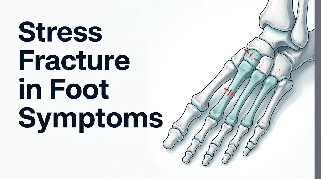
Second and third metatarsal stress fractures produce pain on the top of the forefoot over the middle toes, tenderness along the shaft of the affected metatarsal, and swelling that often appears as a subtle puffiness between the second and third toe rays by the end of the day.
The second metatarsal is the most commonly fractured foot bone in both runners and ballet dancers, according to data reviewed in Foot and Ankle International. Its susceptibility comes from its anatomical position: the second metatarsal is the longest of the five in most people, fixed at its base by the medial cuneiform on both sides, making it relatively immobile and therefore unable to distribute loading forces by shifting position. All that force concentrates at the shaft.
The pain localizes to the dorsum (top) of the forefoot, roughly at the level of the second or third metatarsal shaft, which sits approximately at the midpoint between the base of the toe and the ankle. Pressing directly along this shaft with a thumb reproduces the pain sharply at one specific point. Movement of the second or third toe may be mildly painful because the extensor tendons run adjacent to the fracture site and can become involved in the surrounding soft tissue inflammation.
Ballet dancers develop a specific variant: the Freiberg’s infraction pattern at the metatarsal head, which is slightly more distal than the typical shaft fracture. This requires different imaging and management and warrants evaluation by a board-certified orthopaedic surgeon with foot and ankle subspecialty experience.
Runners who recently increased their weekly mileage, changed running surface (from grass to pavement), or switched to minimal footwear should specifically mention these changes to their sports medicine physician or orthopaedic surgeon, as each factor independently increases second metatarsal loading.
Navicular Stress Fracture Symptoms and Why They Are High Risk
Navicular stress fractures produce pain in the inner midfoot, at the top of the arch, often with tenderness precisely over the “N-spot” (the proximal dorsal surface of the navicular), and they carry a high risk of non-union and avascular necrosis because of the navicular bone’s uniquely poor central blood supply.
The navicular bone sits at the apex of the medial longitudinal arch, directly in the path of compressive and shear forces transmitted from the ankle to the forefoot. Its central third receives blood supply from two directions: branches of the medial and lateral plantar arteries converging toward the center. This convergence creates a watershed zone in the central third of the navicular where blood supply is poorest.
When a fracture line develops in this watershed zone (which is where most navicular stress fractures occur), the already-marginal blood supply to the central navicular fragment is further compromised. The result is a significantly higher rate of delayed union, non-union, and avascular necrosis than is seen with metatarsal fractures. Research published in the Journal of Bone and Joint Surgery has found that navicular stress fractures treated with non-weight-bearing immobilization have substantially better union rates than those treated with partial weight-bearing, a distinction that makes early and accurate diagnosis essential.
Symptoms of a navicular stress fracture are often vague early on, which is part of what makes them dangerous. Pain may feel like a general arch ache, similar to plantar fasciitis or tibialis posterior tendinopathy. The key differentiator is point tenderness directly on the proximal dorsal navicular surface, not along the plantar fascia or the medial ankle.
Any athlete with midfoot arch pain and point tenderness over the navicular warrants urgent imaging, including MRI, within the same week. Waiting for X-ray results alone is not appropriate because plain radiographs miss up to 40 percent of navicular stress fractures, according to data cited in Foot and Ankle International.
Key Takeaway: Navicular stress fractures feel like vague arch pain and are routinely missed on initial X-rays. Point tenderness directly over the navicular requires MRI evaluation, not a “wait and see” approach, because of the bone’s poor central blood supply and high non-union risk.
Calcaneal Stress Fracture Symptoms
Calcaneal stress fractures produce a deep, aching heel pain that differs from plantar fasciitis by its location (the posterior or superior calcaneus rather than the plantar heel), its response to compression (positive squeeze test), and its onset pattern (worsening through the day with any weight-bearing rather than worst at the first morning step).
The calcaneus is the largest tarsal bone, forming the heel. It absorbs the initial impact of every foot strike. Stress fractures of the calcaneus are most common in military recruits and long-distance walkers, particularly those who have recently transitioned to harder surfaces or heavier load-bearing activity. Elderly women with osteoporosis are also at elevated risk because trabecular bone loss within the calcaneus reduces its capacity to absorb compressive loads.
The squeeze test is a specific clinical screen for calcaneal stress fractures: applying medial-to-lateral compression across the calcaneus with both hands produces sharp pain at the fracture site. This is distinct from plantar fasciitis, which produces pain with direct plantar pressure at the medial heel tubercle but not with lateral compression. Clinicians use this distinction routinely in the initial physical examination.
Calcaneal stress fractures are considered low-risk compared to navicular fractures because the calcaneus has generous blood supply through multiple arterial branches. They typically heal with protected weight-bearing in a controlled ankle motion (CAM) boot over 6 to 8 weeks. However, complete fractures of the calcaneus (rare with stress fractures but possible in severe osteoporosis or with continued loading) require orthopaedic surgical evaluation.
Older adults with calcaneal heel pain that does not respond to two to three weeks of reduced activity should be evaluated by a primary care physician, who should order a DEXA scan to assess bone mineral density alongside foot imaging, as calcaneal stress fractures in this age group are frequently the first clinical indicator of previously undiagnosed osteoporosis.
Stress Fracture in Foot Symptoms vs. Plantar Fasciitis and Sprain
Stress fracture symptoms in the foot differ from plantar fasciitis primarily by pain location, tenderness type, and morning pain pattern, and differ from an acute sprain by the absence of a single traumatic event and the progressive rather than immediate onset of symptoms.
These three conditions are the most common sources of diagnostic confusion in foot pain presenting to primary care, sports medicine, and podiatry offices. Getting the distinction right changes the entire management approach.
| Feature | Stress Fracture | Plantar Fasciitis | Acute Ankle or Foot Sprain |
|---|---|---|---|
| Onset | Gradual, over days to weeks | Gradual, often insidious | Sudden, after a single traumatic event |
| Worst pain timing | During and after activity | First steps in the morning | Immediately after injury |
| Morning pain | Mild to absent (early stage) | Severe, first-step pain | Moderate, improves with movement |
| Tenderness location | Directly over a bone shaft or dorsum | Medial plantar heel at fascia origin | Over the injured ligament or tendon |
| Tenderness type | Pinpoint, bony, reproducible with direct bone pressure | Linear along plantar fascia band | Broad, soft tissue, over ligament course |
| Swelling | Mild, localized over bone | Typically absent or minimal | Often significant, diffuse |
| Activity effect | Pain begins during activity and worsens | Pain often improves with warm-up | Pain constant regardless of activity |
| Bruising | Uncommon but possible in advanced cases | Not present | Common, appears within 24 hours |
The most reliable differentiating feature between a stress fracture and plantar fasciitis is that direct bony pressure over the metatarsal shaft or navicular reproduces the pain of a stress fracture but not of plantar fasciitis, while direct pressure along the plantar fascia band reproduces plantar fasciitis pain but not stress fracture pain.
Patients who receive an initial diagnosis of plantar fasciitis and do not improve after 4 to 6 weeks of standard treatment should discuss the possibility of a missed stress fracture with their treating provider and ask specifically about MRI or bone scintigraphy.
Stress Fracture Foot Symptoms in Female Athletes and the Female Athlete Triad
Female athletes with the female athlete triad (the coexistence of low energy availability, menstrual dysfunction, and low bone mineral density) develop stress fractures in the foot at higher rates and at lower training loads than their peers with normal hormonal and nutritional status.
The physiological mechanism is estrogen-dependent. Estrogen promotes osteoblast differentiation through the Wnt/beta-catenin signaling pathway within bone marrow stromal cells. When estrogen falls below functional levels, as occurs in hypothalamic amenorrhea triggered by caloric restriction and high training volume, osteoblast activity decreases. The bone remodeling balance shifts toward net resorption even without increased mechanical loading. Under training loads that a woman with normal estrogen status would tolerate, a woman with hypothalamic amenorrhea sustains accelerated microtrauma accumulation.
A clinical consensus statement published in the British Journal of Sports Medicine on relative energy deficiency in sport (RED-S), the expanded model that includes the female athlete triad, identified stress fractures as one of the primary bone health outcomes of this syndrome, with the foot and lower leg as the most common sites.
Symptoms in this population may be identical to those of any other stress fracture. What is different is the context. A female athlete with absent or irregular menstrual periods, a history of restrictive eating or rapid weight loss, and progressive foot pain should be evaluated not only for the fracture itself but for the underlying hormonal and nutritional status driving increased bone fragility.
Any female athlete under 35 presenting with a foot stress fracture and a history of menstrual irregularity should be evaluated by a primary care physician or sports medicine physician who can assess for RED-S and refer for a DEXA scan, dietary counseling, and endocrine evaluation if indicated.
Stress Fracture Foot Symptoms in Older Adults With Osteoporosis
Older adults with osteoporosis or osteopenia develop stress fractures of the foot at lower activity levels than younger adults, often without the repetitive athletic training typically associated with stress fractures, and frequently present with insufficiency fractures rather than classic fatigue fractures.
The distinction between a fatigue fracture (abnormal load on normal bone) and an insufficiency fracture (normal load on abnormal bone) is clinically meaningful. An older adult who fractures a metatarsal simply from walking their normal daily route has not done anything athletically excessive. Their bone has become too fragile to tolerate ordinary mechanical demands, typically because trabecular bone loss has reduced bone mineral density below the fracture threshold.
According to the NIAMS at the National Institutes of Health, postmenopausal women lose bone mineral density at an accelerated rate due to the drop in estrogen following menopause, with the most rapid loss occurring in the first 5 to 10 years post-menopause. This makes women over 55 disproportionately represented among patients with insufficiency fractures of the foot.
Symptoms in this population may be less dramatic than in younger athletes. The pain may be described as a vague ache or soreness rather than sharp, activity-related pain, because reduced bone pain sensitivity in older adults can blunt the normal nociceptive response. Point tenderness may be present but subtler.
Management in this population requires addressing the underlying bone density, not just the fracture. Any older adult with a foot stress fracture should be referred for a dual-energy X-ray absorptiometry (DEXA) scan to assess bone mineral density, and their primary care physician should evaluate whether bisphosphonate therapy, calcium and vitamin D supplementation, or other bone health interventions are clinically indicated.
Corticosteroid users of any age belong in a similar high-risk category. Chronic glucocorticoid use suppresses osteoblast differentiation through Wnt/beta-catenin pathway inhibition, producing corticosteroid-induced osteoporosis that can develop within 3 to 6 months of regular use. These individuals should report any progressive foot pain to their prescribing physician, who may coordinate orthopaedic evaluation alongside a bone density assessment.
Key Takeaway: Older adults and corticosteroid users can develop foot stress fractures from ordinary daily walking, not just athletic training, because underlying bone fragility lowers the fracture threshold. Any progressive foot pain in this population warrants imaging and a bone density assessment.
When a Stress Fracture in the Foot Requires Urgent Evaluation
A stress fracture in the foot requires urgent evaluation when symptoms include pain at rest, pain that wakes the patient from sleep, inability to bear weight, visible deformity, or a specific high-risk anatomical location such as the navicular, the proximal fifth metatarsal, or the great toe sesamoids.
These presentations indicate either an advanced fracture with a substantial risk of completion and displacement, or a bone site where poor blood supply makes delayed treatment disproportionately harmful. Unlike metatarsal shaft fractures in otherwise healthy adults, these categories do not allow for a “rest and see if it improves in a week” approach.
Go to the emergency room or contact a board-certified orthopaedic surgeon for same-day evaluation if you experience any of the following:
- Foot pain with visible deformity or abnormal bone position (possible complete displaced fracture)
- Sudden inability to bear any weight after a history of progressive foot pain (possible fracture completion during normal activity)
- Pain at rest that wakes you from sleep, combined with a known high-risk fracture site or underlying osteoporosis
- Any foot pain in a person with severe osteoporosis, after a fall, or combined with acute trauma
Contact a board-certified sports medicine physician, board-certified orthopaedic surgeon, or podiatrist within one week (not months) if:
- You have point tenderness over the navicular, the proximal fifth metatarsal base, or the great toe sesamoid bones
- You have progressive foot pain lasting more than 2 weeks with no improvement from rest
- You are a female athlete with irregular or absent periods and progressive foot pain
- You have diabetes with any progressive foot pain, as neuropathy may mask the severity of the injury
These presentations require emergency orthopaedic assessment, not an urgent care appointment or a phone call, because displacement of a stress fracture in a high-risk bone converts a non-surgical injury into one requiring internal fixation surgery.
Diagnosing a Stress Fracture in the Foot: Why X-Rays Miss It and What Imaging Works
Plain X-rays miss stress fractures in the foot during the first 2 to 3 weeks in approximately 50 to 85 percent of cases, depending on the bone site and fracture stage, making MRI the preferred first-line imaging study when clinical suspicion is high.
The reason X-rays miss early stress fractures is straightforward: a stress fracture in its early stage is a crack through cortical bone with no displacement and no callus formation. Callus (new bone deposition bridging the fracture site) is what makes fractures visible on plain radiographs. It takes 2 to 3 weeks of healing for enough callus to form to be detectable on X-ray. Before that, the cortex looks continuous even though microcracks are present.
MRI detects stress fractures early because it images bone marrow edema, the fluid accumulation and inflammatory response within the bone marrow that accompanies cortical microtrauma even before a visible fracture line appears. The Fredericson MRI grading scale classifies bone stress injuries from Grade 1 (periosteal edema only) through Grade 4 (complete fracture line with displacement), allowing the treating physician to understand exactly how far the injury has progressed and calibrate treatment accordingly.
| Imaging Modality | What It Detects | Sensitivity for Early Fracture | Best Used For |
|---|---|---|---|
| Plain X-ray (radiograph) | Callus, displacement, complete fracture | Low in first 2 to 3 weeks (15 to 50%) | Ruling out complete displaced fracture; initial screen |
| MRI | Bone marrow edema, periosteal edema, fracture line | High (up to 99%) | Confirming early stress fracture, grading severity |
| CT scan | Cortical fracture line detail | Moderate to high | Surgical planning, navicular and tarsal fractures |
| Bone scintigraphy (bone scan) | Areas of accelerated bone turnover | High but not specific | When MRI is contraindicated; differentiating from infection |
When you see a primary care physician, board-certified sports medicine physician, or podiatrist for suspected foot stress fracture, ask specifically whether an MRI is indicated given your symptom duration and location. If you have point tenderness over the navicular and a plain X-ray is reported as normal, request an MRI before accepting a diagnosis of plantar fasciitis or soft tissue injury.
Adolescents require additional consideration. MRI is preferred over bone scintigraphy in this age group to avoid radiation exposure, and any imaging that approaches an open growth plate should be reviewed by a clinician experienced with pediatric musculoskeletal injuries to correctly interpret findings near the physis.
Can You Walk on a Stress Fracture in the Foot?
Walking on a stress fracture in the foot risks converting a non-displaced, non-surgical injury into a complete fracture that may require surgical internal fixation, making continued unprotected weight-bearing clinically inadvisable once the diagnosis is confirmed or strongly suspected.
Many people do walk on a stress fracture for days or weeks before diagnosis, because the pain is intermittent early on and full weight-bearing remains possible. But the ability to bear weight does not indicate that weight-bearing is safe. The fracture continues to accumulate damage with each step.
The clinical guidance from the American Academy of Orthopaedic Surgeons is that most low-risk foot stress fractures (second, third, and fourth metatarsal shafts in patients with normal bone density) can be managed with protected weight-bearing in a CAM boot. This means walking is permitted, but only in the boot, which distributes load away from the fracture site. Unprotected activity in regular shoes should stop until a clinician has assessed the fracture type, location, and severity.
High-risk fractures (navicular, proximal fifth metatarsal base, great toe sesamoids) are typically managed with strict non-weight-bearing, meaning crutches or a knee scooter with no foot contact with the ground. Walking on these fractures, even in a boot, risks fracture completion or displacement.
The practical instruction is this: if your foot pain matches the pattern described in this article and you have not yet been evaluated, reduce your weight-bearing activity to walking for essential tasks only and avoid all running, jumping, or extended standing until you have been seen by a primary care physician, board-certified sports medicine physician, board-certified orthopaedic surgeon, or podiatrist who can review imaging and advise on the appropriate level of protected activity.
Frequently Asked Questions About Stress Fracture in Foot Symptoms
How do I know if I have a stress fracture in my foot?
The most reliable indicator is a combination of point tenderness reproducible by pressing on a specific bone, progressive activity-related pain that has worsened over days to weeks, and mild localized swelling over the affected bone.
These three findings together are more specific for a stress fracture than any single symptom alone, but a confirmed diagnosis requires imaging, typically MRI, ordered by a primary care physician, sports medicine physician, or podiatrist.
You cannot confirm a stress fracture through self-assessment; the clinical examination and imaging are necessary because several other foot conditions produce similar pain patterns.
What does a stress fracture in the foot feel like compared to a sprain?
A stress fracture produces pain that builds gradually over days to weeks without a single traumatic event, while a sprain produces immediate, sharp pain at the moment of injury.
Stress fracture pain is localized directly over a bone with pinpoint tenderness, while sprain pain is distributed over the injured ligament and surrounding soft tissue.
Morning pain is typically more prominent with plantar fasciitis than with a stress fracture, whereas stress fracture pain tends to be worse during and after activity, not exclusively at the first step of the day.
Can you walk on a stress fracture in the foot?
Many people can physically walk on a stress fracture, but doing so risks progressing the fracture to a complete, displaced break that may require surgery.
Low-risk metatarsal shaft fractures are often managed with protected weight-bearing in a CAM boot, but high-risk fractures of the navicular, proximal fifth metatarsal, or sesamoids typically require strict non-weight-bearing with crutches.
The appropriate level of weight-bearing can only be determined by a clinician who has reviewed imaging of the specific fracture site and grade.
How long does a foot stress fracture take to heal?
Most low-risk foot stress fractures heal within 6 to 8 weeks of protected weight-bearing when treatment begins promptly, according to clinical guidelines from the American Academy of Orthopaedic Surgeons.
High-risk fractures, particularly navicular and proximal fifth metatarsal stress fractures, can take 10 to 16 weeks of non-weight-bearing, and some require surgical fixation to achieve union.
Individuals with osteoporosis, low estrogen states, or continued weight-bearing activity through the fracture period typically have longer healing timelines and higher complication rates.
Will a stress fracture in the foot show up on an X-ray?
Plain X-rays miss stress fractures in the foot in up to 50 to 85 percent of cases during the first 2 to 3 weeks, because the early fracture produces no callus or displacement visible on radiograph.
MRI is the preferred imaging study for suspected foot stress fractures, with sensitivity approaching 99 percent because it detects bone marrow edema even before a visible fracture line forms.
If your X-ray is reported as normal but your symptoms are consistent with a stress fracture, ask your treating physician specifically about ordering an MRI before accepting an alternative soft tissue diagnosis.
Where exactly does a stress fracture hurt in the foot?
Foot stress fracture pain is most commonly located on the top of the forefoot over the second or third metatarsal shaft, at the inner midfoot over the navicular bone, or at the heel over the posterior calcaneus.
The pain is pinpoint, meaning you can typically reproduce it by pressing with one thumb on a specific spot roughly the size of a fingernail, rather than feeling pain across a broad region.
The exact location changes by population: runners and military recruits most commonly fracture the second metatarsal; female dancers more often fracture the second metatarsal head; military personnel are prone to calcaneal fractures; and older adults with osteoporosis can fracture any of these sites from low-impact activity.
Foot stress fractures are one of the most underdiagnosed overuse injuries in active adults, largely because the early symptoms are easy to attribute to muscle fatigue, poor footwear, or generalized soreness. The pattern that matters is this: pain that arrives progressively earlier in each training session, located precisely over a single bone you can identify with one finger, and eased only by rest.
If that pattern describes what you are experiencing, reducing your activity and seeking evaluation is the correct next step. Request an MRI if your symptoms have been present for more than two weeks or if you have tenderness over the navicular, proximal fifth metatarsal, or sesamoids, since plain X-rays will miss the injury at this stage in many cases. A board-certified sports medicine physician, board-certified orthopaedic surgeon, or podiatrist with sports medicine experience can confirm the diagnosis and determine whether protected weight-bearing or strict non-weight-bearing is appropriate for your specific fracture.
The difference between a six-week recovery and a six-month one often comes down to how quickly you stop loading the fracture. Act on the pattern, not on whether the pain is severe enough to feel like an “emergency.”