Peroneal Tendonitis Symptoms: What Your Ankle Is Telling You in 2026
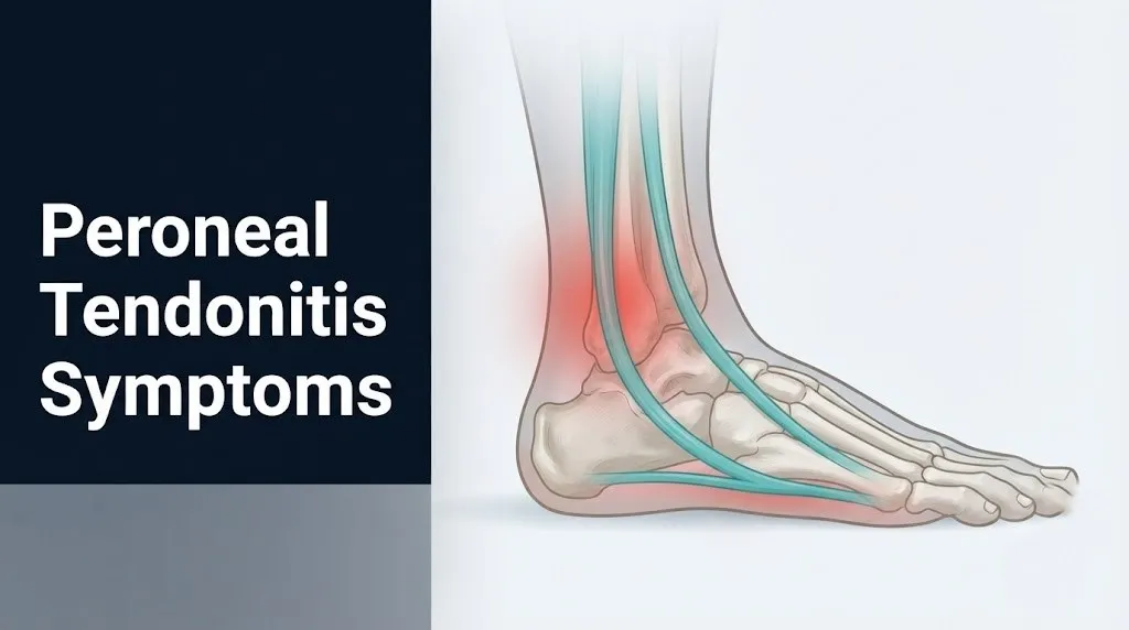
Peroneal tendonitis symptoms center on pain, swelling, and tenderness along the outer edge of the ankle, specifically behind and below the bony prominence known as the lateral malleolus. This pain is produced by inflammation or degeneration within the two main peroneal tendons, the peroneus longus and peroneus brevis, which run side by side through a narrow groove on the back of the fibula before fanning out to stabilize the foot and ankle with every step.
According to the American Academy of Orthopaedic Surgeons, peroneal tendon injuries are among the most frequently missed diagnoses in patients presenting with chronic lateral ankle pain, partly because their symptom pattern overlaps closely with lateral ankle ligament sprains. Understanding the precise location, character, and behavior of these symptoms is the most direct path to getting the right clinical evaluation before the condition progresses from manageable tendinopathy to a structural tear that requires surgery.
This article covers every recognized symptom of peroneal tendonitis with the physiological explanation behind each one, the difference between acute and chronic presentations, what the symptom patterns look like in runners versus older adults versus people with specific foot structures, and exactly when each type of symptom requires professional evaluation.
Peroneal Tendonitis Symptoms: The Clinical Picture
Peroneal tendonitis symptoms are primarily outer ankle pain, localized swelling posterior to the lateral malleolus, tenderness along the peroneal tendon course, and functional weakness in foot eversion, with symptom severity ranging from mild activity-related discomfort to constant pain that limits normal walking.
The two peroneal tendons most commonly affected are the peroneus longus and the peroneus brevis. Both tendons travel through the retromalleolar fibular groove, a shallow channel on the posterior surface of the distal fibula, held in place by the superior peroneal retinaculum, a fibrous restraining band. When these tendons are overloaded or injured, the symptoms they produce reflect both the tendon’s anatomical position and the specific tissue damage that has occurred.
The National Institute of Arthritis and Musculoskeletal and Skin Diseases (NIAMS) describes tendinopathy as an umbrella term covering both acute inflammatory tendinitis and chronic degenerative tendinosis, noting that the distinction matters clinically because the tissue-level pathology, and therefore the symptom character, differs between the two. Acute tendinitis involves active prostaglandin-mediated inflammation in the tendon sheath. Chronic tendinosis involves structural collagen fiber disorganization without active inflammation, which changes how the pain behaves over time.
Key recognized symptoms of peroneal tendonitis include:
- Pain along the outer ankle, posterior to the lateral malleolus
- Localized swelling at or below the bony ankle prominence
- Tenderness to firm touch along the peroneal tendon course
- Warmth over the lateral ankle in acute flares
- Ankle stiffness, particularly after rest
- Weakness or instability during foot-turning movements
- Pain that increases with weight-bearing activities and eases with rest (in early stages)
- A snapping or popping sensation behind the ankle (when tendon subluxation is involved)
Adults over 50 tend to present with more gradual onset and less dramatic acute swelling compared to younger athletes, because age-related decline in tendon collagen turnover produces a more insidious degenerative pattern rather than an acute inflammatory flare.
What Does Peroneal Tendonitis Feel Like?
Peroneal tendonitis typically feels like a dull, aching pain along the outer ankle that sharpens with walking, running, or any activity that requires pushing off the foot, with a burning or throbbing quality during active flares.
Most people describe the pain as sitting just behind or below the outer ankle bone. It rarely radiates far up the leg or across the top of the foot, which helps distinguish it from superficial peroneal nerve irritation or a fifth metatarsal stress fracture. The pain tends to behave predictably: worse during and immediately after exercise, mildly reduced after warming up if the tendinopathy is early-stage, and increasingly constant as the condition progresses.
Think of the peroneal tendons like two cables running through a narrow pulley system. The lateral malleolus acts as the pulley. When the cables are swollen or frayed from overuse, every movement that slides them through the pulley generates friction, pressure, and pain. That friction is most intense at the point where the tendons curve around the fibular groove, which is exactly where most people with this condition feel the sharpest tenderness.
A 2021 study published in the American Journal of Sports Medicine examining patients with surgically confirmed peroneal tendinopathy reported that the most consistently described subjective experience was diffuse lateral ankle aching that became focal point tenderness directly posterior to the lateral malleolus on palpation. Patients with concurrent tendon subluxation more frequently reported a distinct snapping or clunking sensation, which carries a different clinical significance.
Women may experience this pain slightly differently because a wider Q-angle (the angle between the quadriceps and the patellar tendon, which affects the mechanical alignment of the entire lower extremity) can increase lateral loading on the peroneal tendons during walking, producing a more diffuse aching pattern rather than one focal point of tenderness.
Outer Ankle Pain and Swelling: What Is Actually Happening
The outer ankle pain and swelling in peroneal tendonitis result from increased vascular permeability in the synovial tendon sheath, driven by prostaglandin E2 released through the COX-2 inflammatory pathway, which causes fluid to leak into the peritendinous tissue and produces the visible puffiness posterior to the lateral malleolus.
The synovial sheath surrounding the peroneal tendons functions like a lubricated sleeve, reducing friction as the tendons slide through the fibular groove. When tendon loading exceeds the tissue’s repair capacity, damaged tenocytes release pro-inflammatory cytokines, including interleukin-1 beta (IL-1β) and tumor necrosis factor-alpha (TNF-α). These cytokines activate the COX-2 enzyme, which converts arachidonic acid into prostaglandin E2 (PGE2). PGE2 simultaneously sensitizes pain receptors (nociceptors) in the tendon sheath and increases vascular permeability, allowing plasma proteins and fluid to accumulate in the tissue.
The swelling is typically fusiform, meaning it is elongated along the tendon’s course rather than rounded like a fluid-filled cyst. You can often trace its shape from just above the lateral malleolus, curving around the back of the ankle bone, and continuing toward the heel. This distinctive swelling pattern is one of the features that separates peroneal tendinopathy from a lateral ligament sprain, where swelling tends to be broader and less linearly defined.
| Swelling Characteristic | Peroneal Tendonitis | Lateral Ankle Sprain |
|---|---|---|
| Shape | Fusiform, follows tendon course | Diffuse, egg-shaped |
| Location | Posterior to lateral malleolus | Anterior and inferior to lateral malleolus |
| Onset timing | Gradual, post-activity | Immediate, after acute inversion |
| Warmth | Mild in acute phase | Often prominent in first 48 hours |
| Tenderness | Along tendon course | Over ligament attachment points |
Older adults and people taking corticosteroid medications may show less visible swelling even with moderate tendon pathology, because both systemic corticosteroids and age-related changes in skin and subcutaneous tissue reduce the visible inflammatory response.
Key Takeaway: The outer ankle swelling in peroneal tendonitis is fusiform and follows the tendon’s course behind the ankle bone, not the diffuse rounded swelling you get from twisting the ankle, and that shape difference is one of the most reliable ways to tell the two conditions apart before seeing a doctor.
Peroneal Tendon Pain Location: Where It Hurts and Why
The primary pain location in peroneal tendonitis is the posterior lateral ankle, specifically the region directly behind and below the lateral malleolus, extending in a curved arc toward the heel and along the outer border of the foot depending on which tendon is more involved.
The two tendons have slightly different pain locations because they diverge after passing the lateral malleolus. The peroneus brevis tendon inserts at the styloid process of the fifth metatarsal base, the small bony bump on the outer midfoot. Pain from peroneus brevis tendinopathy or a split tear therefore often includes tenderness right at that bony prominence or along the outer edge of the foot between the heel and the base of the little toe. The peroneus longus tendon curves under the foot and travels across the plantar surface to insert at the first metatarsal base and medial cuneiform. Its tendinopathy produces pain that can extend under the arch of the foot, particularly in the region of the cuboid tunnel, where the tendon makes a sharp directional turn.
Knowing which tendon is primarily involved changes where the clinician palpates and which activities are most provocative. Eversion of the foot against resistance (pushing the foot outward against pressure) loads both tendons, but pain that reproduces specifically at the fifth metatarsal base on this test points more strongly toward peroneus brevis pathology.
To locate peroneal tendon tenderness accurately:
- Sit with the affected ankle crossed over the opposite knee so the outer ankle faces upward.
- Place two fingers directly behind the outer ankle bone (the prominent bump on the outside of your ankle).
- Apply firm but not forceful pressure while slowly moving your fingers in a downward arc toward the heel.
- Note whether you feel a cord-like structure and whether pressure on it reproduces the pain you experience during activity.
- Then trace from that point toward the small bony bump on the outer midfoot and check whether pressure there reproduces pain separately.
A positive reproduction of pain along both steps suggests peroneal tendon involvement. Pain only at the outer midfoot bump requires ruling out a fifth metatarsal avulsion fracture, which has a similar location but a very different clinical urgency.
Tendonitis Ankle Symptoms That Worsen With Activity
Ankle tendonitis symptoms from peroneal tendon involvement characteristically worsen with weight-bearing activity and improve with rest in early and moderate stages, a pattern produced by the mechanical loading cycle that exceeds the tendon’s current structural capacity.
During the push-off phase of walking and running, the peroneal muscles contract concentrically to evert the foot and stabilize the subtalar joint. This generates tensile load through the peroneal tendons. In a healthy tendon, this load is distributed evenly across well-organized collagen fibers. In an inflamed or degenerated tendon, the collagen architecture is disrupted, and the same load concentrates unevenly through fewer intact fibers, triggering nociceptor activation and pain.
The activity pattern that produces symptoms is highly specific. Running on banked or cambered roads (where one foot is consistently lower than the other) increases peroneal tendon strain on the lower side. Repeated ankle inversion activities, such as trail running over uneven terrain, generate sudden eccentric loading of the peroneal tendons as they resist the inward roll of the ankle. Stair climbing, hill running, and lateral movement sports like tennis and basketball all load the peroneal tendons in ways that provoke symptoms more reliably than flat walking.
Activities most likely to provoke or worsen peroneal tendonitis symptoms:
- Running, especially on uneven or cambered surfaces
- Uphill and downhill walking with a rigid or high-heel boot
- Lateral cutting movements (tennis, basketball, soccer)
- Extended walking or standing on hard surfaces
- Ballet and dance activities requiring forced plantarflexion
- Wearing unsupportive footwear, particularly flat-soled shoes with no lateral ankle support
People with diabetes-related peripheral neuropathy may not feel the characteristic activity-related pain increase because sensory nerve function is diminished. This makes them particularly at risk of advancing from early tendinopathy to a significant tendon tear without recognizing the warning symptoms, and they should have their feet evaluated by a podiatrist or orthopedic surgeon regularly as part of their diabetes management plan.
Acute Peroneal Tendonitis Symptoms
Acute peroneal tendonitis symptoms appear over days to a few weeks after a specific period of increased activity or a sudden increase in training load, presenting with active inflammation in the tendon sheath and producing sharper, more constant pain than the chronic form.
The American Academy of Orthopaedic Surgeons describes acute peroneal tendinopathy as typically triggered by a sudden change in training volume, a new sport or activity, or an acute ankle inversion injury that compresses or tears the peroneal tendon sheath. In the acute phase, the synovial sheath is actively inflamed, meaning the prostaglandin-mediated vascular response is ongoing, which produces pain even at rest in more severe cases.\
Acute symptoms include warmth and redness over the lateral ankle (not just behind the ankle bone but along the visible tendon path), swelling that appears within 24 to 48 hours of the provoking activity, and pain on passive eversion of the foot, meaning even gentle movement of the foot outward by someone else reproduces pain. This passive pain response distinguishes active tendon sheath inflammation from simple muscle soreness, where passive motion does not typically hurt.
| Symptom | Acute Presentation | Clinical Significance |
|---|---|---|
| Pain at rest | Present in moderate to severe cases | Indicates active synovial sheath inflammation |
| Swelling onset | Within 24-48 hours of trigger event | Prostaglandin-mediated vascular leakage |
| Warmth over tendon | Visible, palpable | Confirms active inflammatory phase |
| Pain with passive eversion | Positive | Distinguishes from muscle soreness |
| Morning stiffness | Prominent, eases within 30 minutes | Gel phenomenon of inflamed synovial tissue |
| Weight-bearing tolerance | Reduced | Axial loading compresses inflamed sheath |
Adolescent athletes deserve specific mention here. Growth-related tightness in the Achilles tendon complex during rapid bone growth phases can increase mechanical demand on the peroneal tendons as compensatory stability mechanisms. Acute peroneal tendinopathy in a teenager who has recently grown several inches should prompt evaluation by a sports medicine physician to rule out a concurrent apophyseal injury at the fifth metatarsal base.
Chronic Peroneal Tendonitis Symptoms
Chronic peroneal tendonitis symptoms reflect tendinosis, a degenerative state of the tendon rather than an actively inflamed one, and present as persistent dull aching with episodic flares, thickening of the tendon, and functional decline that develops over months rather than days.
In tendinosis, the normal tendon architecture has broken down. Histopathological studies published in Foot and Ankle International have documented that chronically diseased peroneal tendons show mucoid degeneration within the collagen matrix, disorganized type I collagen fiber alignment, loss of the normal crimp pattern that allows tendons to absorb shock, and intratendinous neovascularization without corresponding restoration of tensile strength. These structural changes produce a tendon that is thicker and palpably nodular compared to the opposite side, yet weaker and more prone to further injury.
The chronic symptom pattern differs from acute in three important ways. First, pain is less intense at rest and more reliably provoked by specific loading activities. Second, there is often a period of “warm-up pain” where the first 5 to 10 minutes of activity are quite uncomfortable, followed by partial symptom relief mid-activity, followed by a return of pain and stiffness that persists for hours after stopping. Third, the visible swelling is less dramatic than acute tendinitis but the tendon often feels palpably thicker and “ropey” when traced behind the lateral malleolus.
People who have had multiple lateral ankle sprains over their lifetime are at high risk for chronic peroneal tendinopathy because repeated sprains stretch or partially tear the superior peroneal retinaculum, reducing the restraint that keeps the peroneal tendons in their groove. The Journal of Bone and Joint Surgery has documented that chronic lateral ankle instability and peroneal tendinopathy co-exist in a clinically important proportion of patients with recurrent ankle sprains, making a history of ankle sprains one of the most important questions a clinician should ask when evaluating outer ankle pain.
Key Takeaway: Chronic peroneal tendinosis produces a thickened, palpably nodular tendon that hurts most at the start of activity and again after stopping, with less dramatic swelling than acute tendinitis but more structural damage that takes longer to rehabilitate.
Peroneal Tendon Subluxation Symptoms
Peroneal tendon subluxation symptoms include a sharp snapping or popping sensation behind the lateral malleolus, felt most strongly during dorsiflexion of the ankle, caused by one or both tendons slipping out of the fibular groove when the superior peroneal retinaculum is torn or lax.
This symptom pattern is frequently confused with peroneal tendinitis because the outer ankle pain location is nearly identical. The distinguishing feature is the mechanical snapping. Subluxation happens when the superior peroneal retinaculum, the primary restraint holding the tendons in the retromalleolar groove, is disrupted. Without this restraint, the tendons can translate anteriorly over the lateral malleolus when the foot is dorsiflexed (brought upward toward the shin), producing a palpable and sometimes audible snap.
According to clinical classification used in foot and ankle orthopedic surgery, peroneal tendon subluxation is graded from Grade I (retinaculum stretched and elevates off the fibula but remains attached) to Grade III (the retinaculum avulses a fragment of bone from the fibula). Higher grade injuries produce more persistent and painful subluxation episodes and are less likely to resolve without surgical intervention.
Symptoms that suggest peroneal tendon subluxation rather than simple tendinitis:
- A snapping, clunking, or popping sensation behind the outer ankle bone during movement
- The sensation of something “rolling over” the ankle bone during dorsiflexion
- Pain that is provoked specifically by moving the foot up and back (dorsiflexion combined with eversion), rather than by simple walking load
- Ankle instability that seems out of proportion to any ligament injury found on examination
- Onset after an acute injury where the foot was forcefully dorsiflexed and everted simultaneously
People who have experienced this first as an acute injury (often during a ski fall, soccer tackle, or a stumble with the foot caught in an upward position) should be evaluated by a board-certified orthopedic surgeon with foot and ankle specialty training, because Grade II and III subluxation injuries have a high rate of treatment failure with conservative management alone.
Peroneal Tendon Tear Symptoms
Peroneal tendon tear symptoms include severe outer ankle pain, persistent swelling that does not respond to rest, functional weakness in pushing the foot outward, and in the case of a complete rupture, a visible gap or defect along the tendon course detectable on imaging.
The peroneus brevis tendon is the most frequently torn of the two peroneal tendons. It is at particular risk because it lies directly against the fibular groove and is compressed against the groove wall when the peroneus longus contracts forcefully. The characteristic tear pattern is a longitudinal split tear along the length of the brevis tendon, often described in the surgical literature as a “banana peel” tear, where the tendon splits into two leaves along its long axis rather than snapping in half transversely.
A 2019 study in Foot and Ankle International reviewing surgical findings in patients with confirmed peroneal tendon pathology found that longitudinal split tears of the peroneus brevis were present in a substantial proportion of patients who had been clinically diagnosed with “recurrent lateral ankle sprains” and had undergone prior conservative treatment without improvement. This underscores why outer ankle pain that does not respond to standard tendinopathy rehabilitation deserves imaging evaluation.
The clinical symptoms of a partial longitudinal split tear are often indistinguishable from tendinosis without imaging. The distinguishing features that should raise clinical suspicion of a tear rather than simple tendinopathy include:
- Outer ankle pain and swelling that does not improve after 6 to 8 weeks of appropriate physical therapy and activity modification
- Pain that is worse on resisted eversion (pushing the foot outward against manual resistance) than on simple walking or palpation
- Visible or palpable tendon contour irregularity, where the tendon appears thicker at one point and thinner or absent at another
- New onset of ankle instability in someone with a prior history of outer ankle tendon pain
Older adults, particularly those over 65, are more likely to present with advanced degenerative tears that have been present for months or years without being identified, because the gradual onset and lower activity demands of older age can mask the functional decline until the tear becomes nearly complete.
Peroneal Tendonitis Symptoms in Runners and Athletes
Peroneal tendonitis symptoms in runners and athletes tend to be activity-specific and load-dependent, appearing predictably at a certain distance or training intensity and resolving with rest, a pattern that reflects cumulative mechanical overload of the peroneal tendons rather than a discrete tissue failure event.
Runners are at elevated risk for peroneal tendinopathy because of the high-repetition, low-variation loading cycle that long-distance running imposes on the ankle stabilizers. At each heel strike, the peroneal muscles engage eccentrically to control foot inversion and protect the lateral ankle from rolling. Over thousands of repetitions per training session, the cumulative tensile load through the peroneal tendons can exceed the tendon’s adaptive capacity, particularly during periods of rapid training volume increases, a concept the sports medicine community refers to as exceeding the “acute-to-chronic workload ratio.”
According to research in the American Journal of Sports Medicine, runners who increase weekly mileage by more than 10% per week have a substantially higher risk of overuse lower extremity tendon injuries compared to those who follow a gradual progression. Trail runners and cross-country athletes face additional risk from irregular terrain, which generates unpredictable inversion stress and sudden eccentric loading of the peroneal tendons with each stride on uneven ground.
Symptoms in this population often follow a recognizable pattern:
- Lateral ankle aching appears toward the end of long runs, initially resolving within an hour of finishing.
- The aching onset point in runs gradually shortens: what started at mile 10 now appears at mile 5, then mile 3.
- Post-run stiffness persists into the next morning.
- Eventually, pain appears within the first mile and does not warm up through.
- In unaddressed cases, pain begins during normal daily walking.
Basketball players, soccer players, and dancers are at risk through different mechanisms: sudden cutting movements and forced plantarflexion positions load the peroneal tendons in acute high-force events rather than through cumulative repetition.
Key Takeaway: In runners, peroneal tendonitis symptoms reliably worsen at a predictable point in training sessions and progress to affect shorter distances over weeks, which is the clearest early warning that the tendon is not adapting to current training load and needs a structured load-reduction plan before a tear develops.
Peroneal Tendonitis Risk Factors and Causes
Peroneal tendonitis is caused by repetitive mechanical overload of the peroneus longus and peroneus brevis tendons beyond their adaptive capacity, with specific anatomical, biomechanical, and training-related risk factors that significantly increase the probability of tendon pathology in susceptible individuals.
The primary cause is cumulative microtrauma from repetitive ankle eversion and plantarflexion movements that exceed the tendon’s rate of structural repair. This is the biological root of the overuse injury classification. Unlike a traumatic tear, tendinopathy develops over time, with tissue damage accumulating faster than tenocytes can lay down organized replacement collagen.
The NIAMS identifies the following categories of risk factors for tendinopathy across multiple tendon locations, several of which are specifically relevant to the peroneal tendons:
Biomechanical risk factors:
- Pes cavus (high-arch foot): increases lateral loading through the peroneal tendons with every step
- Pes planus (flatfoot): changes the mechanical angle at which the peroneal tendons pull, increasing shear at the fibular groove
- Hindfoot varus alignment: places the ankle in a slightly inverted resting position, increasing peroneal muscle workload to maintain stability
- Prior lateral ankle sprain with residual laxity: disrupts the superior peroneal retinaculum and allows abnormal tendon movement
Training and activity risk factors:
- Sudden increase in training volume (greater than 10% per week increase in running mileage)
- Running on cambered or banked surfaces
- Inadequate footwear without lateral support
- Returning to sport too quickly after an ankle sprain
Individual and systemic risk factors:
- Age over 40: declining collagen turnover rate reduces tendon resilience
- Female sex: wider Q-angle and differences in lower extremity alignment increase lateral tendon loading
- Corticosteroid use (systemic or injected near the tendon): associated with collagen weakening and increased tear risk
- Fluoroquinolone antibiotic use: well-documented in clinical literature as increasing tendinopathy and tendon rupture risk across multiple body locations
How High Arch Affects Peroneal Tendonitis Symptoms
A high-arch foot structure (pes cavus) produces more severe and more persistent peroneal tendonitis symptoms than a neutral foot, because the rigid, supinated position of the cavus foot increases lateral loading through the peroneal tendons with every step and reduces the foot’s natural shock absorption.
In a typical foot during the gait cycle, the arch partially flattens at midstance (pronation) to absorb impact forces, then rises at push-off (supination) to generate propulsion. In a pes cavus foot, the arch is rigidly elevated and the foot tends to remain in a supinated, slightly inverted position throughout the stance phase. This posture increases the mechanical demand on the peroneal muscles and tendons to continuously resist the inward roll of the ankle, generating persistently elevated tensile load on the tendons even during routine walking.
The Mayo Clinic notes that pes cavus is associated with higher rates of lateral ankle instability and lateral ankle tendon pathology compared to neutral or planus foot types. The combination of high arch and hindfoot varus (heel tilted slightly inward) is particularly associated with peroneal tendon problems because it places the foot in the very position that maximally loads these tendons at baseline.
People with pes cavus who develop peroneal tendinopathy typically report:
- Symptoms that appear at lower activity thresholds than runners or athletes with neutral feet
- More pronounced lateral ankle instability between flares, not just during exercise
- A tendency toward recurrence after apparently successful treatment, because the underlying structural loading has not been corrected
- Pain that extends further along the outer foot border, because the rigidity of the high arch places stress on the peroneus longus tendon across the plantar surface, particularly at the cuboid tunnel
Lateral wedge orthotics that slightly tilt the heel outward can reduce peroneal tendon loading in pes cavus patients. This is a device-level intervention that requires fitting by a licensed physical therapist or podiatrist, not a general recommendation for all people with peroneal tendinopathy.
Peroneal Tendonitis vs. Lateral Ankle Sprain
Peroneal tendonitis and a lateral ankle sprain both produce outer ankle pain and swelling, but they differ in the anatomical structures injured, the onset pattern, the point tenderness location, and the way symptoms respond to loading, with sprains causing immediate post-injury pain and tendinopathy producing gradual activity-related pain.
A lateral ankle sprain injures the anterior talofibular ligament (ATFL), the calcaneofibular ligament (CFL), or both. These ligaments sit on the anterior and lateral surface of the ankle. Their tenderness is located slightly more forward and downward compared to the peroneal tendons, which sit posterior to the lateral malleolus. This anatomical difference in tenderness location is the single most reliable clinical examination finding for separating the two conditions.
| Feature | Peroneal Tendonitis | Lateral Ankle Sprain |
|---|---|---|
| Onset | Gradual, activity-related | Sudden, after specific inversion injury |
| Tenderness location | Posterior to lateral malleolus, along tendon course | Anterior to lateral malleolus, ATFL and CFL points |
| Swelling shape | Fusiform, elongated along tendon | Diffuse, periarticular |
| Provoking movement | Resisted foot eversion | Passive inversion stress test |
| Mechanism | Overuse / cumulative microtrauma | Single inversion event |
| X-ray findings | Normal (no fracture) | Normal or tiny avulsion fleck at ATFL |
| MRI findings | Tendon signal change, sheath fluid | ATFL tear, periarticular edema |
The overlap is most confusing in patients who have both conditions simultaneously, which is clinically common. A 2020 study in Foot and Ankle International found that a significant proportion of patients with surgically confirmed peroneal tendon tears had a prior clinical diagnosis of simple lateral ankle sprain, suggesting the two conditions co-exist more frequently than clinical examination alone identifies.
The specific test that best separates the two: resisted ankle eversion (the examiner holds the outer edge of the foot and the patient tries to push against resistance). Pain specifically during this test that is located posterior to the lateral malleolus points strongly to peroneal tendon involvement rather than ligament injury.
Key Takeaway: Tenderness that sits directly behind the outer ankle bone and reproduces with resisted foot-outward movement is peroneal tendon territory; tenderness that sits just below and in front of the ankle bone and reproduces with the foot being turned inward is more likely ligament territory, and many people have both at the same time after an ankle injury.
How Peroneal Tendonitis Is Diagnosed
Peroneal tendonitis is diagnosed through a combination of clinical history, physical examination focused on peroneal tendon palpation and provocative loading tests, and imaging with diagnostic ultrasound or magnetic resonance imaging (MRI) to confirm tendon pathology and rule out tear or subluxation.
The clinical examination begins with a careful history. A sports medicine physician or orthopedic surgeon will ask about the activity that provoked symptoms, whether there was a specific ankle sprain event, how the pain behaves across an activity session, and whether there is a snapping sensation. Physical examination then includes palpation along the entire course of both peroneal tendons from their fibular origin to their distal insertions, testing resisted ankle eversion strength, checking for a subluxation provocation sign (asking the patient to actively dorsiflex and evert the foot while the examiner observes whether the tendons snap forward over the lateral malleolus), and assessing overall ankle stability.
Plain radiographs (weight-bearing X-rays) are typically ordered first to rule out fifth metatarsal fracture, avulsion fracture at the ATFL attachment, and lateral malleolus fracture, none of which are visible on a clinical examination alone.
The diagnostic imaging pathway for confirmed or suspected peroneal tendinopathy:
- Weight-bearing anteroposterior and lateral radiographs: exclude fracture and assess foot alignment.
- If radiographs are normal and symptoms are consistent with tendinopathy: diagnostic ultrasound provides real-time visualization of the tendons in dynamic movement, can detect sheath fluid, tendon thickening, and longitudinal split tears.
- If ultrasound is inconclusive or clinical suspicion for a complete tear, subluxation, or concurrent ligament injury is high: MRI of the ankle provides the most detailed soft-tissue characterization.
- In cases where symptoms suggest nerve entrapment rather than tendinopathy (burning, tingling, numbness): nerve conduction velocity (NCV) testing may be ordered to assess superficial peroneal nerve function.
- If surgical intervention is being considered: MRI findings guide surgical planning for groove deepening, retinaculum repair, or tendon debridement.
According to the American Academy of Orthopaedic Surgeons, MRI remains the reference standard for definitive characterization of peroneal tendon pathology before surgical decision-making, while high-resolution diagnostic ultrasound performed by a skilled musculoskeletal sonographer is increasingly used for initial evaluation and for guiding injections.
When to See a Doctor for Peroneal Tendonitis
See a sports medicine physician or board-certified orthopedic surgeon for peroneal tendonitis if your outer ankle pain has not improved after 2 to 4 weeks of relative rest and activity modification, if you feel a snapping or popping behind your ankle bone, if you cannot bear weight comfortably, or if the pain came on immediately after a significant ankle injury.
Many people with mild peroneal tendinopathy initially manage symptoms appropriately at home: reducing the activity that caused the problem, applying ice for 15 to 20 minutes two to three times daily during the first week, avoiding footwear without lateral support, and switching to low-impact activity while the tendon recovers. This approach is appropriate for early-stage symptoms rated 3/10 or below on a pain scale during activity, with no snapping, no significant swelling, and a clear overuse activity as the cause.
The picture changes when any of the following are present:
- Pain rated 5/10 or above during daily walking: this level of pain with normal activity suggests more than superficial tendon irritation and warrants evaluation by a sports medicine physician.
- A snapping or clunking sensation behind the lateral malleolus: this specific symptom requires orthopedic evaluation for potential tendon subluxation.
- Outer ankle pain that came on immediately after an ankle injury: the possibility of a concurrent peroneal tendon tear or avulsion fracture requires same-week imaging evaluation, not a wait-and-see approach.
- Symptoms that have been present for more than 6 weeks without improvement: chronic tendinosis does not resolve with rest alone and requires a structured physical therapy rehabilitation program.
- Sudden severe pain and inability to push the foot outward: this pattern, particularly in someone with a prior history of outer ankle tendinopathy, warrants urgent evaluation for a peroneal tendon rupture.
The American Physical Therapy Association recommends that patients with peroneal tendinopathy who proceed to physical therapy receive a program incorporating eccentric strengthening of the peroneal muscles, progressive loading through a graduated return-to-activity protocol, and neuromuscular training for ankle proprioception, not passive treatments like ultrasound therapy or massage alone.
Emergency Symptoms: When to Call 911 or Go to the ER
Certain symptoms associated with acute ankle and lower leg injury require immediate emergency evaluation. Do not wait to see if these resolve on their own.
Call 911 or go to the nearest emergency room immediately if you experience:
- Severe, rapidly escalating pain and tightness throughout the calf and lower leg after an ankle injury, particularly if accompanied by the leg feeling firm or “wooden” to the touch: this pattern can indicate acute compartment syndrome, a surgical emergency caused by pressure buildup within the fascial compartment of the lower leg that can cause permanent muscle and nerve damage within hours.
- Complete inability to bear any weight on the leg after a significant ankle injury, combined with visible deformity of the ankle or lower leg: this pattern warrants emergency evaluation for ankle fracture or dislocation.
- Sudden onset of severe calf pain, swelling, warmth, and redness after a period of immobility or in a person with recent surgery, combined with new breathlessness or chest pain: this symptom combination can indicate deep vein thrombosis (DVT) with or without pulmonary embolism, requiring emergency medical assessment.
- A pulsating, rapidly expanding hematoma behind the ankle after acute injury, particularly in a person on blood-thinning medications: this warrants emergency evaluation for vascular injury.
These presentations can indicate surgical emergencies or life-threatening vascular events and require emergency medical assessment, not an urgent care appointment or a phone call.
Frequently Asked Questions About Peroneal Tendonitis Symptoms
Where exactly does peroneal tendonitis hurt?
Peroneal tendonitis pain is located primarily along the outer ankle, specifically in the region directly behind and below the lateral malleolus (the bony prominence on the outside of the ankle).
The peroneus brevis tendon also produces tenderness along the outer foot border toward the base of the little toe, while peroneus longus tendinopathy can cause discomfort under the arch near the cuboid bone.
Tenderness that is anterior to (in front of) the outer ankle bone points more toward a lateral ankle ligament injury than peroneal tendinopathy.
Can peroneal tendonitis symptoms come and go?
Yes, peroneal tendonitis symptoms commonly follow an activity-dependent pattern, present during or after exercise and easing with rest in early and moderate stages of tendinopathy.
As the condition progresses to tendinosis, the intermittent pattern often becomes more persistent, with a characteristic morning stiffness and post-activity aching that outlasts the exercise session.
Symptoms that cycle with training load are a reliable indicator that the tendon is being loaded beyond its current adaptive capacity, and a structured reduction in activity is the appropriate first response.
How do I know if I have peroneal tendonitis or a sprained ankle?
The most reliable distinguishing feature is the tenderness location: peroneal tendinopathy produces tenderness directly behind the outer ankle bone, while a lateral ankle sprain produces tenderness below and in front of the outer ankle bone over the ligament attachment points.
Peroneal tendinopathy is also more likely to have developed gradually over weeks, while a sprained ankle typically begins with a specific inversion event and immediate pain.
Because both conditions can be present simultaneously after an ankle injury, evaluation by a sports medicine physician with imaging is often necessary to confirm which structures are damaged.
What makes peroneal tendonitis worse?
Running on uneven or cambered terrain, sudden increases in training volume, lateral cutting movements, and footwear without lateral ankle support all increase mechanical loading on the peroneal tendons and consistently worsen symptoms.
Activities that require repeated foot eversion (pushing the foot outward) or forced plantarflexion (pointing the foot downward) place the highest tensile demands on the peroneal tendon-muscle unit.
In people with high-arch feet, even routine walking on hard surfaces generates elevated peroneal tendon loading that sustains and worsens symptoms even without specific athletic activity.
Can you walk with peroneal tendonitis?
Many people with mild to moderate peroneal tendinopathy can walk without severe pain, though they will typically notice increased outer ankle discomfort after extended walking periods.
Walking tolerance depends on the severity of the tendon pathology: early-stage tendinopathy usually permits comfortable short-distance walking, while more advanced tendinosis or a partial tear can make sustained weight-bearing consistently painful.
If walking produces pain rated above 4 to 5 out of 10, this level of discomfort suggests meaningful tendon loading stress, and a sports medicine physician or orthopedic surgeon should evaluate the tendon before normal walking is used as a rehabilitation activity.
How long do peroneal tendonitis symptoms last?
Mild peroneal tendinopathy identified early and managed with appropriate load reduction and physical therapy typically shows meaningful symptom improvement within 6 to 12 weeks, according to clinical rehabilitation guidelines from the American Physical Therapy Association.
Chronic tendinosis, or tendinopathy that has been present for several months before treatment begins, typically requires 3 to 6 months of structured rehabilitation before consistent symptom resolution.
Cases involving a partial longitudinal split tear of the peroneus brevis may require surgical debridement and repair if conservative management fails after 3 to 6 months, with recovery from surgery extending an additional 3 to 6 months of supervised rehabilitation before return to full athletic activity.
What the Symptoms Are Telling You
Peroneal tendonitis symptoms follow a consistent anatomical logic once you understand the tendons’ course behind the outer ankle. Pain behind and below the lateral malleolus that worsens with running or lateral movement and eases with rest is the early signal that these tendons are being loaded faster than they can repair. A snapping sensation, persistent swelling, or pain that arrives earlier and earlier in activity sessions are the tendon’s way of communicating that the process has progressed beyond the early stage.
The most actionable takeaway from this clinical picture is timing. Peroneal tendon injuries that are addressed at the early tendinopathy stage, with structured load management and targeted rehabilitation under a licensed physical therapist, have a strong track record of full recovery without surgery. The same injuries left to progress through to chronic tendinosis or a longitudinal split tear require significantly longer treatment and sometimes surgical intervention.
If your outer ankle pain has been present for more than 2 to 4 weeks, is not responding to rest, or includes any snapping behind the ankle bone, schedule an evaluation with a sports medicine physician or board-certified orthopedic surgeon with foot and ankle experience. Bring a clear description of when the pain started, what activities provoke it, and whether you have noticed any snapping or instability. That clinical detail gives the examining physician the clearest possible starting point for an accurate diagnosis.