Cholesteatoma Symptoms: Full Guide to Signs and Stages
Cholesteatoma symptoms begin quietly, often as nothing more than a mildly foul-smelling ear discharge and gradual hearing loss in one ear, but without surgical treatment the condition progresses through a predictable and potentially devastating sequence that can reach the facial nerve, the inner ear, and ultimately the brain. The danger is not the cholesteatoma itself but the enzymatic bone erosion it drives continuously and silently, which is why recognizing the full symptom picture at every stage matters.
The National Institute on Deafness and Other Communication Disorders (NIDCD) identifies cholesteatoma as a serious, non-cancerous but destructive growth of keratinizing skin tissue in the middle ear that requires surgical removal and cannot be treated with medications alone. Research published in the Laryngoscope has documented that the long process of the incus (one of the three hearing bones) is the ossicular structure most commonly eroded in cholesteatoma, producing the conductive hearing loss that is frequently the patient’s first functional complaint.
This guide covers every recognized cholesteatoma symptom from the earliest presentations through the most advanced and dangerous complications, the specific anatomical mechanism behind each symptom, how to distinguish cholesteatoma from other ear conditions, what the diagnostic process involves, and the precise threshold that makes a scheduled ENT appointment insufficient and an emergency room visit necessary.
What Is Cholesteatoma and How Does It Develop
A cholesteatoma is a self-expanding mass of keratinizing squamous epithelium (skin-like tissue) that develops within the middle ear (tympanic cavity) and, without treatment, grows progressively into the mastoid air cells, the temporal bone, and potentially the intracranial space.
The name is historically misleading. “Cholesteatoma” derives from the Greek words for bile, fat, and tumor, reflecting an early (incorrect) belief that the lesion contained fatty material. The lesion contains no fat: it is a sac of accumulating keratin debris produced by desquamating squamous epithelial cells, surrounded by a matrix layer of living keratinocytes that continue to proliferate and expand the sac.
Three distinct types of cholesteatoma exist, each with a different development mechanism:
- Congenital cholesteatoma: Develops from epithelial rests (small islands of squamous epithelium) left behind during embryological development of the middle ear, behind an intact tympanic membrane in a person with no history of ear infection, perforation, or surgery.
- Primary acquired cholesteatoma: Develops from a retraction pocket in the pars flaccida (Shrapnell’s membrane, the superior portion of the tympanic membrane), drawn inward by chronic negative pressure from Eustachian tube dysfunction, accumulating keratin in the Prussak’s space (the epitympanic recess above the tympanic membrane).
- Secondary acquired cholesteatoma: Develops through migration of squamous epithelium from the ear canal through an existing tympanic membrane perforation, typically after chronic suppurative otitis media.
Children with cleft palate and those with Down syndrome have substantially elevated rates of chronic Eustachian tube dysfunction, placing them at higher risk for primary acquired cholesteatoma than the general pediatric population. The Eustachian tube’s abnormal orientation and reduced opening function in these conditions creates the sustained negative middle ear pressure that pulls the pars flaccida inward.
Cholesteatoma Symptoms: The Complete Clinical Picture
Cholesteatoma symptoms encompass hearing loss, foul-smelling ear discharge, ear pressure and pain, dizziness, tinnitus, and in advanced cases, facial nerve weakness, all driven by the progressive bone erosion that the keratin-accumulating mass produces within the confined temporal bone.
Here is the complete symptom inventory with mechanism and clinical significance:
| Symptom | Anatomical Structure Involved | Mechanism | Clinical Significance |
|---|---|---|---|
| Foul-smelling ear discharge | Middle ear, cholesteatoma sac | Keratin debris infected with anaerobic bacteria | Often the first persistent symptom; distinguishes from simple otitis media by odor character |
| Conductive hearing loss | Ossicular chain (incus most commonly) | Ossicular discontinuity from MMP-mediated erosion | Progressive; correlates with ossicular destruction extent |
| Ear pressure or fullness | Middle ear cavity | Accumulating mass effect reducing middle ear compliance | Often precedes measurable hearing loss |
| Tinnitus | Cochlea, ossicular chain | Mass altering resonance; cochlear involvement in advanced disease | Accompanies hearing loss; worsens with progression |
| Earache or pain | Middle ear, mastoid | Secondary infection, increased pressure from expanding mass | Often indicates active infection rather than uncomplicated growth |
| Dizziness or vertigo | Lateral semicircular canal (labyrinthine fistula) | Bony labyrinth erosion exposing perilymph | Suggests advanced disease; warrants urgent evaluation |
| Facial weakness | Facial nerve (CN VII), Fallopian canal | Erosion of bony facial nerve canal | Surgical emergency when acute |
| Headache or neurological signs | Tegmen, sigmoid sinus plate | Intracranial extension from tegmen or sinus erosion | Life-threatening emergency |
Older adults with pre-existing sensorineural hearing loss from presbycusis may underestimate progressive cholesteatoma-related hearing loss because the baseline impairment masks new conductive loss. Any older adult with a known history of tympanic membrane disease who notices a change in hearing should specifically mention this to their primary care physician.
Early Cholesteatoma Symptoms
Early cholesteatoma symptoms are deceptively mild, which is precisely what makes the condition dangerous: the lesion can grow for months or years before producing symptoms severe enough to prompt medical evaluation.
The earliest recognizable symptom in most acquired cholesteatoma cases is a sensation of fullness or pressure in one ear. This reflects the initial accumulation of keratin debris in Prussak’s space and the earliest expansion of the cholesteatoma sac into the epitympanum. At this stage, the ossicular chain is typically intact and hearing is only minimally or not yet affected.
A small amount of intermittent ear discharge may appear, easily confused with an ordinary ear infection. The critical distinguishing feature even at this early stage is the character of the discharge: cholesteatoma produces a discharge that has a distinctly foul, fetid odor unlike the odorless or mildly medicinal smell of a simple otitis media discharge. This difference in odor reflects the anaerobic bacterial colonization of the keratin debris within the cholesteatoma sac.
Early symptoms that warrant a scheduled evaluation by a board-certified otolaryngologist within 2 to 4 weeks include:
- Persistent foul-smelling ear drainage that does not fully clear with antibiotic ear drops
- A sensation of ear fullness or pressure in one ear lasting more than 2 weeks
- A small white or pearly mass visible through the eardrum when examined with an otoscope by a clinician
- New mild hearing reduction in one ear without obvious cause (cold, foreign body, cerumen impaction)
- A retraction pocket or crust visible in the superior portion of the eardrum on examination
Children tend to present at a more advanced stage than adults because early symptoms are often attributed to recurrent otitis media without examining the specific characteristics of the tympanic membrane anatomy. A pediatrician who notices a pars flaccida retraction or a white mass behind an intact eardrum should refer to a pediatric otolaryngologist promptly.
Cholesteatoma Ear Discharge and Odor
Cholesteatoma ear discharge is one of its most clinically recognizable symptoms, and the specific character of that discharge separates it from the ordinary purulent drainage of a bacterial ear infection.
The discharge produced by cholesteatoma is foul-smelling, viscous, and often described by patients as having a particularly offensive, musty, or putrid quality. This odor comes from the anaerobic bacterial fermentation of keratin debris within the enclosed cholesteatoma sac. Organisms including Pseudomonas aeruginosa, anaerobic streptococci, and Bacteroides species colonize the necrotic keratin, producing the volatile sulfur compounds and organic acids responsible for the characteristic smell.
Normal ear infections (acute otitis media or acute otitis externa) do not typically produce this degree of offensive odor. Chronic suppurative otitis media can produce malodorous discharge, but the discharge of cholesteatoma tends to be more persistently foul and is often accompanied by a white, cheesy, or pearly material in the drainage, which is the extruded keratin debris from within the sac.
According to the American Academy of Otolaryngology-Head and Neck Surgery (AAO-HNS), persistent unilateral ear discharge that does not resolve with standard antibiotic therapy should always prompt examination of the tympanic membrane for retraction, perforation, or evidence of cholesteatoma, because persistent unilateral otorrhea is a clinical hallmark of the condition.
Key clinical details to report to your otolaryngologist about ear discharge:
- Which ear is affected
- How long the discharge has been present
- Whether the smell has changed over time (worsening odor can indicate increasing infection of the cholesteatoma)
- Whether you can see any white material in the discharge
- Whether the discharge is continuous or episodic
Patients who have received multiple courses of antibiotic ear drops for “ear infections” without lasting resolution should specifically ask their treating physician whether the tympanic membrane has been fully visualized under magnification, because routine otoscopy without a microscope or otomicroscope can miss a pars flaccida retraction pocket.
Cholesteatoma Hearing Loss: How and Why It Progresses
Cholesteatoma hearing loss is almost always progressive, affecting the same ear as the cholesteatoma, and it becomes one of the most reliable indicators of ossicular chain involvement as the lesion expands.
The hearing loss of cholesteatoma is initially conductive hearing loss: sound reaches the outer ear normally but cannot be transmitted efficiently through the middle ear because the ossicular chain (the chain of three tiny bones: malleus, incus, and stapes) is being disrupted. The expanding cholesteatoma sac fills the middle ear space, physically impeding ossicular movement. As the lesion grows, the matrix metalloproteinases it secretes begin eroding the ossicular bones directly.
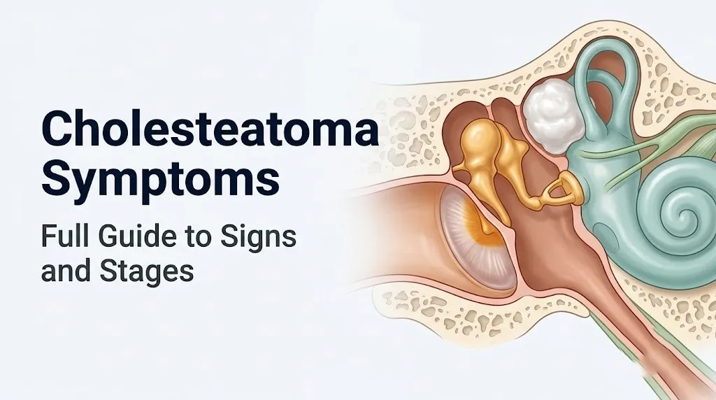
The long process of the incus is the ossicular structure most commonly eroded in cholesteatoma, a finding consistently documented across multiple surgical case series published in the Laryngoscope and Otolaryngology-Head and Neck Surgery journal. The long process of the incus has a relatively thin cortical bone and is in direct contact with the expanding cholesteatoma in the epitympanum, making it the path of least resistance for the enzymatic erosion front.
Once the incus long process is significantly eroded, ossicular discontinuity produces a substantial conductive hearing loss of typically 30 to 60 decibels, equivalent to hearing at the threshold of a normal conversational voice only at very close range.
Sensorineural hearing loss develops when the cholesteatoma erodes through the bony labyrinth to the level of the oval window or round window, or forms a labyrinthine fistula exposing the perilymph of the inner ear. This sensorineural component reflects direct cochlear injury and is often irreversible even after surgical removal of the cholesteatoma.
Adults over 60 with bilateral pre-existing age-related hearing loss may find that cholesteatoma-related conductive loss in one ear is particularly difficult to isolate without formal audiological testing, because they attribute worsening hearing to aging rather than new pathology. Formal audiometry (pure tone audiogram with bone conduction testing) comparing both ears is the appropriate tool for quantifying which component of hearing loss is conductive versus sensorineural.
Key Takeaway: Cholesteatoma hearing loss begins as conductive loss from ossicular disruption and sac-filling of the middle ear, progresses as the long process of the incus is eroded, and becomes irreversible if cochlear involvement through labyrinthine fistula occurs, making early surgical intervention the only way to preserve serviceable hearing.
Cholesteatoma Ear Pain and Pressure
Ear pain in cholesteatoma is not always present, and its absence should not reassure a patient that the condition is mild or benign, because the enzyme-driven bone erosion can proceed silently.
When ear pain does occur in cholesteatoma, it typically has one of two causes. The first is increased pressure from the expanding mass within the confined bony cavity of the middle ear and epitympanum. As the cholesteatoma grows, it compresses surrounding structures including the ossicular chain and the mucosal lining of the tympanic cavity, producing a deep aching pressure sensation rather than the sharp, acute pain of acute otitis media.
The second cause of ear pain in cholesteatoma is secondary bacterial infection. When the keratin debris within the sac becomes heavily colonized, the resulting infection produces local inflammatory edema, elevated middle ear pressure, and pain similar in character to an acute ear infection, typically described as a throbbing or aching pain that may radiate to the mastoid process (the bony prominence behind the ear).
Mastoid tenderness, felt as pain or tenderness when pressing on the mastoid process (the bone directly behind and below the ear lobe), indicates that the infection has extended from the middle ear into the mastoid air cells. This presentation warrants prompt evaluation by an otolaryngologist rather than watchful waiting, because mastoid involvement means the cholesteatoma or its infectious complications have spread beyond the tympanic cavity.
Patients with immunosuppressive conditions (diabetes mellitus, HIV, those on chronic corticosteroids or chemotherapy) may experience more severe infection-related ear pain from cholesteatoma because their impaired immune response allows secondary bacterial infection to spread more aggressively. These patients warrant lower threshold for same-day evaluation rather than waiting for a routine appointment.
Cholesteatoma Dizziness and Balance Symptoms
Dizziness associated with cholesteatoma is a red flag symptom that indicates the lesion has eroded close to or through the bony wall of the inner ear (labyrinth), and it should always prompt urgent rather than routine evaluation.
The most specific dizziness-producing complication of cholesteatoma is a labyrinthine fistula, most commonly involving the lateral (horizontal) semicircular canal. A labyrinthine fistula occurs when the cholesteatoma’s enzymatic erosion breaks through the bony covering of the semicircular canal, exposing the membranous labyrinth within. When the remaining membranous barrier is intact (the most common scenario), the patient experiences episodic vertigo triggered by pressure changes in the ear canal, such as pressing on the tragus, blowing the nose, or straining.
Think of the labyrinthine fistula like a small break in the shell of a gyroscope. The gyroscope inside (the membranous labyrinth) continues to function, but it is now vulnerable to any external force that produces a pressure wave. Pressing on the ear or blowing the nose transmits pressure through the fistula to the labyrinthine fluid, producing a sudden, brief spinning sensation.
This pressure-induced vertigo is called a positive fistula test (Hennebert’s sign), and when it is present, it provides a clinical clue that the cholesteatoma has reached the labyrinth. Any patient with cholesteatoma who develops new dizziness, particularly dizziness triggered by nose blowing or pressing on the ear, should be evaluated by an otolaryngologist within days rather than weeks.
Sudden onset of severe, persistent vertigo combined with significant hearing loss in a patient with known cholesteatoma indicates possible rupture of the membranous labyrinth (labyrinthine rupture), which is an otological emergency. Go to the emergency room immediately if this combination of symptoms develops.
Cholesteatoma Tinnitus: Why Ringing Occurs
Tinnitus (ringing, buzzing, or hissing sounds in the ear) is a frequent accompanying symptom of cholesteatoma, and its mechanism differs depending on the stage of disease.
In the early to intermediate stages, cholesteatoma tinnitus arises primarily from ossicular chain disruption. The ossicular chain normally acts as an acoustically efficient mechanical system transmitting vibration from the tympanic membrane to the oval window. When the chain is interrupted or loaded by an expanding mass, the altered mechanical resonance of the partially damaged system produces low-frequency tinnitus that often correlates with the degree of conductive hearing loss.
As the cholesteatoma progresses to involve the inner ear, tinnitus changes in character. Inner ear involvement produces high-pitched tinnitus more consistent with cochlear hair cell injury, often accompanied by the onset of sensorineural hearing loss. A shift in tinnitus character from low-pitched to high-pitched in a patient with known cholesteatoma is clinically relevant and suggests advancing disease.
According to the NIDCD, tinnitus associated with progressive unilateral hearing loss should prompt audiological evaluation and imaging to exclude a structural cause, because unilateral tinnitus with hearing loss has a broader differential diagnosis that includes cholesteatoma, acoustic neuroma (vestibular schwannoma), and other middle ear pathology.
Tinnitus in cholesteatoma is typically unilateral (affecting only the ear with the lesion). Bilateral tinnitus is less likely to represent cholesteatoma and more likely to reflect other causes including noise-induced hearing loss, Meniere’s disease, or systemic conditions. Patients should clearly communicate to their treating physician whether the tinnitus is perceived in one ear or both, and whether it has changed in pitch or intensity over time.
People with prior occupational noise exposure or age-related sensorineural hearing loss in both ears may find it difficult to isolate unilateral tinnitus changes that suggest cholesteatoma progression. Formal audiometric evaluation with comparison of both ears provides the objective data needed to separate cholesteatoma-associated change from the pre-existing baseline.
Cholesteatoma Facial Nerve Symptoms
Facial nerve involvement from cholesteatoma is one of the most feared complications because it can produce permanent facial paralysis when the facial nerve (cranial nerve VII) is significantly damaged before surgical decompression occurs.
The facial nerve travels through the facial nerve canal (also called the Fallopian canal), a bony channel running through the medial wall of the middle ear, the mastoid, and the temporal bone before exiting the skull through the stylomastoid foramen. In approximately 0.5% to 1% of the general population, the bony canal covering the facial nerve in the middle ear is naturally dehiscent (partially absent), making the nerve directly vulnerable to any pathological process in the middle ear space.
When cholesteatoma erodes the bony facial nerve canal, it exposes the nerve sheath to direct pressure from the expanding mass and to the inflammatory mediators released by the cholesteatoma perimatrix, including IL-1beta and TNF-alpha. The result is progressive facial nerve conduction impairment, initially presenting as subtle facial asymmetry that progresses to frank weakness and ultimately to complete peripheral facial palsy if untreated.
Peripheral facial palsy from cholesteatoma produces weakness of the entire ipsilateral (same side) half of the face, including the forehead. This distinguishes it from a central (stroke-related) facial palsy, which typically spares the forehead due to bilateral cortical representation of the frontalis muscle.
Specific facial nerve symptoms to recognize:
- Subtle asymmetry when smiling or showing teeth: one corner of the mouth moves less than the other
- Difficulty closing the eye fully on the affected side (lagophthalmos): a sign of orbicularis oculi weakness
- Food or liquid leaking from the corner of the mouth during eating
- Asymmetric eyebrow position (affected side brow lower than the unaffected side)
- Inability to whistle or puff out cheeks from buccinator weakness
Acute onset of facial weakness in any person with known cholesteatoma or chronic ear disease is a surgical emergency. Go to the nearest emergency room immediately. Facial nerve decompression surgery must be performed promptly to prevent permanent nerve damage.
Key Takeaway: Facial weakness in a patient with cholesteatoma indicates erosion of the facial nerve’s bony canal and is a surgical emergency requiring immediate emergency room evaluation, not a next-day ENT appointment.
Late Stage and Complicated Cholesteatoma Symptoms
Late stage cholesteatoma produces its most dangerous symptoms when the eroding lesion breaches the boundaries of the temporal bone and reaches intracranial structures or produces severe inner ear complications.
The intracranial complications of cholesteatoma arise through specific anatomical pathways. Erosion of the tegmen tympani (the thin bony plate forming the roof of the middle ear and mastoid) allows infection from cholesteatoma to reach the middle cranial fossa, producing epidural abscess, subdural empyema, or meningitis. Erosion of the sigmoid sinus plate allows infection to reach the sigmoid sinus, producing sigmoid sinus thrombophlebitis (lateral sinus thrombosis), a serious condition involving septic thrombus formation within the venous sinus, which can seed pulmonary emboli or spread to produce brain abscess.
Temporal lobe brain abscess and cerebellar abscess are the most feared intracranial complications. A study published in the Journal of Laryngology and Otology documented that intracranial complications of cholesteatoma, while uncommon in high-income countries with access to early surgical intervention, carry mortality rates that vary from under 5% with prompt treatment to substantially higher rates when diagnosis is delayed.
The Bezold abscess is a specific complication where infection from cholesteatoma and mastoiditis tracks through the tip of the mastoid process into the neck, producing a tender, fluctuant swelling below the mastoid tip and deep to the sternocleidomastoid muscle.
Late stage symptoms that are immediate medical emergencies include:
- Severe headache, fever, and neck stiffness (meningism)
- New fever and spiking temperature in a patient with known cholesteatoma or chronic ear disease
- Altered consciousness, confusion, or seizure in a patient with chronic ear disease
- Swelling or fluctuance below the mastoid process
- Sudden complete hearing loss in the affected ear combined with severe persistent vertigo
Congenital Cholesteatoma Symptoms and How They Differ
Congenital cholesteatoma produces an entirely different symptom presentation from acquired types, and this difference is precisely what leads to diagnostic delay in affected children.
Congenital cholesteatoma develops from epithelial cell rests left behind during embryological development of the middle ear, behind a completely intact tympanic membrane. Because it does not arise from a perforation or retraction pocket, it produces no ear discharge. Because it develops slowly in a closed space, it may produce no pain. Because many children’s hearing loss is not detected until formal audiological screening, the hearing loss it causes may not be noticed for months or years.
The typical congenital cholesteatoma is discovered as an incidental finding: a white, pearly mass visible through the intact tympanic membrane on routine otoscopic examination, most commonly in the anterosuperior quadrant of the tympanic membrane where the embryological epithelial rests most frequently persist. The child has no history of ear infections, no history of tympanic membrane perforation, and no ear discharge.
Because the condition is often discovered before significant bone erosion has occurred, congenital cholesteatoma can be treated with smaller surgical procedures than advanced acquired cholesteatoma, with better hearing preservation outcomes. The American Academy of Otolaryngology-Head and Neck Surgery position on early identification emphasizes that any white mass seen through an intact tympanic membrane in a child should be evaluated by a pediatric otolaryngologist promptly, regardless of how well the child seems to hear.
The absence of classic symptoms (no discharge, no pain, no obvious hearing complaint) in congenital cholesteatoma is the feature that most reliably distinguishes it from acquired types, not the presence of any specific positive symptom.
Cholesteatoma Symptoms in Children vs Adults
Children and adults with cholesteatoma share the same fundamental symptom categories, but the disease behaves more aggressively in children and presents at more advanced stages for reasons rooted in anatomy and diagnostic access.
Pediatric cholesteatoma grows more rapidly than adult disease, a finding supported by surgical series data in Otolaryngology-Head and Neck Surgery. Children have more cellular cholesteatoma matrices with higher keratinocyte proliferation rates, higher MMP expression per unit area of matrix, and less ossified temporal bone structure, which offers less resistance to the expanding lesion. As a result, children at surgical presentation frequently have more extensive disease involving the mastoid than adults at comparable symptom duration.
The recurrence rate after surgical removal is also higher in children than adults, estimated at 20% to 40% in pediatric series compared to lower rates in adult series, depending on the surgical technique used and how recurrence is defined.
Adults typically present with more chronic, gradual symptom onset and often have a longer history of ear disease (prior perforations, multiple ear infections, previous ear surgeries) that created the conditions for cholesteatoma development. Adults may also have concurrent presbycusis (age-related hearing loss) complicating the assessment of cholesteatoma-related conductive hearing loss.
| Feature | Children | Adults |
|---|---|---|
| Growth rate | More rapid | Slower and more variable |
| Recurrence post-surgery | 20% to 40% | Lower in most surgical series |
| Bilateral disease | More frequent | Less frequent |
| Presenting stage | Often more advanced | Variable; often earlier if incidental otoscopy |
| Congenital type prevalence | Higher | Rare in adults |
| Concurrent hearing baseline | Normal baseline makes new loss more apparent | May have pre-existing sensorineural loss |
| Eustachian tube dysfunction | More pronounced | Less central in older adults |
Children with Down syndrome or cleft palate who have chronic Eustachian tube dysfunction are at substantially elevated risk and should have regular otological examination with pneumatic otoscopy by a pediatric otolaryngologist as part of their ongoing medical care.
How Cholesteatoma Erodes Bone: The Mechanism Behind the Symptoms
Every cholesteatoma symptom traces back to one fundamental biological process: the keratinizing epithelium of the cholesteatoma actively secretes enzymes and activates bone-resorbing cells, producing progressive, irreversible destruction of the bony structures surrounding it.
This is categorically different from a simple cyst that passively presses on surrounding structures. Cholesteatoma is an active, enzymatically aggressive lesion.
The mechanism works in two parallel pathways. First, the cholesteatoma keratinocytes (skin cells forming the matrix layer) secrete matrix metalloproteinases, particularly MMP-2 (gelatinase A), MMP-9 (gelatinase B), and MMP-13 (collagenase 3). These enzymes degrade type I and type IV collagen, fibronectin, and other extracellular matrix proteins in the surrounding bone’s organic matrix, breaking down the protein scaffold that gives bone its structural integrity.
Second, and equally destructive, the cholesteatoma matrix and perimatrix cells produce RANKL (receptor activator of nuclear factor kappa-B ligand), which binds to RANK receptors on osteoclast precursors and drives their differentiation into active osteoclasts, the cell type responsible for normal bone resorption. This recruitment of osteoclasts transforms the bone surrounding the cholesteatoma into an active zone of accelerated bone resorption, not merely passive pressure erosion.
Research published in Laryngoscope has specifically identified elevated MMP-9 expression as correlating with more aggressive disease behavior and higher rates of ossicular chain erosion in surgical specimens.
Think of the cholesteatoma as a lesion that brings its own demolition crew. The MMP enzymes are the chemical solvent, dissolving the mortar between the bricks. The RANKL-activated osteoclasts are the wrecking crew, systematically removing the structural elements. Together, they can hollow out the temporal bone’s interior with a thoroughness that no passive pressure process could achieve.
Cholesteatoma vs Chronic Otitis Media: Distinguishing the Symptoms
Cholesteatoma and chronic otitis media (COM) share several symptoms, making clinical differentiation necessary and sometimes difficult without direct otoscopic or otomicroscopic examination.
Both conditions can produce chronic ear discharge, hearing loss, and intermittent ear discomfort. The clinical distinction between them determines the urgency of surgical referral and the type of surgery required.
Key differentiating features:
| Feature | Cholesteatoma | Chronic Otitis Media (without cholesteatoma) |
|---|---|---|
| Discharge odor | Distinctly foul, fetid, putrid | Mildly purulent or odorless |
| Discharge character | May contain white cheesy material (keratin) | Purulent without visible keratin debris |
| Tympanic membrane | Retraction pocket in pars flaccida, or attic perforation with crust or debris | Central perforation, often without retraction |
| Hearing loss type | Conductive; may become mixed with inner ear involvement | Predominantly conductive |
| Associated white mass | White pearly mass visible in attic or through perforation | No white mass; inflamed mucosal appearance |
| Response to antibiotics | Discharge may reduce but condition does not resolve | May respond partially to antibiotic drops |
| Bone erosion on CT | Present; ossicular chain changes, mastoid opacity | Present only in advanced disease; typically mucosal thickening |
| Risk of intracranial complication | Higher due to enzymatic erosion pathway | Lower; requires advanced osteitis |
The distinction cannot be made reliably by symptom history alone. Direct examination under magnification (otomicroscopy) by a board-certified otolaryngologist, supplemented by high-resolution computed tomography (CT) of the temporal bone, is required for definitive differentiation.
Any patient with chronic unilateral ear drainage that does not fully resolve with antibiotics should be referred to a board-certified otolaryngologist for otomicroscopic examination specifically to exclude cholesteatoma, because the presence of a cholesteatoma fundamentally changes the treatment approach from medical management to surgical intervention.
Key Takeaway: The combination of persistently foul-smelling discharge, white or cheesy material in the drainage, and a retraction pocket or crust in the superior part of the eardrum (pars flaccida region) distinguishes cholesteatoma from chronic otitis media far more reliably than any single symptom alone.
How Cholesteatoma Is Diagnosed
Cholesteatoma diagnosis requires a structured approach combining clinical examination, audiological testing, and imaging, because the severity of disease and surgical planning both depend on knowing which structures have been eroded and to what degree.
The diagnostic pathway for suspected cholesteatoma:
- Otoscopy or otomicroscopy: The examining otolaryngologist inspects the tympanic membrane under magnification (using an operating microscope or an otoendoscope) looking for retraction pockets in the pars flaccida, attic perforations, crusting or white material in the superior ear canal, aural polyps, or pearly masses. Pneumatic otoscopy (applying gentle pressure to the ear canal) may trigger a positive Hennebert’s sign (pressure-induced vertigo), suggesting labyrinthine fistula.
- Pure tone audiometry with bone conduction: Formal hearing testing quantifies the degree and type (conductive, sensorineural, or mixed) of hearing loss, establishes a baseline for comparison after treatment, and helps localize the extent of disease. A large conductive gap suggests significant ossicular chain discontinuity.
- High-resolution computed tomography (CT) of the temporal bone: CT provides the best imaging of bony structures. It can identify soft tissue density filling the middle ear and mastoid, ossicular erosion, tegmen erosion, semicircular canal involvement, facial nerve canal involvement, and sigmoid sinus plate erosion. CT is the standard pre-operative planning tool.
- MRI with non-echo-planar diffusion-weighted imaging (non-EPI DWI): This specialized MRI sequence has transformed the non-invasive detection of cholesteatoma. Cholesteatoma exhibits restricted diffusion (appears bright on DWI sequences) because keratin debris restricts water molecule movement. Non-EPI DWI has approximately 80% to 90% sensitivity and specificity for cholesteatoma detection in published series, and it is particularly useful for detecting residual or recurrent cholesteatoma after surgery without requiring a second-look surgical procedure.
- Culture of ear discharge: In cases with active infection, culture identifies the specific organisms and guides antibiotic selection for pre-operative infection control.
The appropriate specialist for diagnosis and management is a board-certified otolaryngologist (ENT surgeon), with referral to a neurotologist (a fellowship-trained subspecialist in ear and skull base surgery) for complex cases involving extensive disease, labyrinthine fistula, or intracranial complications.
Cholesteatoma Treatment and What Happens After Surgery
Surgery is the only definitive treatment for cholesteatoma. Antibiotics, ear drops, and other medications cannot remove the keratin-accumulating mass or stop its enzymatic bone erosion; they can temporarily control secondary infection but do not address the underlying pathological process.
The surgical options and what each involves:
| Surgical Approach | Description | Indication | Recurrence Risk |
|---|---|---|---|
| Canal wall up (intact canal wall) mastoidectomy with tympanoplasty | Removes cholesteatoma while preserving the posterior ear canal wall; better hearing preservation | Localized disease; favorable anatomy | Higher; requires planned second-look surgery at 12 to 18 months |
| Canal wall down (modified radical) mastoidectomy | Removes the posterior ear canal wall to create an open mastoid cavity; better disease access and visualization | Extensive disease, revision surgery, labyrinthine fistula | Lower recurrence rate; requires lifelong cavity care |
| Endoscopic ear surgery | Uses thin telescopes through the ear canal; can access hidden recesses | Selected cases of limited cholesteatoma | Emerging data; comparable to microscopic approaches in experienced hands |
Post-operative symptom expectations that patients should understand:
- Hearing may initially be worse after surgery while the middle ear heals, then may improve if ossicular reconstruction was performed.
- A watery or bloody discharge from the ear in the first weeks after surgery is expected.
- Dizziness in the first days after surgery is normal; persistent or severe dizziness warrants contact with the surgical team.
- Any facial weakness developing after surgery should be reported to the surgical team immediately.
- Second-look surgery (for canal wall up procedures) is typically planned 12 to 18 months after the initial procedure to inspect for residual or recurrent cholesteatoma.
Patients with post-operative recurrent discharge, new hearing loss, or new dizziness months to years after initial surgery should request evaluation by their otolaryngologist for possible recurrent cholesteatoma, and should ask specifically whether MRI with non-EPI DWI imaging would be appropriate for surveillance.
Emergency Symptoms: When to Call 911 or Go to the ER
Certain symptoms associated with cholesteatoma require immediate emergency evaluation. Do not wait to see if these resolve on their own.
Call 911 or go to the nearest emergency room immediately if you experience:
- Any new weakness, drooping, or asymmetry of one side of the face in a person with known cholesteatoma or chronic ear disease: this indicates possible facial nerve (cranial nerve VII) compression or injury from cholesteatoma erosion of the Fallopian canal, which is a surgical emergency requiring decompression
- Sudden severe vertigo (a violent spinning sensation) combined with complete hearing loss in the affected ear: this suggests possible labyrinthine rupture or complete labyrinthine fistula breach, which is an otological emergency
- Severe, escalating headache combined with fever and neck stiffness in anyone with chronic ear disease: this presentation indicates possible meningitis from intracranial extension of cholesteatoma or its infectious complications
- Confusion, altered consciousness, or new seizure activity in a person with known or suspected chronic ear disease: this suggests possible temporal lobe or cerebellar brain abscess from advanced cholesteatoma-related intracranial complication
- Swelling, redness, or fluctuance (fluid-filled softness) below the mastoid process (behind and below the ear) with fever: this suggests possible Bezold abscess (deep neck space abscess from mastoid infection)
- High fever with ear pain and spiking daily temperature pattern in a person with chronic ear disease: this pattern suggests possible sigmoid sinus thrombophlebitis requiring immediate imaging and intervention
These presentations can indicate intracranial extension of infection, brain abscess, meningitis, sigmoid sinus thrombophlebitis, facial nerve injury, or labyrinthine rupture and require emergency medical assessment, not an urgent care appointment or a telephone call to an answering service.
Frequently Asked Questions About Cholesteatoma Symptoms
What are the first signs of cholesteatoma?
The earliest signs of acquired cholesteatoma are typically a sensation of ear fullness or pressure in one ear and a small amount of foul-smelling discharge that does not fully clear with standard antibiotic ear drops.
The characteristic odor, which is distinctly more offensive than ordinary ear infection discharge, reflects anaerobic bacterial colonization of the keratin debris accumulating within the cholesteatoma sac.
A retraction pocket or white crust visible in the superior portion of the eardrum on examination is often the first otoscopic finding, which is why any persistent unilateral ear discharge warrants formal examination by a board-certified otolaryngologist rather than repeated empirical antibiotic prescriptions.
Does cholesteatoma always cause hearing loss?
Cholesteatoma does not always cause hearing loss in its earliest stages, particularly when the mass is confined to the epitympanum and has not yet physically disrupted the ossicular chain.
As the lesion expands and erodes the ossicular chain (typically beginning with the long process of the incus), progressive conductive hearing loss develops that worsens in proportion to the degree of ossicular destruction.
Sensorineural hearing loss develops only in advanced disease when the cholesteatoma reaches the cochlea through a labyrinthine fistula, and this cochlear component may be irreversible even after successful surgical removal.
What does cholesteatoma discharge smell like?
Cholesteatoma discharge has a distinctly foul, fetid, or putrid odor that most patients describe as unlike any ordinary ear discharge they have experienced.
The odor comes from anaerobic bacterial fermentation of keratin debris within the cholesteatoma sac, producing volatile sulfur compounds and organic acids from organisms including Pseudomonas aeruginosa and Bacteroides species.
The presence of this specific offensive smell, particularly combined with white or cheesy material visible in the drainage, is one of the most clinically distinctive features separating cholesteatoma from ordinary chronic otitis media.
Can cholesteatoma cause dizziness?
Cholesteatoma can cause dizziness when it erodes through the bony covering of the inner ear (labyrinth), most commonly affecting the lateral semicircular canal and producing a labyrinthine fistula.
The resulting dizziness is often pressure-sensitive, meaning it is triggered or worsened by pressing on the ear, blowing the nose, or straining, because these actions transmit pressure through the fistula to the inner ear fluid.
New dizziness in a patient with known cholesteatoma is a red flag symptom requiring evaluation by an otolaryngologist within days rather than at the next available routine appointment.
How do I know if my cholesteatoma has spread to the brain?
Spread of cholesteatoma infection to the intracranial space produces specific warning symptoms including severe escalating headache, fever, and neck stiffness, which together suggest meningitis.
Confusion, altered consciousness, or new seizure activity in a person with chronic ear disease suggests possible brain abscess from intracranial extension.
Any of these neurological symptoms combined with known or suspected cholesteatoma or chronic ear disease require immediate emergency room evaluation, not a scheduled outpatient appointment.
Can cholesteatoma go away without surgery?
Cholesteatoma cannot resolve without surgical removal, because it is a self-expanding lesion that actively produces enzymes to erode surrounding bone and will not stop growing without physical removal.
Antibiotics and antibiotic ear drops can temporarily control the secondary infection within the cholesteatoma, reducing discharge and discomfort, but they cannot remove the keratin mass, stop the matrix metalloproteinase-mediated bone erosion, or prevent the lesion from continuing to grow.
Delaying surgery allows continued bone erosion that progressively destroys the ossicular chain, risks the inner ear, the facial nerve, and ultimately the intracranial compartment, making early surgical treatment the only path that prevents irreversible structural damage.
Closing
Cholesteatoma is not a condition that rewards watchful waiting. Every week without surgical treatment represents more enzymatic erosion of the temporal bone, more ossicular destruction, and a progressively higher probability of facing the serious complications that make this condition genuinely dangerous.
If you have persistent foul-smelling discharge from one ear, progressive hearing loss in one ear, or a chronic history of ear infections in the same ear that never fully resolve, request referral to a board-certified otolaryngologist and ask specifically for otomicroscopic examination of the tympanic membrane. Do not accept repeated antibiotic prescriptions as a long-term management plan for unilateral ear drainage without that examination.
If you develop any facial weakness, sudden severe dizziness with hearing loss, severe headache with fever, or any neurological symptom alongside ear disease, go to the emergency room that day. Those symptoms change the clinical situation from a scheduled surgical case to an emergency one, and acting on that distinction promptly is what prevents permanent damage.