Tailbone Cancer Symptoms: What to Know in 2026
Tailbone cancer symptoms include deep, persistent aching pain at the base of the spine, neurological changes in the lower body, and bowel or bladder disruption that does not fit the pattern of a common musculoskeletal injury. These symptoms arise because tumors in the sacrococcygeal region, most often a rare primary bone cancer called sacrococcygeal chordoma, grow in a confined anatomical space where even a slow-expanding mass presses against nerve roots, pelvic viscera, and bone before it produces a clear clinical picture.
According to the National Cancer Institute, chordoma is diagnosed in roughly 1 in 1,000,000 people annually in the United States, making it rare but not negligible. The Chordoma Foundation reports that the average time from symptom onset to confirmed diagnosis is 2 to 4 years, often because early symptoms are attributed to benign conditions like coccydynia or lumbar disc disease, which delays access to appropriate imaging and specialist care.
This article covers every clinically recognized symptom category for tailbone cancer, explains the anatomical and physiological reason each symptom occurs, identifies which features distinguish a potentially malignant presentation from common tailbone injury, and specifies the exact clinical thresholds that warrant urgent evaluation or emergency care.
Tailbone Cancer Symptoms: The Clinical Overview
Tailbone cancer symptoms are produced by tumor growth within or adjacent to the coccyx and distal sacrum, a narrow anatomical region packed with nerve roots, ligaments, pelvic floor muscles, and visceral structures that all respond to compression or bony destruction.
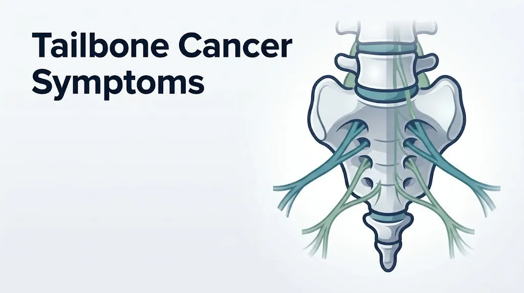
The symptom pattern differs depending on the tumor type, its size at the time of diagnosis, and the direction of its growth. Tumors expanding anteriorly into the presacral retroperitoneal space affect the rectum and bladder first. Tumors expanding posteriorly erode through the coccygeal bone and may eventually become palpable as a mass under the skin of the buttock crease. Tumors pressing laterally into the sacral plexus (the nerve network formed from the L4 through S4 ventral rami) produce radicular pain, sensory changes, and weakness that travel down the legs.
What sets tailbone cancer symptoms apart from common tailbone pain is a combination of features that, individually, might seem manageable, but together form a pattern that clinical guidelines and the American Cancer Society describe as requiring imaging evaluation rather than conservative musculoskeletal management.
| Symptom Category | Produced By | Clinical Significance |
|---|---|---|
| Deep, aching tailbone pain | Periosteal stretch, tumor expansion | Present in nearly all cases; often the first symptom |
| Night pain or rest pain | Tumor-mediated inflammation, not mechanical loading | Key red flag distinguishing malignant from benign |
| Bowel dysfunction | Rectal displacement, S4-S5 parasympathetic disruption | Present in 30-50% of chordoma cases at diagnosis |
| Bladder dysfunction | Pelvic splanchnic nerve compression (S2-S4) | Present in 20-40% of advanced cases |
| Leg pain or numbness | Sacral plexus or S4-S5 nerve root compression | Indicates tumor has reached the nerve roots |
| Palpable presacral mass | Posterior bony erosion with soft tissue extension | Late sign; indicates substantial tumor volume |
| Unexplained weight loss | Systemic tumor burden, cachexia | More common in metastatic disease than primary tumors |
Older adults, people with a personal or family history of colorectal cancer, and people with prior pelvic malignancy face a higher clinical risk that tailbone pain represents metastatic disease rather than a primary tumor.
What Is Tailbone Cancer
Tailbone cancer is a malignant tumor arising in or spreading to the coccyx (the os coccygis, a series of 3 to 5 small fused vertebral segments at the base of the spine) and the adjacent distal sacrum.
The coccyx sits at the terminal point of the vertebral column, connected to the sacrum at the sacrococcygeal joint and surrounded by a dense network of ligaments, pelvic floor muscles (including the levator ani and coccygeus muscles), and branches of the coccygeal plexus (formed from S4, S5, and Co1 nerve fibers). Immediately anterior to the coccyx lies the retrorectal or presacral space, a narrow fatty compartment containing lymphatic tissue, autonomic nerve fibers, and embryological remnants that can give rise to tumors.
Think of the presacral space like a hidden room behind the lowest drawer of a filing cabinet. Tumors that develop there have plenty of room to grow slowly before pressing against anything that produces obvious symptoms, which is part of why tailbone cancer goes undetected for years.
The most common form of primary tailbone cancer in adults is chordoma, a malignant tumor that arises from remnant cells of the embryonic notochord, a flexible rod-like structure that guides vertebral development in the fetus and normally disappears before birth. In roughly 50% of all chordoma cases, the tumor develops at the sacrococcygeal region. It is categorized under ICD-10 code C41.4 (malignant neoplasm of the coccyx) and grows slowly but destructively, invading surrounding bone and soft tissue over months to years.
According to the Mayo Clinic, the 5-year relative survival rate for localized sacrococcygeal chordoma treated at a specialty center ranges from 60% to 80%, but survival drops sharply when the tumor has spread to lymph nodes or distant organs, underscoring why early recognition of symptoms matters.
Types of Cancer That Affect the Tailbone
Several distinct cancer types can involve the tailbone, and their symptom patterns, age distributions, and clinical urgency differ enough that treating them as a single category leads to diagnostic errors.
Sacrococcygeal chordoma is the most common primary malignancy of the coccyx in adults. It arises from notochordal remnant cells and is locally aggressive but slow-growing, with a low rate of distant metastasis at initial presentation. It accounts for approximately 40% of all chordomas, which collectively represent about 0.2% of all bone tumors according to the National Cancer Institute SEER database.
Giant cell tumor of bone (GCTB) affecting the sacrum is the second primary bone tumor that must be considered. It is locally aggressive, classified as intermediate malignancy (rarely metastasizes but frequently recurs), and peaks in adults between ages 20 and 40. Its symptom pattern overlaps substantially with chordoma but tends to present at a younger age and with faster symptom progression because giant cell tumors grow more rapidly.
Sacrococcygeal teratoma is the most common solid tumor in neonates (occurring in approximately 1 in 35,000 to 40,000 live births, according to the American Cancer Society), but it also occurs in adolescents and young adults as a presacral mass that may undergo malignant transformation. In adults, it typically presents as a slowly enlarging presacral mass.
Metastatic cancer to the coccyx from distant primary tumors is actually more common than primary coccygeal malignancy in people over age 50. The primary cancers most likely to seed the coccyx via hematogenous spread include colorectal carcinoma, prostate adenocarcinoma, cervical carcinoma, lung adenocarcinoma, and breast carcinoma. Metastatic lesions produce osteolytic bone destruction through RANKL-mediated osteoclast activation, causing pain that often has a sharper, more rapid onset than slow-growing primary tumors.
People with a known history of any of these primary cancers who develop new or changing tailbone pain should speak with their oncologist promptly rather than attributing the pain to a musculoskeletal cause.
Key Takeaway: Tailbone cancer includes several distinct tumor types, the most common of which in adults is sacrococcygeal chordoma arising from notochordal remnant cells; distinguishing it from metastatic disease requires imaging because the symptom patterns overlap but the treatment paths diverge completely.
Early Symptoms of Tailbone Cancer
Early symptoms of tailbone cancer are often subtle and nonspecific, which is the primary reason diagnosis is delayed an average of 2 to 4 years according to research published in the Spine journal.
At the earliest stage, the dominant symptom is a dull, deep aching pain localized to the coccyx and the lowest part of the sacrum. This pain does not have a clear mechanical trigger. It is not reliably provoked by a specific posture change or aggravated only by sitting, the way traumatic coccydynia typically is. Instead, it has a persistent baseline quality: present even during light activity, sometimes waking the person from sleep, and not substantially relieved by common analgesics.
Early tailbone cancer symptoms to recognize:
- Persistent low-grade aching at the tailbone with no remembered injury
- Pain that begins in the coccyx region and feels “deep” rather than superficial or skin-level
- Mild, intermittent perianal or perineal fullness or pressure
- Discomfort with prolonged sitting that does not resolve fully with a coccyx cushion or positional adjustment
- A vague sense of rectal pressure or difficulty with complete bowel emptying, even without a confirmed gastrointestinal diagnosis
- Low-grade tingling or altered sensation in the saddle area (the inner thighs, perineum, and perianal skin) that is not explained by a lumbar spine diagnosis
The physiological basis for these early symptoms is the tumor mass beginning to exert pressure within the presacral space before substantial bony erosion has occurred. The periosteum (the dense fibrous membrane covering the bone surface) is richly innervated with nociceptors. As the tumor expands against the periosteal surface of the coccyx and distal sacrum, it activates these pain fibers through mechanical stretch and tumor-mediated prostaglandin and bradykinin release. This produces deep aching that is present even when no external force is applied to the bone.
People who attribute these symptoms to hemorrhoids, pelvic floor dysfunction, or chronic coccydynia and receive treatments that provide no sustained relief should consider raising the possibility of imaging with their primary care physician.
Tailbone Pain That Won’t Go Away
Tailbone pain that does not resolve within 6 to 8 weeks of conservative management, or that recurs and progressively worsens over months, is one of the most clinically reliable distinguishing features between a benign musculoskeletal cause and a structural lesion of the coccyx or sacrum.
Traumatic coccydynia (bruised or fractured coccyx from a fall, childbirth, or prolonged hard sitting) typically improves within 4 to 12 weeks with conservative care: donut cushions, anti-inflammatory medication, and activity modification. Pain from a coccygeal tumor does not follow this trajectory. It either remains unchanged or gradually worsens over weeks to months despite standard conservative measures.
The reason is structural: a tumor growing within the presacral space or eroding the coccyx is adding mass and pressure with each passing week. There is no mechanical rest position that eliminates the source of pain because the source is not a posture-dependent mechanical load but a biological process of tissue expansion and bony destruction driven by tumor cell proliferation.
Research published in the Journal of Bone and Joint Surgery found that in retrospective series of surgically treated sacral chordoma patients, the most common reason for delayed diagnosis was that primary care providers and even initial specialist evaluations attributed the pain to lumbar disc disease or coccydynia without ordering cross-sectional imaging. Patients whose plain radiographs (X-rays) were initially interpreted as normal or showing only degenerative change had their diagnosis delayed significantly compared to patients who received MRI early.
To describe this symptom accurately when speaking with a physician:
- Note the exact date when you first noticed the pain.
- Describe the trajectory: better over time, unchanged, or gradually worsening.
- State specifically whether it responds to rest, heat, ice, or common analgesics.
- Note whether the pain is ever absent, or whether it is present in some form every day.
- State whether it has changed in character: from dull ache to sharper, or from localized to spreading.
People who have had an MRI of the lumbar spine but not specifically the sacrococcygeal region should know that standard lumbar MRI sequences may not adequately image the distal sacrum and coccyx. Request clarification from your primary care physician about whether the imaging obtained actually covers this region.
Sacral Nerve Compression Symptoms From Tailbone Tumors
Sacral nerve compression symptoms from coccyx tumors appear when tumor growth reaches the S4 and S5 nerve roots, the coccygeal plexus (Co1), or the broader sacral plexus, and they represent a qualitative shift in the clinical picture from localized pain to a multi-system neurological presentation.
The S4 and S5 nerve roots exit the sacrum through the sacral foramina and supply sensory and autonomic innervation to the perineum, perianal skin, coccyx region, and pelvic floor. When these roots are compressed, the first neurological symptom is typically a tingling, burning, or numbness in the saddle area: the perineum, inner thighs, and skin around the anus. Clinicians call this saddle anesthesia when sensory loss in this distribution is complete.
The sacral plexus, formed from the L4 through S4 ventral rami, also carries motor and sensory fibers to the lower extremities through the sciatic nerve and pudendal nerve. When tumor compression extends to the plexus itself, symptoms extend into the buttocks, posterior thighs, and calves.
Neurological symptoms from sacral nerve compression to recognize:
- Tingling, burning, or numbness in the perineum or perianal region
- Reduced sensation in the inner thighs and buttocks (saddle distribution)
- Weakness in the muscles of the hip, thigh, or calf on one or both sides
- Altered sexual sensation or function (pudendal nerve involvement)
- Difficulty sensing the urge to urinate or defecate (S2-S4 afferent disruption)
According to the Chordoma Foundation, neurological deficits are present in approximately 50% of patients with sacrococcygeal chordoma at the time of diagnosis, and in most cases these deficits developed gradually over months before the diagnosis was confirmed.
People with bilateral neurological symptoms, meaning symptoms affecting both legs or both sides of the saddle area simultaneously, face a more urgent clinical picture than those with unilateral symptoms, as bilateral involvement suggests the tumor has compressed the midline sacral nerve roots or the cauda equina rather than a single root unilaterally.
Key Takeaway: Saddle area numbness, perianal tingling, or leg weakness in combination with tailbone pain indicates that a tumor may have reached the sacral nerve roots, which moves the clinical picture from pain management to urgent imaging and specialist evaluation.
Bowel and Bladder Problems Caused by Coccyx Tumors
Bowel and bladder dysfunction caused by coccyx tumors arises from two simultaneous mechanisms: direct anatomical compression of the rectum by an anteriorly expanding presacral mass, and disruption of the parasympathetic pelvic splanchnic nerves (S2, S3, and S4) that control bowel motility, rectal sphincter tone, bladder emptying, and urethral sphincter function.
The parasympathetic fibers responsible for bladder contraction and rectal propulsion travel from the S2-S4 spinal levels through the pelvic splanchnic nerves (nervi erigentes) to the pelvic organs. When a sacrococcygeal tumor compresses these fibers at their origin, it produces a neurogenic bladder pattern: difficulty initiating urination, incomplete bladder emptying, urinary retention, or, in some cases, loss of the normal urge to void. The same mechanism applied to the rectum produces reduced rectal sensation, difficulty with defecation, incomplete evacuation, or fecal incontinence.
The rectal displacement effect is separate from the neurological one. A large presacral tumor pushes the rectum anteriorly, mechanically narrowing the rectal lumen. This produces a sensation of rectal pressure or fullness, altered stool caliber (narrower stools), or difficulty with complete bowel evacuation that may be mistaken for a primary colorectal condition.
A 2021 review published in JAMA Oncology examining sacral chordoma outcomes across multiple specialty centers found that bowel or bladder dysfunction was present in approximately 35% of patients at initial diagnosis and that the degree of autonomic deficit correlated strongly with tumor volume at the time of presentation, reinforcing why early symptom recognition matters.
Quick Tip:
- Bowel or bladder symptoms in combination with tailbone pain are not a sign of two separate conditions; they are likely a single neurological event involving the S2-S4 parasympathetic pathway.
- A person experiencing urinary retention or urinary incontinence alongside tailbone pain should not wait for a scheduled appointment. Contact a primary care physician or urgent care center the same day.
- Older adults and people who have previously undergone pelvic radiation or pelvic surgery may have pre-existing bowel and bladder changes that make this symptom pattern harder to attribute correctly. Inform your physician of this history explicitly.
Tailbone Cancer Symptoms in the Legs and Buttocks
Tailbone cancer symptoms spreading to the legs and buttocks occur when tumor growth compresses or invades the sciatic nerve (formed from L4-S3 nerve roots) or the broader sacral plexus, producing a pattern of pain, numbness, or weakness that travels from the buttock down the posterior thigh and potentially into the calf and foot.
This symptom pattern is frequently misattributed to lumbar disc herniation or piriformis syndrome because the distribution overlaps with common sciatic nerve root irritation from lumbar spine pathology. The key anatomical distinction is that sacrococcygeal tumor-related leg symptoms originate from nerve root compression at the sacral level rather than the lumbar level, meaning standard lumbar MRI sequences do not capture the causative lesion.
Symptoms in the legs and buttocks from sacral plexus involvement:
- Deep, aching buttock pain that is not provoked by specific hip movements
- Posterior thigh pain that does not clearly trace to a lumbar disc level
- Calf or foot numbness or tingling that appears alongside tailbone pain
- Leg weakness, particularly in hip extension (gluteus maximus, innervated by the inferior gluteal nerve from L5-S2) or knee flexion (hamstrings, innervated by the sciatic nerve from L4-S3)
- Difficulty walking distances without the leg becoming fatigued or heavy
The physiological basis is direct mechanical compression of the nerve fibers within the sacral plexus by the expanding tumor mass. Unlike lumbar disc herniation, in which nerve root compression is typically positional and may improve with specific movements, sacral plexus compression from a tumor is constant and often progressive, producing symptoms that do not fluctuate predictably with posture.
Teenagers and young adults with these symptoms warrant particular clinical attention because giant cell tumor of bone affecting the sacrum and presacral teratoma with malignant transformation both occur in this age group and can produce a similar symptom complex that is easily attributed to sports injuries or growing pains.
Tailbone Pain Worse at Night or at Rest
Tailbone pain that is worse at night or at rest rather than during activity is one of the most clinically recognized red flags for a bone tumor, and it operates through a mechanism entirely different from the mechanical load-dependent pain of coccydynia or lumbar disc disease.
Mechanical tailbone pain from injury or degenerative change is typically provoked by sitting, transitioning from sitting to standing, or direct pressure on the coccyx. It is relieved by lying down or removing the mechanical load. This is the expected pain pattern of benign coccydynia.
Tumor-related tailbone pain is driven not by mechanical load but by the prostaglandin-mediated inflammatory process within the tumor microenvironment and the periosteal stretch produced by tumor expansion against the coccygeal bone surface. These processes continue at night regardless of body position, producing pain that wakes the patient from sleep, is present first thing in the morning, and is not substantially relieved by common positional adjustments.
The American Cancer Society lists bone pain that is worse at night or present at rest as one of the primary warning signs that a bone pain complaint requires imaging evaluation to exclude a malignant cause. This guidance applies to all bone regions, including the coccyx.
| Pain Characteristic | Benign Coccydynia | Tumor-Related Tailbone Pain |
|---|---|---|
| Worse with sitting | Yes, typically | Variable; often present but not the dominant trigger |
| Relieved by lying down | Usually | Often no; may be worse at night |
| Wakes from sleep | Rare | Common; a key distinguishing feature |
| Progresses over weeks/months | No; typically improves | Yes; gradual worsening is typical |
| Clear traumatic trigger | Often present | Usually absent or unclear |
| Response to NSAIDs | Moderate to good | Often poor or partial |
People who take anti-inflammatory medication (NSAIDs such as ibuprofen or naproxen) and find that their tailbone pain responds minimally or not at all over multiple weeks should raise this finding specifically with their physician, as poor analgesic response to NSAIDs is consistent with the deeper inflammatory and nociceptive mechanisms involved in tumor-related bone pain.
Key Takeaway: Tailbone pain that wakes you from sleep or is present and unrelieved when lying down is a recognized red flag for bone pathology and warrants imaging, not a longer trial of conservative musculoskeletal care.
Palpable Mass or Lump Near the Tailbone
A palpable mass or lump near the tailbone represents a late-stage symptom of coccygeal tumor growth, occurring when the tumor has eroded through the posterior cortex of the coccyx or expanded enough in the presacral space to become detectable through rectal examination or as a visible soft tissue prominence beneath the skin of the gluteal cleft.
Not all coccygeal masses are malignant. Benign presacral cysts (epidermoid cysts, dermoid cysts, anterior meningoceles) and pilonidal cysts can all produce a palpable mass in this region. However, any new, firm, or enlarging mass in the sacrococcygeal region in an adult that is associated with pain and neurological symptoms warrants immediate imaging rather than watchful waiting.
The presacral mass produced by a chordoma has a characteristically lobulated, semi-firm texture on rectal examination, and it is often large by the time it becomes externally palpable, sometimes reaching 10 to 15 centimeters in diameter before diagnosis. This is because the presacral space accommodates significant tumor volume before symptoms escalate.
Research published in the Journal of Bone and Joint Surgery found that mean tumor diameter at time of diagnosis for sacrococcygeal chordoma is approximately 8 to 11 centimeters in most retrospective surgical series, reflecting the anatomical room available for growth before symptoms become severe enough to prompt cross-sectional imaging.
What to do if you or a clinician identifies a mass near the tailbone:
- Request magnetic resonance imaging (MRI) of the sacrococcygeal region with contrast. Do not accept a plain X-ray as a sufficient evaluation for a new palpable mass.
- Do not agree to biopsy at a general surgery center or interventional radiology facility without first consulting an orthopedic oncologist at a sarcoma specialty center.
- Report any recent changes in mass size, skin changes over the mass, or associated systemic symptoms (fever, night sweats, weight loss) to your physician at the same appointment.
- Provide a complete history of prior cancers, radiation treatment, and family cancer history to the physician evaluating the mass.
- Ask specifically whether the proposed biopsy approach will not compromise the surgical resection margin if the mass proves to be a chordoma, as poorly planned biopsy tracks can seed tumor cells along the needle path.
The fifth point above is particularly important because inappropriate biopsy technique for chordoma is a recognized contributor to higher local recurrence rates. The Chordoma Foundation recommends that all biopsies of suspected chordoma be planned in consultation with the surgical team that will perform the definitive resection.
Unexplained Weight Loss and Tailbone Pain
Unexplained weight loss occurring alongside tailbone pain shifts the differential diagnosis toward either a large primary tumor with significant metabolic burden or metastatic disease involving the coccyx from a distant primary cancer.
Weight loss is not a feature of early-stage, localized sacrococcygeal chordoma. It becomes relevant when the tumor has reached a size that produces measurable systemic effects or when the malignancy represents widespread metastatic disease. The physiological mechanism is tumor-induced cachexia, driven by inflammatory cytokines, particularly tumor necrosis factor-alpha (TNF-a) and interleukin-6 (IL-6), which disrupt normal anabolic signaling, reduce appetite, increase muscle protein breakdown, and elevate resting energy expenditure.
The clinical definition of unexplained weight loss that warrants investigation is a loss of more than 5% of body weight over 6 to 12 months without an intentional dietary change or identified medical explanation, according to National Institutes of Health clinical references. In the context of tailbone pain, any weight loss meeting this threshold should be treated as a clinical red flag requiring workup rather than reassurance.
Unexplained weight loss with tailbone pain is particularly concerning in people over age 50 with a prior diagnosis of colorectal, prostate, lung, breast, or gynecological cancer, as the coccyx and sacrum are common sites of late bone metastasis from all of these primary tumors. In this population, new or changing tailbone pain with systemic symptoms should prompt oncology evaluation, not a wait-and-see approach.
Additional systemic symptoms that, combined with weight loss and tailbone pain, increase the clinical concern for malignancy:
- Night sweats (particularly drenching sweats requiring a change of clothing)
- Persistent fatigue not explained by sleep disruption or other causes
- Low-grade fever without an identified infectious source
- New anemia identified on routine blood work
People managing active cancer who develop new tailbone pain should contact their oncologist the same day rather than waiting for a scheduled visit, as new bone pain in a person with a known primary malignancy is a recognized presentation of skeletal metastasis.
Tailbone Cancer Symptoms in Women
Tailbone cancer symptoms in women present within a pelvic anatomical context that is distinct from men in ways that affect both symptom pattern and diagnostic timing.
Women have a broader pelvic outlet, a more horizontal sacrococcygeal complex, and a shorter coccyx on average than men. The female pelvic floor sits in closer anatomical proximity to the rectum, vaginal vault, and uterus in the space anterior to the sacrococcygeal complex. This means that a presacral tumor in a woman has a higher likelihood of producing vaginal pressure, dyspareunia (pain with sexual intercourse), or menstrual irregularity as early symptoms, in addition to the core tailbone pain pattern.
Women who have given birth vaginally have an additional complicating factor: obstetric coccygeal injury occurs in a measurable proportion of vaginal deliveries, producing post-partum coccydynia that can be mistakenly attributed to birth trauma for months or years. A 2019 study published in the Spine journal found that coccygeal abnormalities on imaging are present in up to 20% of women who delivered vaginally, which creates a background noise problem: a real tumor can be masked by the expectation that postpartum tailbone pain is benign.
Women of reproductive age who develop tailbone pain that does not resolve after an appropriate recovery period from childbirth, or that appears for the first time without a clear obstetric trigger, should specifically request MRI of the sacrococcygeal region rather than accepting a postpartum coccydynia explanation without imaging confirmation.
Postmenopausal women face a higher background risk of metastatic bone disease from prior or current breast, ovarian, or endometrial malignancy, meaning that new tailbone pain in this population deserves earlier imaging evaluation than the same symptom in a premenopausal woman with no cancer history.
The appropriate specialist for a woman with suspected coccygeal tumor is an orthopedic oncologist working within a multidisciplinary team that includes a gynecologic oncologist and colorectal surgeon, depending on the extent of tumor involvement.
Key Takeaway: Women face a specific diagnostic challenge because obstetric trauma creates a plausible alternative explanation for tailbone pain; any tailbone pain in a woman that does not follow the expected recovery trajectory of postpartum coccydynia warrants sacrococcygeal MRI before a musculoskeletal diagnosis is accepted.
Tailbone Cancer Symptoms by Age Group
The type of tailbone cancer and its specific symptom presentation vary substantially by age, making age-specific awareness an important clinical tool for both patients and the physicians evaluating them.
Neonates and infants (birth to 1 year): Sacrococcygeal teratoma is the dominant diagnosis in this group. According to the American Cancer Society, it presents as a visible external mass protruding from the lower back and buttock area at birth. Fetal and neonatal presentation is often detected on prenatal ultrasound. Malignant transformation is rare in neonates but increases with age if the mass is not surgically removed.
Children and adolescents (ages 1 to 20): Sacrococcygeal teratoma that was not identified at birth may present as a growing presacral mass with constipation, urinary frequency, or visible buttock asymmetry. Giant cell tumor of bone begins to appear in late adolescence. Primary Ewing sarcoma, while more common in the diaphyses of long bones, occasionally affects the sacrum in this age group and produces rapid-onset pain, swelling, and systemic symptoms (fever, fatigue).
Young to middle-aged adults (ages 20 to 50): Giant cell tumor of bone peaks in this range. Early-stage sacrococcygeal chordoma begins to appear from age 40 onward. Symptoms in this group are most commonly misattributed to lumbar disc disease, sports injuries, or occupational musculoskeletal strain.
Adults over age 50: Sacrococcygeal chordoma peaks in incidence between ages 55 and 65 in men and slightly later in women, according to National Cancer Institute SEER data. Metastatic bone disease to the coccyx from colorectal, prostate, lung, or breast primary cancers becomes the most common malignant cause of tailbone pain in this age group.
| Age Group | Most Likely Malignant Cause | Key Distinguishing Symptom |
|---|---|---|
| Neonates and infants | Sacrococcygeal teratoma | Visible external mass at birth |
| Ages 1 to 20 | Teratoma (delayed), Ewing sarcoma | Growing buttock mass, systemic symptoms |
| Ages 20 to 45 | Giant cell tumor of bone | Localized sacral pain, rapid progression |
| Ages 45 to 70 | Sacrococcygeal chordoma | Slow progressive pain, neurological deficit |
| Ages 50 and older | Metastatic disease | Prior cancer history, systemic symptoms |
A primary care physician evaluating any person in the over-50 age group with new tailbone pain and a prior cancer diagnosis should consider bone metastasis as the primary differential until imaging excludes it.
How Tailbone Cancer Is Diagnosed
Tailbone cancer is diagnosed through a sequential process that begins with clinical evaluation, progresses to cross-sectional imaging, and is confirmed by pathological analysis of tumor tissue obtained through image-guided biopsy.
Plain radiographs (X-rays) are frequently the first imaging obtained but are inadequate for detecting early-stage coccygeal tumors. A 2020 retrospective analysis published in the Spine journal found that plain radiographs were reported as normal or showing only degenerative change in a substantial proportion of chordoma patients at initial evaluation, even when MRI subsequently demonstrated a large mass. This is because chordoma infiltrates bone and soft tissue in ways that are subtle on plain film until significant bony destruction has occurred.
Magnetic resonance imaging (MRI) with and without gadolinium contrast of the sacrococcygeal region is the gold-standard initial diagnostic study for any suspected coccygeal or sacral tumor. MRI provides superior soft tissue contrast, defining tumor boundaries, nerve root involvement, presacral extension, and the relationship of the mass to the rectum and pelvic floor muscles. T2-weighted sequences show chordoma as a bright, lobulated mass with characteristic signal intensity.
Computed tomography (CT) scan of the pelvis and sacrum is complementary to MRI, providing superior bone detail and quantifying the extent of cortical destruction, which is essential for surgical planning.
CT-guided core needle biopsy is the standard method for obtaining tissue for pathological diagnosis, but it must be planned in consultation with the surgical oncology team before execution. Biopsy tract contamination along the needle path can seed tumor cells and complicate the surgical resection margin, which affects local recurrence risk.
Immunohistochemistry of the biopsy specimen for the brachyury protein (encoded by the T-box transcription factor gene), which is a highly specific molecular marker for chordoma, confirms the diagnosis when histological features are ambiguous. This test distinguishes chordoma from other notochordal lesions and from chondrosarcoma, which can appear similar histologically.
Staging evaluation for distant metastasis typically includes CT of the chest, abdomen, and pelvis, and may include whole-body PET scan, particularly for chordoma with atypical histological features (dedifferentiated chordoma).
The physician best positioned to coordinate this diagnostic pathway is an orthopedic oncologist at a sarcoma specialty center. A primary care physician who suspects a coccygeal tumor based on clinical and imaging findings should refer directly to a sarcoma center rather than to a general orthopedic surgeon, as surgical resection margins for sacrococcygeal chordoma are the single most important predictor of local recurrence and long-term survival.
Emergency Symptoms: When to Call 911 or Go to the ER
Certain symptoms associated with tailbone cancer or sacrococcygeal tumors require immediate emergency evaluation. Do not wait to see if these resolve on their own.
Call 911 or go to the nearest emergency room immediately if you experience:
- Sudden loss of control of the bladder or bowel (inability to prevent urination or defecation), particularly in combination with tailbone or sacral pain, which indicates possible cauda equina syndrome, a surgical emergency
- Saddle anesthesia (complete or near-complete loss of sensation in the perineum, genitals, inner thighs, and buttocks, bilaterally) alongside worsening low back or tailbone pain
- Sudden, severe leg weakness affecting one or both legs that prevents weight-bearing, in the context of existing sacral or tailbone pain
- Inability to urinate (acute urinary retention) for more than several hours with accompanying lower abdominal or pelvic pressure and sacral pain
- Sudden severe escalation in tailbone or sacral pain to a level that is completely different from prior pain, particularly if accompanied by fever, chills, or septic appearance, which may indicate tumor necrosis, infection, or pathological fracture
- Pathological fracture: an audible crack or sudden collapse sensation in the sacrococcygeal region following minimal force in a person with known sacral bone involvement
These presentations can indicate acute cauda equina syndrome, pathological fracture through a tumor-invaded coccyx or sacrum, or neurological compromise requiring emergency surgical decompression or oncological stabilization. These are not presentations appropriate for urgent care centers, telephone advice lines, or next-day appointments.
Tailbone Cancer vs. Coccydynia: How to Tell the Difference
The distinction between tailbone cancer and benign coccydynia (non-malignant tailbone pain) is one of the most clinically important differentiations a physician must make when evaluating tailbone pain, because the two conditions share their most prominent symptom: pain at the base of the spine.
Benign coccydynia is common. It affects an estimated 1 in 250 patients presenting to musculoskeletal specialists with low back or pelvic pain, according to clinical observation data. It has identifiable mechanical causes: trauma (falls, childbirth, repetitive seated pressure), postural factors, and idiopathic causes related to coccygeal hypermobility or hypomobility. It follows a predictable course: worse with sitting and transitions, better with lying down and removing pressure, and responsive to conservative management over 4 to 12 weeks in most cases.
Coccygeal tumor pain is uncommon but follows a starkly different clinical trajectory that a well-informed patient or clinician can recognize.
| Clinical Feature | Benign Coccydynia | Tailbone Cancer |
|---|---|---|
| Onset | Often after identifiable trauma or event | Gradual, no clear precipitating event |
| Pain with sitting | Primary trigger; often immediate | Variable; sitting aggravates but not the only trigger |
| Pain at rest or night | Uncommon | Common; frequently wakes from sleep |
| Neurological symptoms | Absent | Present in 50% at diagnosis |
| Bowel/bladder symptoms | Absent | Present in 35% at diagnosis |
| Palpable mass | Absent | Present in advanced cases |
| Weight loss | Absent | Present with large or metastatic tumors |
| Response to conservative care | Improves over 4 to 12 weeks | Does not improve; worsens over time |
| Imaging (X-ray) | Normal or minor degenerative change | May be normal early; MRI shows mass |
| Age of concern | Any age (most common in adults 40-60) | Depends on tumor type; chordoma peaks age 55-65 |
The single most important decision point is this: any tailbone pain that does not improve with 6 to 8 weeks of appropriate conservative management, or that is accompanied by any of the neurological, visceral, or systemic symptoms described in this article, warrants MRI of the sacrococcygeal region rather than continued conservative care.
The clinical cost of missing a coccygeal tumor is high. A 2018 analysis in the Chordoma Foundation clinical literature review found that patients with chordoma who received wide surgical resection with negative margins had substantially better 5-year and 10-year local recurrence-free survival than those who received intralesional or marginal resection, and resection margins are directly affected by how early in the tumor’s growth the diagnosis is made. Earlier imaging equals larger surgical margins equals better outcomes.
The primary care physician is the first critical decision-maker. A physician evaluating persistent tailbone pain in a patient with one or more of the distinguishing features above should order MRI of the sacrococcygeal region as the next step, not a longer trial of NSAIDs or physical therapy.
Key Takeaway: The clearest clinical distinction between benign coccydynia and tailbone cancer is the combination of night pain, neurological symptoms, and failure to improve with conservative care; if your tailbone pain checks any of these boxes, push for sacrococcygeal MRI rather than accepting a musculoskeletal label without imaging.
Frequently Asked Questions About Tailbone Cancer Symptoms
What does tailbone cancer feel like?
Tailbone cancer typically feels like a deep, persistent aching pain at the base of the spine that does not have a clear mechanical trigger and is present even at rest or during sleep.
Unlike the sharp, position-dependent pain of a bruised coccyx, tumor-related pain is driven by periosteal stretch and tumor-mediated inflammation, producing a constant baseline ache that progressively worsens over weeks to months.
As the tumor grows, additional feelings may include perianal tingling, a sense of rectal fullness or pressure, and pain that radiates into the buttocks or posterior thighs.
Can tailbone pain be a sign of cancer?
Tailbone pain can be a sign of cancer, though the most common cause of coccyx pain is benign musculoskeletal injury rather than malignancy.
The features that raise concern for a tumor include pain that does not improve with conservative management after 6 to 8 weeks, pain that wakes you from sleep, neurological symptoms in the legs or saddle area, and bowel or bladder changes alongside the pain.
Any tailbone pain accompanied by these features warrants MRI of the sacrococcygeal region rather than continued empirical conservative treatment, according to clinical guidance from the Chordoma Foundation and Mayo Clinic.
How long does tailbone cancer take to show symptoms?
Sacrococcygeal chordoma, the most common primary tailbone cancer, is a slow-growing tumor that may be present for 2 to 4 years before symptoms become severe enough to prompt medical evaluation, according to research published in the Spine journal.
Early symptoms are often subtle (deep aching pain, mild perianal pressure, mild rectal fullness) and are frequently attributed to benign conditions.
Metastatic cancer to the coccyx from a distant primary tumor typically produces symptoms more rapidly, often over weeks to a few months, because osteolytic bone destruction progresses at a faster rate than chordoma growth.
What is the survival rate for tailbone cancer?
The 5-year relative survival rate for localized sacrococcygeal chordoma treated with wide surgical resection at a sarcoma specialty center ranges from approximately 60% to 80%, according to the Mayo Clinic and published surgical series.
Survival drops substantially when the tumor has metastasized to distant organs, with 5-year survival for metastatic chordoma estimated at less than 30% in most published series.
Achieving negative surgical resection margins is the single most important predictor of long-term local recurrence-free survival, which is why early diagnosis and treatment at an experienced sarcoma center are the most impactful factors in outcomes.
What type of doctor do you see for tailbone cancer?
The primary specialist for suspected or confirmed tailbone cancer is a board-certified orthopedic oncologist at a sarcoma specialty center with experience in sacropelvic tumors.
Management of sacrococcygeal chordoma requires a multidisciplinary team that typically includes the orthopedic oncologist, a neurosurgeon (for cases involving sacral nerve roots), a colorectal surgeon (for cases involving rectal involvement), a musculoskeletal radiologist for imaging interpretation and biopsy planning, and a radiation oncologist for proton beam or carbon ion radiotherapy where indicated.
A primary care physician who suspects coccygeal tumor based on clinical findings and initial imaging should refer directly to a sarcoma center rather than a general orthopedic surgeon, as expertise in surgical resection margins for this specific tumor type is not uniformly available at community orthopedic practices.
How do doctors tell the difference between coccydynia and a tailbone tumor?
Clinicians distinguish benign coccydynia from a tailbone tumor primarily through imaging: MRI of the sacrococcygeal region with gadolinium contrast is the gold-standard study, and plain radiographs (X-rays) alone are insufficient to exclude a mass.
Clinical features that prompt imaging rather than continued empirical conservative management include tailbone pain lasting beyond 6 to 8 weeks without improvement, pain that is worse at night, any neurological symptoms (leg numbness, saddle area sensory changes, bowel or bladder dysfunction), a palpable mass, or unexplained weight loss.
Once imaging confirms a mass, tissue biopsy planned in consultation with an orthopedic oncologist is required for definitive diagnosis, as several different tumor types can produce a similar radiological appearance at the sacrococcygeal level.
Persistent tailbone pain that does not follow the expected recovery trajectory of a common musculoskeletal injury deserves a precise clinical evaluation, not reassurance and a longer trial of conservative care. The symptoms described in this article, particularly night pain, saddle area neurological changes, bowel or bladder dysfunction, and failure to improve with standard treatment, are the signals that should prompt you to ask your primary care physician for MRI of the sacrococcygeal region specifically.
If that imaging identifies a mass, the next step is a referral to an orthopedic oncologist at a sarcoma specialty center before any biopsy or surgical intervention is planned. Biopsy technique and surgical resection margins in sacrococcygeal tumors directly affect long-term outcomes, and those decisions belong in experienced hands.
You have the information you need to describe your symptoms precisely and to ask the right questions. The trajectory from first symptom to confirmed diagnosis in tailbone cancer is shaped by how soon the right imaging gets ordered. That starts with the conversation you have with your physician.