Cervical Stenosis Symptoms: Full Warning Sign Guide 2026
Cervical stenosis symptoms span from intermittent neck pain and arm tingling to spinal cord compression signs that can permanently disable, and the difference between these presentations is not just a matter of severity: it reflects two anatomically distinct conditions, nerve root compression and spinal cord compression, that require completely different levels of medical urgency. Knowing which category your symptoms fall into is the most practically important thing this article can give you.
The National Institute of Neurological Disorders and Stroke (NINDS) identifies cervical spinal stenosis as one of the most frequently encountered causes of spinal cord dysfunction in adults over 55, with degenerative cervical myelopathy representing the leading cause of spinal cord impairment in the developed world. The condition is underdiagnosed because many of its earliest neurological signs are subtle enough to be mistaken for normal aging.
This article covers every recognized cervical stenosis symptom in clinical medicine, explains the precise spinal anatomy and nerve pathway producing each one, maps the specific symptom pattern of each compressed nerve root level, and gives you an unambiguous framework for identifying when your symptoms require an emergency room visit tonight versus a spine specialist appointment next week.
Cervical Stenosis Symptoms: What Is Actually Happening in Your Neck
Cervical stenosis symptoms are produced when the bony and ligamentous canal that surrounds the spinal cord and nerve roots in the neck narrows enough to compress these structures, disrupting the electrical and chemical signaling that controls sensation, movement, and organ function below the level of compression.
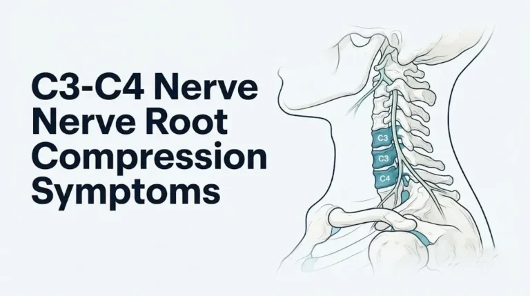
The cervical spine consists of seven vertebrae (C1 through C7) stacked in a lordotic curve. Each vertebra has a central hole (the vertebral foramen), and these holes align to form the spinal canal, through which the cervical spinal cord passes. At each vertebral level, two nerve roots exit through small side openings called intervertebral foramina, one on each side, carrying motor and sensory fibers to and from the arms, shoulders, and upper chest.
When this system functions normally, the spinal cord and nerve roots have adequate space, blood supply, and freedom from mechanical irritation. Canal narrowing from any structural source, whether disc herniation, bone spur formation, or ligament thickening, reduces this space. Below a critical threshold, compression produces clinical symptoms.
The clinical picture depends on where the narrowing occurs. Think of the cervical spine as a highway with a main tunnel (the central spinal canal, carrying the spinal cord) and multiple exit ramps (the intervertebral foramina, carrying the nerve roots). Narrowing that affects the exit ramps produces symptoms only in the specific arm territory that ramp serves. Narrowing that blocks the main tunnel affects everything traveling below that point, including legs, bowel, bladder, and balance.
Symptom pattern by compression type:
| Compression Type | Structure Affected | Symptom Distribution | Urgency |
|---|---|---|---|
| Foraminal (nerve root) | One nerve root, one side | One arm: pain, numbness, weakness | Urgent, not usually emergency |
| Central canal (spinal cord) | Spinal cord, bilateral | Both arms, both legs, bowel, bladder | Often urgent to emergency |
| Combined | Nerve roots and cord | Mixed arm and bilateral leg symptoms | High urgency |
Women who are postmenopausal experience accelerated cervical disc degeneration due to the loss of estrogen’s protective effect on intervertebral disc hydration, making them a higher-risk group for developing foraminal stenosis after age 55.
What Is Cervical Stenosis and How Does It Compress the Spinal Cord and Nerves
Cervical stenosis is the pathological narrowing of the cervical spinal canal or intervertebral foramina to a degree that mechanically compresses the spinal cord, individual nerve roots, or both, disrupting their function and producing the clinical symptoms that define the condition.
The most common cause of cervical stenosis is cervical spondylosis, the degenerative process of disc and joint aging in the cervical spine. As intervertebral discs dehydrate with age, they lose height. The narrowing disc space changes the load distribution on the adjacent facet joints and uncovertebral joints (joints of Luschka), which respond by forming osteophytes (bone spurs). These bony outgrowths project into the intervertebral foramen from the sides and into the central canal from the front.
Simultaneously, the ligamentum flavum, a thick elastic ligament connecting adjacent laminae on the posterior aspect of the canal, undergoes hypertrophy with age. Instead of remaining flat and taut, it buckles inward when the neck is extended, projecting into the posterior canal and reducing available cord space. Disc herniation adds a third compression vector: when the nucleus pulposus extrudes posteriorly through a tear in the annulus fibrosus, it compresses either the exiting nerve root (posterolateral herniation) or the cord itself (central herniation).
According to the North American Spine Society (NASS) clinical practice guidelines for degenerative cervical myelopathy, the critical canal diameter threshold at which spinal cord compression typically produces clinical signs is approximately 10 to 13 millimeters, compared to the normal adult cervical spinal canal diameter of 17 to 18 millimeters. Individuals with congenitally narrower canals (the Torg-Pavlov ratio below 0.8) develop symptoms from a smaller degree of superimposed degeneration than those with normal baseline canal dimensions.
Young competitive athletes with congenital cervical stenosis face a distinct risk: a single hyperextension injury during contact sports can produce acute central cord syndrome in a cervical canal that has inadequate reserve space. This population-specific risk is why some sports medicine organizations require cervical canal assessment before return-to-play decisions after neck injuries in contact sport athletes.
Neck Pain as a Cervical Stenosis Symptom
Neck pain from cervical stenosis is produced by inflammation and mechanical irritation of the facet joints, uncovertebral joints, and surrounding soft tissues in the cervical spine, and it characteristically differs from neck pain caused by muscle strain or cervicogenic headache.
The facet joints and joints of Luschka are richly innervated by the medial branch of the dorsal ramus of each spinal nerve. When osteophytes form on these joints or when degenerating discs change the load distribution across them, the synovial membrane and joint capsule become inflamed, generating nociceptive signals through these medial branch fibers. The pain is typically described as a deep, aching, poorly localized discomfort in the posterior and lateral neck, sometimes radiating to the base of the skull (causing cervicogenic headache) or across the top of the shoulder.
Neck stiffness accompanies the pain because cervical paraspinal muscles, including the semispinalis cervicis, multifidus cervicis, and splenius capitis, reflexively contract in response to facet joint inflammation. This protective guarding limits range of motion and produces the characteristic “stiff neck” that worsens in the morning after sustained nighttime postures.
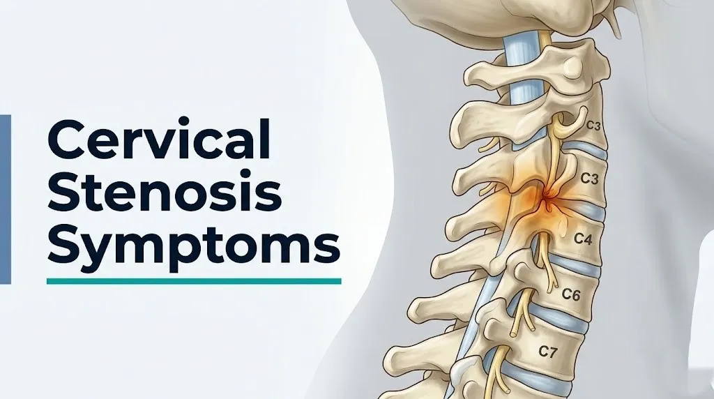
Neck pain from cervical stenosis tends to worsen with neck extension (looking upward) and rotation toward the affected side. Extension narrows the intervertebral foramina and causes the ligamentum flavum to buckle posteriorly, increasing compression. Flexion, by contrast, often provides temporary relief because it opens the foramina slightly and flattens the ligamentum flavum.
According to Mayo Clinic, not all people with cervical stenosis on imaging have symptoms, and neck pain alone, without arm or leg neurological symptoms, does not confirm that stenosis is the cause. A clinician must correlate imaging findings with symptoms before attributing neck pain specifically to stenosis.
Older adults over 70 frequently have cervical osteophytes visible on imaging that are entirely asymptomatic. Neck pain in this age group warrants evaluation to determine whether the stenotic changes are causing the symptoms or are incidental findings from age-related degeneration.
Arm Pain, Numbness, and Tingling as Cervical Stenosis Symptoms
Arm pain, numbness, and tingling from cervical stenosis are the hallmark symptoms of cervical radiculopathy: compression of a nerve root exiting the cervical spine, producing symptoms that travel from the neck down the arm in a pattern determined by which nerve root is affected.
The mechanism is specific. Each cervical nerve root carries sensory fibers from a defined skin territory (a dermatome) and motor fibers to a defined muscle group (a myotome). When the root is compressed by an osteophyte or herniated disc within the intervertebral foramen, sensory signals from that dermatome are disrupted, producing numbness, tingling (paresthesia), or burning pain along the arm, forearm, or hand in the distribution that root serves. Motor fibers simultaneously lose conduction efficiency, producing weakness in the muscles that root controls.
The arm pain of radiculopathy is distinct from the localized aching of neck pain. Radicular arm pain typically feels sharp, shooting, burning, or electric, and it travels from the neck down the arm following a specific path. It often worsens with certain neck positions (particularly extension and lateral rotation toward the affected side) and may be briefly relieved by placing the hand on top of the head, a finding called the Bakody sign or shoulder abduction relief sign, because this position reduces tension on the compressed nerve root.
A 2023 review published in the Journal of Bone and Joint Surgery confirmed that cervical radiculopathy affects approximately 83 per 100,000 adults annually in the United States, with the peak incidence in the fifth and sixth decades of life.
Men between ages 45 and 60 have the highest incidence of cervical radiculopathy from degenerative disc disease, though women have comparable rates. Laborers and people with occupations requiring sustained neck flexion or vibration exposure (drivers, dentists, surgeons) have higher rates of early cervical disc degeneration that predisposes to foraminal stenosis.
Cervical Stenosis Symptoms by Level: C5, C6, C7, and C8 Nerve Roots
The specific arm and hand symptom pattern in cervical radiculopathy is determined by which nerve root is compressed, and understanding the level-specific pattern is the single most practically useful information for describing your symptoms accurately to a physician.
The C5 nerve root exits between C4 and C5. When compressed, it produces pain and numbness over the lateral shoulder and upper arm (the deltoid region). Weakness affects the deltoid muscle (shoulder abduction), producing difficulty raising the arm to the side. The biceps reflex (tapped at the antecubital fossa) is diminished. There is typically no numbness in the hand.
The C6 nerve root exits between C5 and C6. Compression produces pain, numbness, and tingling in the thumb, index finger, and radial (thumb-side) forearm. Weakness affects the biceps brachii and wrist extensors, producing reduced elbow flexion strength and wrist extension weakness. The brachioradialis reflex is diminished. C6 is the most frequently compressed root in cervical disc disease.
The C7 nerve root exits between C6 and C7, the most commonly involved level in cervical disc herniation. Compression produces pain, numbness, and tingling in the middle finger and index finger, and along the dorsal (back) of the forearm. Weakness affects the triceps brachii (elbow extension) and wrist flexors. The triceps reflex (tapped at the olecranon) is diminished.
The C8 nerve root exits between C7 and T1. Compression produces pain and tingling in the ring and small fingers, and the ulnar (little finger) side of the hand. Weakness affects the intrinsic hand muscles, producing grip weakness and difficulty with fine motor tasks.
Cervical nerve root level symptoms at a glance:
| Root | Pain/Numbness Region | Weakness | Diminished Reflex |
|---|---|---|---|
| C5 | Lateral shoulder, upper arm | Deltoid, shoulder abduction | Biceps |
| C6 | Thumb, index finger, radial forearm | Biceps, wrist extension | Brachioradialis |
| C7 | Middle finger, dorsal forearm | Triceps, wrist flexion | Triceps |
| C8 | Ring/small finger, ulnar hand | Intrinsic hand muscles, grip | None prominent |
Bilateral arm symptoms at multiple levels simultaneously suggest central canal stenosis with cord involvement rather than simple nerve root compression, and warrants a different diagnostic and management approach.
Key Takeaway: If your thumb and index finger are numb, suspect C6. If your middle finger is numb and your triceps feels weak, suspect C7. Level-specific symptom patterns are the map your neurologist or spine surgeon uses to correlate your clinical findings with the MRI.
Hand Weakness and Fine Motor Loss as Cervical Stenosis Symptoms
Hand weakness and loss of fine motor coordination are among the earliest functional symptoms of cervical myelopathy, the spinal cord compression syndrome that represents the more serious of the two clinical presentations of cervical stenosis.
Unlike radiculopathy, which produces weakness in specific muscles served by one compressed root, myelopathy produces hand dysfunction through disruption of the lateral corticospinal tract within the spinal cord itself. This tract carries descending voluntary motor commands from the motor cortex to the anterior horn cells at each spinal level. When the lateral corticospinal tract is compressed at a cervical level, voluntary motor control below that level is disrupted, producing weakness that is not localized to one nerve root distribution but is instead diffuse and bilateral.
The earliest functional complaints of myelopathy in the hands are often subtle. Patients report difficulty buttoning shirts or blouses, trouble typing accurately, problems with handwriting deteriorating in control and legibility, difficulty picking up small objects like coins, and reduced ability to open jars. These tasks all require the precise coordination of intrinsic hand muscles controlled by upper motor neuron pathways in the lateral corticospinal tract.
Think of the lateral corticospinal tract like a fiber optic cable carrying finely coded signals from the brain to the hand muscles. When that cable is progressively pinched in the neck, the signals arrive degraded and some fail to arrive at all. The hand’s precision movements are the first to suffer because they require the highest resolution of motor control.
According to published research in the journal Spine, fine motor dysfunction of the dominant hand, particularly impaired dexterity on grip-and-release testing, is one of the most sensitive early indicators of degenerative cervical myelopathy and often predates gait disturbance by months to years.
People who perform fine motor tasks professionally, such as surgeons, musicians, and dentists, may notice hand coordination changes from cervical myelopathy earlier than the general population, because their professional work reveals subtle dysfunction that casual daily activities would not expose.
Cervical Myelopathy Symptoms: Gait Disturbance and Leg Weakness
Gait disturbance and leg weakness in cervical stenosis are produced by cervical myelopathy, and they represent the transition point between a spine condition managed conservatively and a neurological emergency that may require urgent surgical intervention.
When the lateral corticospinal tract and spinothalamic tract within the cervical spinal cord are compressed, the disruption affects not just the arms but every body region below the compression level. The legs, which are controlled by motor pathways running through the compressed cord levels, develop upper motor neuron weakness: spasticity (increased muscle tone and resistance to passive movement), hyperreflexia (exaggerated knee and ankle jerks), and clonus (rhythmic, involuntary muscle contractions at the ankle).
The resulting gait disturbance is characteristic. Patients with cervical myelopathy walk with a spastic, stiff-legged pattern. The legs move in a scissor-like fashion with reduced knee flexion during the swing phase. Turns are made slowly and with difficulty. Steps become smaller and wider-based as the patient unconsciously increases the base of support to compensate for proprioceptive loss from dorsal column dysfunction.
Dorsal column compression adds a second gait component: proprioceptive failure. The dorsal columns carry joint position sense (proprioception) from the lower extremities to the brain. When they are compressed, the patient cannot reliably sense where their feet are in space without looking down. Walking in the dark or on uneven terrain becomes unsafe. This produces a pattern of falls, particularly at night or on stairs.
According to the North American Spine Society (NASS), gait disturbance from cervical myelopathy should be evaluated urgently, ideally within days of onset, by a neurologist or board-certified orthopedic spine surgeon or neurosurgeon, because progressive myelopathy can produce permanent cord injury if compression is not relieved.
Older adults over 65 with cervical myelopathy are at particularly high fall risk, because the myelopathic gait disturbance compounds existing age-related vestibular and proprioceptive decline. A fall assessment and home safety evaluation should accompany any cervical myelopathy workup in this age group.
Lhermitte’s Sign and Other Specific Cervical Myelopathy Symptoms
Lhermitte’s sign (also called Lhermitte’s phenomenon) is a specific and diagnostically important cervical myelopathy symptom: a brief, intense electric shock sensation that travels down the spine and into the arms or legs when the neck is flexed forward, and it indicates direct mechanical irritation of the spinal cord’s posterior columns at a cervical level.
The physiological mechanism is specific. When the neck is flexed, the cervical spinal cord moves slightly upward and forward within the canal. In a patient with cervical myelopathy, this movement stretches an already compressed and mechanically sensitized posterior cord against the structural abnormality causing the stenosis. The dorsal columns, which carry large-diameter myelinated sensory fibers, generate a burst of ectopic electrical discharges in response to this mechanical stretch. The patient experiences these discharges as a sudden electric shock that follows the course of the spinal cord, lasting only seconds.
Lhermitte’s sign is not exclusive to cervical stenosis; it also occurs in multiple sclerosis, cervical radiation myelopathy, and vitamin B12 deficiency. However, in a patient with neck pain, arm symptoms, and gait changes, Lhermitte’s sign points strongly to structural cervical cord involvement and warrants MRI evaluation.
Hoffmann’s sign is a reflex test performed by the examining clinician: the middle fingernail is flicked downward, and a positive sign is when the thumb and index finger flex involuntarily in response. Hoffmann’s sign indicates upper motor neuron pathway dysfunction in the corticospinal tract, consistent with cervical myelopathy. It is analogous to the Babinski sign in the lower extremities.
Other specific myelopathy signs to report to your physician:
- Electric shock sensation traveling to the arms or legs when the neck bends forward (Lhermitte’s sign)
- Exaggerated knee-jerk or ankle-jerk reflexes noticed by a clinician during examination
- Difficulty with tandem gait (walking heel-to-toe in a straight line)
- Unsteadiness when standing with eyes closed (positive Romberg test)
- Involuntary foot or ankle rhythmic movements when ankle is dorsiflexed (clonus)
Patients with multiple sclerosis (MS) who also have cervical stenosis may have overlapping Lhermitte’s sign, making the differentiation between MS-related demyelination and structural cord compression a diagnostic priority requiring MRI with and without gadolinium contrast.
Key Takeaway: Lhermitte’s sign (electric shock down the spine when you flex your neck) is a red flag for spinal cord involvement in the cervical spine and should be reported to your physician at the next available appointment, not dismissed as a muscle cramp or positional discomfort.
Bowel and Bladder Dysfunction as Cervical Stenosis Symptoms
Bowel and bladder dysfunction as cervical stenosis symptoms indicate that the spinal cord compression has reached a severity affecting the descending voluntary motor pathways that control sphincter function, and these symptoms place cervical myelopathy firmly in the serious-to-emergency category.
Bladder and bowel sphincters are controlled by both voluntary (descending cortical) and autonomic pathways. Voluntary control of urination is mediated by the descending corticospinal pathways running through the lateral funiculus of the cervical cord. When these pathways are compressed at a cervical level, the supraspinal inhibition of the sacral micturition reflex is reduced, producing detrusor hyperreflexia: the bladder contracts spontaneously without normal cortical override. The patient experiences urinary urgency, frequency, and in more severe cases, urinary incontinence.
Urinary retention (inability to void despite a full bladder) is a less frequent but more alarming presentation, indicating more severe corticospinal pathway disruption that prevents the voluntary initiation of the micturition reflex. Any new onset of urinary retention in a patient with cervical stenosis symptoms should be treated as an emergency.
Bowel dysfunction follows a similar mechanism: loss of descending inhibitory control over the sacral defecation reflex produces fecal urgency or, in more advanced disease, fecal incontinence. Constipation from neurogenic bowel dysfunction (reduced peristalsis from impaired autonomic coordination) is an earlier and subtler finding that is often underreported.
According to the Cleveland Clinic, bladder symptoms occur in a meaningful proportion of patients with moderate to severe cervical myelopathy, and their onset typically indicates a level of cord compromise that warrants urgent consultation with a neurosurgeon or orthopedic spine surgeon specializing in cervical myelopathy.
Men over 60 with benign prostatic hyperplasia (BPH) may have overlapping urinary symptoms from prostatic obstruction and from cervical myelopathy simultaneously, making attribution of urinary symptoms to the correct cause a clinical priority. Both conditions require evaluation and management.
What Are the Worst Symptoms of Spinal Stenosis in the Cervical Spine
The worst symptoms of spinal stenosis in the cervical spine are those that indicate significant spinal cord compression: bilateral leg weakness, gait failure requiring assistive devices, bowel or bladder incontinence, and acute paralysis from cord injury, all of which can progress to permanent disability without timely intervention.
Acute central cord syndrome is the single most severe presentation of cervical stenosis and the one most often associated with catastrophic functional loss. It occurs when a person with pre-existing cervical stenosis sustains a hyperextension injury, typically a fall or a motor vehicle accident. The already narrowed canal cannot tolerate the additional compression from the backward extension of the neck, and the cervical cord is acutely compressed from both the anterior disc-osteophyte complex and the buckling ligamentum flavum posteriorly simultaneously.
Central cord syndrome produces weakness that is disproportionately greater in the arms than the legs (because the cervical enlargement is most directly compressed), combined with variable sensory loss and, frequently, urinary retention. Recovery is variable. Outcomes depend heavily on the patient’s age, the degree of cord compression on MRI, the severity of the initial neurological deficit, and the speed of surgical decompression.
The severity spectrum of cervical myelopathy:
| Severity Level | Functional Impact | mJOA Score Range | Clinical Action |
|---|---|---|---|
| Mild | Mild hand clumsiness, minor gait change | 15 to 17 | Specialist evaluation, monitor or treat |
| Moderate | Significant gait disturbance, moderate hand dysfunction | 12 to 14 | Urgent specialist evaluation, surgical discussion |
| Severe | Gait failure, requiring assistance; marked hand weakness | Below 12 | Urgent surgical consultation |
| Emergency | Acute bilateral paralysis, bowel/bladder loss | Acute onset | 911 or ER immediately |
The Modified Japanese Orthopaedic Association (mJOA) score rates cervical myelopathy severity across upper extremity motor function, lower extremity motor function, sensory function, and bladder function on a scale of 0 to 18. A score below 12 is classified as severe myelopathy and typically prompts surgical consultation.
Cervical Stenosis Symptoms That Progress Over Time
Cervical stenosis symptom progression is not uniform across all patients, and understanding the typical natural history helps distinguish cases where watchful waiting is reasonable from cases where progressive neurological deterioration demands earlier intervention.
For cervical radiculopathy (nerve root compression), the natural history is generally favorable without surgical intervention. According to a landmark review published in Spine, approximately 75 to 90% of patients with cervical radiculopathy from disc herniation improve substantially with conservative management over 4 to 12 weeks, as the herniated disc material is resorbed by the body’s immune response and inflammatory mediators subside. Foraminal stenosis from osteophytes is less likely to resolve because the bone spur does not resorb, meaning patients may experience intermittent recurrences over years rather than one-time improvement.
Cervical myelopathy from cord compression has a more variable and less reassuring natural history. Research published in the Journal of Neurosurgery: Spine indicates that while some patients with mild myelopathy remain stable for years without intervention, approximately 20 to 36% of patients with conservatively managed myelopathy experience neurological deterioration within 3 to 5 years. Stepwise deterioration is more common than linear decline: long periods of stability are followed by sudden worsening, often triggered by a minor neck injury or systemic illness.
This stepwise pattern has a clinically important implication. A patient whose myelopathy appears stable for 18 months may still experience a sudden deterioration from a low-impact event that a person without stenosis would not even notice. This is why myelopathy patients are often counseled to avoid high-risk activities like contact sports, trampolines, and roller coasters regardless of current symptom stability.
Patients with diabetes mellitus have accelerated cervical disc degeneration and may experience faster symptom progression from stenosis, because hyperglycemia damages the vascular supply to the intervertebral disc, accelerating dehydration and structural failure of the nucleus pulposus and annulus fibrosus.
Cervical Stenosis Symptoms in Specific Populations
Cervical stenosis symptoms present differently, progress more rapidly, and carry different emergency thresholds in several specific populations compared to the general adult with degenerative cervical disease.
Rheumatoid arthritis patients face a categorically different cervical stenosis risk at the C1-C2 level (atlantoaxial joint). Rheumatoid synovitis erodes the transverse atlantoaxial ligament that holds the C1 vertebra’s ring against the C2 odontoid process. When this ligament fails, C1 can sublux anteriorly on C2, compressing the upper cervical cord between the posterior arch of C1 and the odontoid. The cord at this level is only millimeters from the odontoid. According to the American College of Rheumatology, atlantoaxial instability occurs in approximately 25 to 30% of rheumatoid arthritis patients over the disease course. Symptoms include occipital headache, neck instability, and in severe cases, myelopathy from an entirely different mechanism than degenerative stenosis. Any rheumatoid arthritis patient who develops new neurological symptoms warrants cervical spine evaluation before any procedure requiring neck positioning.
Down syndrome (trisomy 21) patients have ligamentous laxity affecting the atlantoaxial ligaments in approximately 10 to 30% of cases, producing radiographic atlantoaxial instability. The American Academy of Pediatrics recommends that Down syndrome patients receive cervical spine evaluation before participation in contact sports and before general anesthesia procedures requiring neck manipulation.
Patients with ossification of the posterior longitudinal ligament (OPLL) experience a form of cervical stenosis that is disproportionately prevalent in East Asian populations (Japanese, Korean, Chinese) and produces more severe anterior canal narrowing than degenerative disc disease alone. OPLL-related myelopathy tends to progress faster and is more likely to require surgical intervention.
Pregnant women with pre-existing cervical stenosis may experience worsening symptoms in the third trimester as increased circulating relaxin produces ligamentous laxity that slightly increases spinal instability. Prone positioning for sleep or procedure-related neck extension should be discussed with the managing physician.
Key Takeaway: If you have rheumatoid arthritis and develop new neck instability, occipital headache, or any arm or leg neurological symptom, your cervical spine at C1-C2 requires evaluation before any procedure, including a dental appointment requiring neck extension.
How a Physician Diagnoses Cervical Stenosis Symptoms
A physician diagnoses cervical stenosis through a structured combination of clinical history, neurological examination, and imaging, because symptoms alone cannot confirm the diagnosis or determine the severity of cord or nerve root involvement.
The diagnostic process typically unfolds as follows:
- Clinical history: The clinician characterizes the duration, location, quality, and provocation pattern of symptoms. Specific questions include whether pain is dermatomal (following a nerve root pattern), whether hand function has changed, whether gait has changed, and whether bowel or bladder function has changed.
- Neurological examination: Clinicians test upper extremity strength by muscle group, sensory testing in each dermatome, deep tendon reflexes (biceps C5-C6, brachioradialis C6, triceps C7), and upper motor neuron signs (Hoffmann’s sign, finger escape sign, Romberg test, tandem gait).
- MRI of the cervical spine: The primary imaging study for cervical stenosis. MRI shows the canal diameter, degree of cord compression, presence of cord signal change (T2-weighted hyperintensity within the cord indicating edema or myelomalacia), disc herniations, and osteophyte location. Cord signal change on MRI correlates with symptom severity and surgical prognosis.
- CT myelogram: When MRI is contraindicated (pacemaker, certain implants), CT myelography provides detailed bony and soft tissue canal anatomy with contrast material injected into the subarachnoid space.
- Electromyography (EMG) and nerve conduction velocity (NCV): Used to confirm and localize nerve root compression and to distinguish cervical radiculopathy from peripheral nerve entrapment (such as carpal tunnel syndrome, which can mimic C6 or C7 radiculopathy).
- Somatosensory evoked potentials (SSEPs) and motor evoked potentials (MEPs): Electrophysiological tests that measure the conduction speed of sensory and motor signals through the cord, used to quantify myelopathy severity and to monitor cord function during surgical decompression.
A board-certified orthopedic spine surgeon or neurosurgeon is the appropriate specialist for surgical consultation when myelopathy or refractory radiculopathy is confirmed.
Cervical Stenosis Symptoms Versus Lumbar Stenosis Symptoms
Cervical stenosis symptoms and lumbar stenosis symptoms share some features but differ in their most serious presentations in ways that reflect the different anatomy of the spinal cord and nerve roots at each region of the spine.
The most important distinguishing point: the spinal cord ends at the L1-L2 vertebral level in adults, where it tapers into the cauda equina (the bundle of nerve roots descending to the lumbar and sacral levels). This anatomical difference means that compression below L1-L2 affects nerve roots only, not the spinal cord. Lumbar stenosis, therefore, never produces true myelopathy: no upper motor neuron signs, no spasticity, no Hoffmann’s sign, no Babinski sign. Cervical stenosis is the only common spinal stenosis that can compress the spinal cord directly.
Leg pain from lumbar stenosis is characteristically provoked by walking and standing (neurogenic claudication) and relieved by sitting or forward bending, because lumbar flexion opens the lumbar foramina and central canal. Leg symptoms from cervical myelopathy are not relieved by sitting or forward bending; the spastic leg weakness from cord compression is present regardless of position.
Cervical versus lumbar stenosis symptom comparison:
| Feature | Cervical Stenosis | Lumbar Stenosis |
|---|---|---|
| Arm symptoms | Yes, radiculopathy or myelopathy | Rare, only if cervical coexists |
| Leg symptoms | Yes (myelopathy), spastic weakness | Yes, neurogenic claudication |
| Symptom relief with sitting | No improvement from sitting | Typically improves with sitting |
| Upper motor neuron signs | Present in myelopathy | Absent |
| Bowel/bladder risk | Yes, from cervical cord compression | Yes, from lumbar cauda equina syndrome |
| Emergency threshold | Lower (cord compression) | Present (cauda equina syndrome) |
The most serious lumbar stenosis emergency is cauda equina syndrome: acute compression of the cauda equina producing bilateral leg weakness, saddle anesthesia (numbness of the inner thighs, perineum, and genitals), and bowel/bladder incontinence. Both cauda equina syndrome and acute cervical myelopathy require emergency evaluation, but through anatomically distinct mechanisms.
When Cervical Stenosis Symptoms Require Emergency Evaluation
Certain cervical stenosis symptoms indicate a spinal cord emergency that cannot wait for a scheduled appointment. Do not wait to see if these resolve on their own.
Call 911 or go to the nearest emergency room immediately if you experience:
- Sudden onset of bilateral arm or leg weakness: Rapid development of weakness in both arms or both legs simultaneously indicates acute cord compression, possibly from central cord syndrome following an injury in a person with pre-existing stenosis.
- New onset of bladder or bowel incontinence combined with neck or arm symptoms: Loss of sphincter control from cord compression requires emergency imaging and surgical consultation within hours, not days.
- Inability to walk or sudden severe gait failure: Acute loss of the ability to ambulate from cervical cord compression is a spinal emergency requiring same-day evaluation.
- Acute onset of bilateral numbness from the neck down: Sudden sensory loss at a clear level from the neck downward indicates acute cord compression.
- Neck injury followed by new neurological symptoms in a person with known cervical stenosis: Even a low-impact hyperextension injury in a person with pre-existing stenosis can produce central cord syndrome. Any new neurological symptom after a neck injury in this context is an emergency.
- Progressive worsening of myelopathy symptoms over hours to days: Rapidly escalating hand weakness, gait failure, and bowel/bladder symptoms indicate dynamic cord injury requiring urgent surgical decompression assessment.
Call 911 immediately if you or someone you are with develops sudden inability to move arms or legs, sudden loss of bladder or bowel control combined with new neurological symptoms, or any presentation suggesting acute spinal cord injury. These presentations indicate acute cervical cord compression and require emergency medical assessment, not an urgent care appointment or a phone call to a primary care office.
What to Tell Your Doctor About Cervical Stenosis Symptoms
Communicating your cervical stenosis symptoms precisely and completely to a physician dramatically improves the accuracy of the initial clinical assessment and the efficiency of the diagnostic process.
To describe your cervical stenosis symptoms effectively at your appointment:
- Map the exact location of your symptoms: State whether pain, numbness, or tingling is in the neck only, radiating into one arm or both, and if arm, name the specific fingers, hand region, or forearm area affected. Use the anatomical nerve root map from this article to describe the distribution.
- Describe what makes symptoms better or worse: Note whether neck extension (looking up) worsens symptoms, whether flexion relieves them, whether walking worsens leg symptoms, and whether placing your hand on your head temporarily relieves arm pain.
- Report any hand function changes: Describe specifically whether buttoning, typing, handwriting, or fine manipulation has changed, noting when this started and whether it is progressive.
- Report any gait changes: State whether you have noticed stumbling, widened stance, difficulty with stairs, balance problems, or falls, and when these began.
- Report bowel and bladder changes immediately: Any new urinary urgency, frequency, retention, or incontinence, and any bowel urgency or incontinence, must be stated explicitly regardless of embarrassment. These symptoms change the urgency tier of the evaluation.
- Bring a symptom timeline: Write down when each symptom started, whether each has improved, worsened, or stayed the same, and any event (injury, new activity, illness) that preceded a change.
Bring a list of all medications, including NSAIDs, anticoagulants, and any supplements, to your appointment, and specifically note any prior neck surgery or injections, as these affect imaging interpretation and treatment options.
Emergency Symptoms: When to Call 911 or Go to the ER
Certain cervical stenosis symptoms represent a spinal cord emergency. Do not wait to see if these resolve or try to manage them at home.
Call 911 or go to the nearest emergency room immediately if you experience:
- Sudden bilateral arm or leg weakness: Indicates acute cervical cord compression, possibly central cord syndrome, requiring emergency imaging and surgical assessment.
- New bowel or bladder incontinence combined with neck or arm symptoms: Sphincter loss from cord compression at the cervical level requires emergency evaluation within hours.
- Inability to walk from new onset of leg spasticity or weakness: Acute gait failure from cervical cord compression is a neurological emergency.
- Sudden widespread numbness from the neck down: A clear sensory level at the chest or abdomen indicates acute cord compression.
- Any new neurological symptom developing after a neck injury in a person with known cervical stenosis: Even a fall or minor motor vehicle accident can produce central cord syndrome when the cervical canal is already narrowed.
- Rapidly progressive myelopathy symptoms worsening over hours: Escalating weakness, hand dysfunction, and balance failure over hours indicates dynamic cord injury requiring emergency surgical assessment.
These presentations indicate acute cervical myelopathy, central cord syndrome, or progressive spinal cord compression requiring emergency medical assessment at a hospital emergency department, not a phone consultation or urgent care visit.
Frequently Asked Questions About Cervical Stenosis Symptoms
What does cervical stenosis feel like?
Cervical stenosis can feel like deep neck aching, electric or burning arm pain following a specific path down the arm, tingling or numbness in specific fingers, or a heavy, stiff, clumsy sensation in the hands and legs.
The specific feeling depends on whether a nerve root or the spinal cord is compressed; nerve root compression produces arm symptoms in a pattern that follows one specific nerve distribution, while spinal cord compression produces a broader pattern including arm and leg symptoms simultaneously.
Any symptom that feels like an electric shock traveling from the neck into the arms or legs when the head bends forward is called Lhermitte’s sign and indicates cord involvement, warranting evaluation by a neurologist or spine specialist.
How do I know if my neck pain is from spinal stenosis?
Neck pain alone does not confirm cervical stenosis; the defining feature that distinguishes stenosis-related neck pain from muscle strain or postural pain is the presence of neurological symptoms in the arms, hands, or legs.
Neck pain that radiates down one arm in a shooting, burning, or electric pattern, particularly when worsened by neck extension or rotation, is highly suggestive of cervical radiculopathy from foraminal stenosis and warrants evaluation by a primary care physician who can order an MRI of the cervical spine.
If neck pain is accompanied by hand clumsiness, balance changes, or leg stiffness, a spine specialist evaluation is warranted urgently, not after a prolonged trial of conservative treatment.
What is the difference between cervical radiculopathy and cervical myelopathy?
Cervical radiculopathy is compression of a single nerve root exiting through the intervertebral foramen, producing symptoms in one arm following the dermatomal pattern of that root (pain, numbness, weakness in a specific finger or arm region).
Cervical myelopathy is compression of the spinal cord itself within the central canal, producing symptoms in both arms, both legs, and potentially the bowel and bladder, along with upper motor neuron signs such as hyperreflexia, Hoffmann’s sign, and spastic gait.
Myelopathy is the more serious condition, carries a higher risk of permanent neurological disability if untreated, and requires urgent evaluation by a neurosurgeon or orthopedic spine surgeon, while radiculopathy is often managed conservatively.
Can cervical stenosis cause leg symptoms?
Cervical stenosis can cause leg symptoms when spinal cord compression (myelopathy) occurs at a cervical level, because the lateral corticospinal tract (voluntary motor control) and dorsal columns (proprioception) controlling the legs pass through the cervical cord.
Leg symptoms from cervical myelopathy include spasticity (stiffness and increased muscle tone), heaviness, weakness, wide-based or scissor gait, balance problems, and in severe cases, inability to walk.
Leg symptoms from a cervical spine problem, rather than a lumbar spine problem, are identified by the concurrent presence of arm symptoms and upper motor neuron signs, which a neurologist or spine specialist can confirm on clinical examination.
What are the most serious cervical stenosis symptoms?
The most serious cervical stenosis symptoms are acute bilateral arm and leg weakness, loss of bowel or bladder control, inability to walk from new neurological deficit, and sudden onset of numbness from the neck downward.
These symptoms indicate acute spinal cord compression, potentially central cord syndrome from a hyperextension injury in a person with pre-existing stenosis, and require immediate emergency evaluation rather than a scheduled appointment.
Even without acute trauma, progressively worsening gait failure, escalating hand dysfunction, and new bowel or bladder symptoms in a patient with known cervical myelopathy warrant same-day or next-day surgical consultation.
When should cervical stenosis symptoms send you to the emergency room?
Go to the emergency room immediately if you develop sudden bilateral arm or leg weakness, new bowel or bladder incontinence combined with neurological symptoms, sudden inability to walk, or any new neurological symptom after a neck injury.
These presentations indicate acute cervical cord compression requiring emergency imaging, stabilization, and surgical consultation within hours, not days.
Call 911 immediately if weakness or paralysis develops rapidly and you cannot safely transport yourself, as acute central cord syndrome from cervical stenosis is a time-sensitive spinal emergency.
The Clinical Bottom Line
Cervical stenosis symptoms occupy a wide clinical spectrum, from the uncomfortable but manageable arm tingling of a compressed C6 nerve root to the spinal emergency of acute central cord syndrome. The difference between those two ends of that spectrum is not subtle once you understand what each symptom means anatomically.
If your symptoms are confined to neck pain and intermittent arm tingling in one arm without weakness, gait changes, or hand function loss, schedule an evaluation with your primary care physician, request an MRI of the cervical spine, and ask for a referral to a neurologist or orthopedic spine surgeon if the MRI shows significant stenosis. If your symptoms include hand clumsiness, gait instability, leg spasticity, or any bowel or bladder change, schedule that appointment urgently, within days rather than weeks.
Any sudden bilateral neurological change, loss of sphincter control, or acute motor deficit goes directly to the emergency room. The cervical spinal cord does not recover well from delayed decompression of acute compression, and the window for optimal neurological recovery narrows with every hour.