Astigmatism Symptoms: What Your Eyes Are Telling You
Astigmatism symptoms include blurry vision at multiple distances, distorted images, halos around lights, eye strain, and headaches, all produced by an irregularly curved cornea or lens that prevents light from focusing cleanly on the retina. Recognizing these symptoms accurately is the first step toward getting the correction that eliminates them entirely.
According to the National Eye Institute, astigmatism affects approximately one in three people in the United States to some degree, making it one of the most common refractive errors worldwide. Most cases are either present from birth or develop gradually during childhood, and a substantial portion go undiagnosed for years because mild presentations are easy to attribute to tiredness, screen overuse, or tension headaches.
This article explains the full clinical picture of astigmatism symptoms, covering what each symptom is, why it occurs in the optics of the eye, how symptoms change with severity and age, and what differentiates astigmatism in one eye from a bilateral presentation. It also covers the specific signs that warrant a same-day or urgent evaluation.
What Is Astigmatism and How Does It Affect Vision
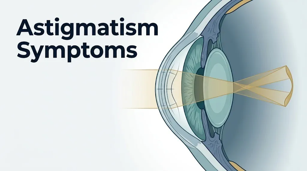
Astigmatism is a refractive error caused by an irregular curvature of the cornea or the crystalline lens, which prevents light from converging at a single focal point on the retina’s fovea centralis.
In a normal spherical eye, the cornea’s anterior surface curves uniformly in all directions, bending light evenly toward one point on the retina. Think of it like a perfectly round ball: light hits every part of the surface at a consistent angle and converges cleanly. In astigmatism, the cornea or lens is shaped more like a football or the back of a spoon. The curvature is steeper along one meridian than along the perpendicular one, so horizontal and vertical light rays refract at different angles.
This creates what optical physicists call Sturm’s conoid, a spread-out interval of focal points rather than one clean focal point. The visual cortex receives a blurred, elongated, or distorted signal from the fovea regardless of viewing distance, which is why astigmatism, unlike simple myopia, cannot be corrected by squinting.
Two primary anatomical sources produce this irregular curvature. Corneal astigmatism involves the anterior or posterior surface of the cornea itself. Lenticular astigmatism involves uneven curvature of the crystalline lens inside the eye. Corneal astigmatism is far more common and accounts for the majority of clinically relevant cases.
The American Academy of Ophthalmology classifies astigmatism as “regular” when the two principal meridians are perpendicular to each other, which is the typical correctable type, and “irregular” when the meridians are not perpendicular, which is harder to correct with standard glasses and is associated with conditions like keratoconus.
| Astigmatism Type | Location of Irregularity | Correctable with Glasses? | Common Association |
|---|---|---|---|
| Corneal (regular) | Anterior corneal surface | Yes | Most common type; often congenital |
| Lenticular | Crystalline lens curvature | Yes | Age-related lens changes, diabetes |
| Irregular | Cornea, non-perpendicular meridians | Not fully | Keratoconus, corneal scarring, surgery |
Individual variation note: Astigmatism that is present at birth (congenital) often remains stable through childhood, while astigmatism that develops or worsens in the late teen years or early 20s may be the early sign of keratoconus, a progressive corneal thinning condition. People with a family history of keratoconus should be screened with corneal topography rather than standard autorefraction.
Core Symptoms of Astigmatism
The core symptoms of astigmatism are blurry or distorted vision at one or more distances, eye strain, headaches, halos around lights, and difficulty seeing clearly at night, all of which trace back to the eye’s failure to produce a single focused image on the retina.
Most people with uncorrected astigmatism experience more than one symptom at the same time. The pattern and severity depend on the degree of refractive error, measured in diopters of cylinder power, whether it is combined with myopia or hyperopia, and whether it affects one eye or both.
Key symptoms, in order of clinical frequency, include:
- Blurred vision at near, intermediate, or far distances (present in virtually all clinically relevant astigmatism; the blur is not distance-dependent in the way nearsightedness is)
- Distorted or “smeared” images, particularly with letters or lines appearing stretched or tilted
- Eye strain (asthenopia), a fatigued, aching, or heavy feeling behind or around the eyes after visual tasks
- Headaches, typically frontal or temporal in location, worsened by reading, screen work, or driving
- Halos or starbursts around light sources, especially at night
- Difficulty reading or sustaining near-focus tasks, often confused with early presbyopia in adults over 40
- Squinting to temporarily improve clarity by changing the effective aperture of the pupil
- Monocular diplopia, seeing two images with one eye alone, a hallmark of optical aberration from astigmatism
According to a 2019 study published in Investigative Ophthalmology and Visual Science, patients with uncorrected astigmatism of one diopter or greater report statistically higher rates of asthenopia and headache compared to emmetropic individuals (those with no refractive error), confirming that even moderate astigmatism produces measurable functional impairment beyond just blurry vision.
Individual variation note: Adults with hyperopia (farsightedness) combined with astigmatism often experience more severe eye strain and headache than those with pure myopic astigmatism, because the ciliary muscle must work harder to compensate for the combined refractive demand of both errors simultaneously.
Blurry or Distorted Vision from Astigmatism
Blurry or distorted vision is the defining symptom of astigmatism, produced when two or more focal planes compete on the retina simultaneously, preventing the fovea centralis from receiving a sharp, coherent image signal.
The specific character of astigmatic blur distinguishes it from myopia. In myopia, distant objects are blurry while near objects are clear. In astigmatism, blur tends to occur at multiple distances and, critically, the image often appears smeared or stretched in a specific direction rather than uniformly unfocused. Letters may appear to have “ghost” edges. A straight line may look slightly tilted. A circle may look like an oval.
This directional distortion reflects the orientation of the steeper corneal or lens meridian. If the vertical meridian refracts light more strongly than the horizontal one (the most common pattern, called with-the-rule astigmatism), vertical line elements of letters and objects tend to appear sharper than horizontal ones, or vice versa. The brain processes these inconsistent signals and produces the perception of overall blur or smear.
Squinting temporarily narrows the pupillary aperture, which reduces the spread of Sturm’s conoid and produces a brief improvement in acuity. This is why squinting is a behavioral marker of uncorrected refractive error, including astigmatism, though it does not correct the underlying optical problem and increases the burden on the orbicularis oculi and periorbital muscles, worsening fatigue and headache over time.
According to the American Optometric Association, uncorrected astigmatism consistently ranks among the leading causes of reduced distance and near visual acuity in school-age children and working-age adults, with many affected individuals unaware that their visual standard is below normal because blurry vision has been their baseline from childhood.
Individual variation note: In people with mixed astigmatism, where the cylinder power creates one meridian that is myopic and one that is hyperopic simultaneously, the blur is often more disorienting than in pure myopic or hyperopic astigmatism. Mixed astigmatism is associated with greater difficulty reading and stronger complaints of visual fatigue.
Halos Around Lights and Night Vision Problems
Halos around lights and impaired night vision in astigmatism occur because the optical spread of Sturm’s conoid scatters point sources of light into elongated or ring-shaped patterns on the retina, a process that becomes dramatically more noticeable in low-light conditions when the pupil dilates.
In bright conditions, the pupil constricts. A small pupil aperture reduces the amount of peripheral corneal curvature that participates in refraction and limits the spread of the focal interval. Low-light conditions reverse this. The pupil dilates to allow more light in, exposing a larger area of the irregularly curved cornea. That larger area contributes more optical aberration, spreading the focal interval across a wider portion of the retinal surface.
A point source of light, such as a streetlamp, a car headlight, or an overhead ceiling light at night, enters the dilated pupil across multiple meridians with different refractive powers. Instead of converging to one point, those rays land at different positions on the retina. The visual cortex interprets this spread as a radiating halo, a starburst pattern, or a comet-like smear around the light source.
The starburst pattern is directionally oriented in regular astigmatism: the spikes or rays project perpendicular to the steeper meridian of curvature. An experienced optometrist or ophthalmologist can sometimes estimate the axis of astigmatism from a patient’s description of their starburst orientation before performing formal refraction.
Key night-vision symptoms associated with astigmatism:
- Halos (ring-shaped glow around light sources)
- Starburst or comet-like streaks from point lights
- Reduced contrast sensitivity in dim conditions
- Difficulty reading instrument panels, road signs, or menus in low ambient light
- Increased visual fatigue when driving after dusk
Individual variation note: People with larger natural pupils, which tend to be more common in younger adults and in people with lightly pigmented irides, experience more pronounced halos from astigmatism because the larger aperture exposes more of the irregularly curved peripheral cornea. Post-LASIK patients can experience similar halos if the treatment zone does not extend to the full optical zone activated during nighttime pupil dilation.
Key Takeaway: Astigmatism produces blurry vision, directional image distortion, and halos around lights because the irregularly curved cornea or lens creates multiple focal planes on the retina instead of one, a problem that worsens significantly in low light when the pupil dilates.
Astigmatism Eye Strain and Headaches
Astigmatism causes eye strain and headaches because the ciliary muscle, the ring of smooth muscle that controls lens curvature for focusing, must continuously shift its tension to attempt clarity in different meridians, a sustained and often unconscious effort that produces muscular fatigue and referred pain.
Think of the ciliary muscle like someone holding a heavy object slightly to the side of their body rather than directly in front. A healthy eye holding focus directly (emmetropia) keeps the ciliary muscle at an even, balanced tension. In astigmatism, the muscle must bias its contraction toward one meridian of the lens while the other remains blurry, a positional compromise that taxes the muscle over time.
This sustained accommodative effort is called asthenopia. The fatigue is cumulative over the course of a day’s worth of near and intermediate visual tasks. Symptoms typically worsen in the afternoon and evening, after extended screen time or reading, and improve with rest or eye closure. The headache associated with uncorrected astigmatism is most often located across the forehead, temples, or behind the eyes, and it is specifically worsened by visually demanding tasks.
The American Academy of Ophthalmology identifies asthenopia as one of the most common presenting complaints in patients with undiagnosed refractive error, including astigmatism, and notes that patients frequently attribute it to dehydration, stress, or screen time before their refractive error is identified and corrected.
How to describe your eye strain and headache to an optometrist or ophthalmologist:
- Note what time of day the headache typically begins (morning or afternoon)
- Identify whether the headache is worsened by specific tasks (reading, screens, driving)
- Describe whether the pain is behind the eyes, across the forehead, or at the temples
- Note whether closing your eyes or resting for 20 minutes reduces the pain
- Mention whether the headache is accompanied by any visual disturbance at the same time
Individual variation note: In patients with hyperopic astigmatism, the eye must accommodate constantly even for distant targets, making eye strain and headache more severe and more constant than in myopic astigmatism. These patients often report headaches that begin within the first hour of their workday and persist through the evening, sometimes misattributed to chronic tension-type headache or migraine.
Double Vision and Astigmatism
Astigmatism can cause monocular diplopia, which means seeing a doubled or “ghost” image with one eye alone, a symptom that is specifically produced by the optical spread of the focal interval rather than by a misalignment of the two eyes.
This is an important clinical distinction. Binocular diplopia, the double vision that occurs only when both eyes are open together, is caused by a misalignment of the extraocular muscles or a neurological problem affecting eye movement control. Monocular diplopia, by contrast, persists even when the other eye is covered, which is the defining test: if closing one eye eliminates the double image, the cause is binocular. If the double image remains with one eye covered, the cause is optical, most commonly astigmatism or, less frequently, an irregularity of the lens or corneal surface.
In astigmatism, the two principal meridians each form a separate line focus within Sturm’s conoid. The visual cortex receives two slightly separated image signals from the same retina and interprets them as two overlapping images. Patients describe this as a shadowed letter, a “ghost” of a character to one side, or a slightly doubled number on a page.
The degree of ghosting correlates broadly with the cylinder power of the astigmatism. Higher diopter values in the cylinder component tend to produce more noticeable monocular diplopia, though the axis of astigmatism also determines the direction of the ghost image.
Individual variation note: In older adults experiencing early posterior subcapsular cataract changes in the crystalline lens, the combination of lenticular astigmatism and early cataracts can produce monocular diplopia that is more variable and harder to correct with glasses alone. An ophthalmologist should evaluate any monocular diplopia that is new in onset, sudden in character, or accompanied by changing visual acuity in a patient over 55.
Light Sensitivity and Astigmatism
Light sensitivity in astigmatism, known clinically as photophobia, occurs because the visual cortex must process a constant stream of degraded, defocused retinal signals, a neurological processing burden that makes the brain more reactive to bright light stimuli than it would be with clear, well-focused input.
The connection between refractive errors and light sensitivity is not purely optical. Research published in JAMA Ophthalmology has documented that uncorrected refractive errors, including significant astigmatism, increase cortical visual processing load, which correlates with heightened sensitivity to luminance changes. The visual cortex effectively becomes fatigued by the continuous effort of interpreting low-quality retinal input and responds with increased reactivity to bright or rapidly changing light environments.
People with uncorrected astigmatism often report discomfort in brightly lit stores, when stepping outside from indoor environments, or when looking at high-contrast screens. This discomfort is distinct from the photophobia associated with migraine or uveitis, which tends to be more intense and is accompanied by other symptoms.
Light sensitivity symptoms associated with astigmatism:
- Discomfort in fluorescent lighting environments
- Squinting or flinching when moving from dim to bright environments
- Difficulty looking at screens without adjusting brightness
- Increased discomfort when reading under direct overhead lighting
- Worsened light sensitivity at the end of the day when visual fatigue is highest
Individual variation note: In people who have astigmatism combined with dry eye disease, light sensitivity is often more pronounced because the irregular tear film over the corneal surface compounds the optical aberration from the corneal curvature irregularity. Both conditions impair the tear-film-to-corneal optical interface, and treating dry eye can partially reduce light sensitivity even before the underlying astigmatism is corrected.
Key Takeaway: Eye strain, headaches, and light sensitivity from astigmatism share a common root: the ciliary muscle and visual cortex work continuously to compensate for a refractive error that standard corrective lenses can fully neutralize, which is why these symptoms often resolve completely after proper glasses or contact lens correction.
Astigmatism Symptoms at Night and While Driving
Astigmatism symptoms at night and while driving are typically the most functionally disabling presentation of the condition, because low-light conditions remove the pupillary constriction that normally limits aberration, and dynamic visual targets like oncoming headlights push the optical spread to its maximum.
Driving at night requires rapid processing of high-contrast point sources (headlights, traffic signals, streetlamps) against a dark background. For someone with uncorrected astigmatism, each light source fans out into a starburst or comet-shaped aberration. When multiple light sources are present simultaneously, as in urban traffic, these aberrations can overlap and obscure pedestrians, road markings, and distance information.
The U.S. National Eye Institute notes that nighttime visual impairment from refractive errors contributes meaningfully to road safety risk. Drivers who report difficulty with nighttime glare, halos, or reduced contrast sensitivity should have a formal eye examination that includes both manifest refraction and low-contrast acuity testing.
Nighttime astigmatism-specific driving symptoms:
- Starburst or halo patterns from headlights of oncoming vehicles
- Difficulty reading road signs at night despite ability to read them in daylight
- Reduced ability to distinguish lane markings in wet or unlit road conditions
- Fatigue setting in earlier on nighttime drives than daytime drives
- Tendency to reduce driving speed more than other drivers to compensate for visual uncertainty
People with myopic astigmatism experience the additional disadvantage of reduced distance acuity at night combined with the optical aberration of astigmatism. Their nighttime driving performance is compounded by both errors simultaneously.
Individual variation note: Adults over 40 face an additional nighttime challenge. The natural aging process causes the lens to stiffen (the beginning of presbyopia) and the pupil to become somewhat less reactive to light. When age-related lenticular changes occur alongside pre-existing astigmatism, nighttime vision can deteriorate faster than daytime vision, creating a growing gap between daytime and nighttime functional performance that is often the first symptom prompting an eye examination in this age group.
Mild Versus High Astigmatism Symptoms
Mild and high astigmatism produce the same categories of symptoms, but the clinical experience differs substantially in intensity, frequency, and functional impact based on the diopter value of the cylinder refractive error.
Astigmatism is typically classified by cylinder power: below 1.00 diopter is mild, 1.00 to 2.00 diopters is moderate, and above 2.00 diopters is high, though some clinical references extend the high-astigmatism threshold to above 3.00 diopters. These are not rigid diagnostic categories but practical guides to expected symptom severity.
| Severity | Cylinder Power | Common Symptoms | Functional Impact |
|---|---|---|---|
| Mild | Under 1.00 D | Occasional eye strain, mild blur at specific distances | Minimal; often subclinical |
| Moderate | 1.00 to 2.00 D | Regular eye strain, clear blur, headaches after visual tasks | Affects reading, screen work, driving |
| High | Above 2.00 D | Constant blur at multiple distances, pronounced halos, monocular diplopia, significant asthenopia | Affects all visual tasks; may impair daily function |
People with mild astigmatism may be entirely asymptomatic in childhood when the visual system’s accommodative flexibility can partially compensate for the refractive error. Symptoms often become apparent in early adulthood when reading and screen demands increase and accommodative reserve begins to decline.
High astigmatism that is left uncorrected in childhood carries an additional clinical risk: meridional amblyopia. This is a form of amblyopia in which one orientation of visual detail is chronically undertreated during the sensitive period of visual cortex development, leaving a permanent reduction in acuity for lines and edges oriented along the poorly focused meridian even after the refractive error is corrected.
A 2022 study published in Investigative Ophthalmology and Visual Science found that adults with a childhood history of high uncorrected astigmatism showed measurably reduced orientation-specific contrast sensitivity compared to adults whose astigmatism was corrected before age seven, illustrating the long-term developmental consequence of delayed correction.
Individual variation note: In elderly patients with high astigmatism, the combination of reduced corneal sensitivity from aging and reduced accommodative reserve from presbyopia can paradoxically reduce the symptom burden. These patients may report fewer headaches than younger patients with equivalent cylinder power, not because the optical aberration is less, but because the aging visual system adapts its processing expectations to a lower acuity standard.
Astigmatism in One Eye Symptoms
Astigmatism in one eye, a condition called unilateral astigmatism, produces a distinct clinical picture from bilateral astigmatism because the brain often suppresses the blurry input from the affected eye and relies predominantly on the clearer input from the unaffected eye, which can mask the refractive error for years.
This compensatory suppression is the key feature that makes unilateral astigmatism diagnostically tricky. A person with moderate or even high astigmatism in one eye and normal or near-normal vision in the other may pass a basic binocular visual acuity screening because the good eye carries the test. Closing one eye and comparing visual quality between eyes is the simplest self-assessment step, though formal refraction is required to characterize the error.
Symptoms specific to astigmatism in one eye:
- Monocular blur noticed only when the fellow eye is covered
- A subtle depth perception difficulty or reduced three-dimensional visual quality in daily tasks
- Fatigue or aching localized to the affected eye, rather than the bilateral frontal headache typical of bilateral astigmatism
- Difficulty with fine motor tasks requiring binocular depth judgment, such as threading a needle, parking a vehicle, or catching a moving object
- In children: tilting the head to align the better eye with the visual target, or closing one eye when reading
The most serious consequence of untreated unilateral astigmatism in children is amblyopia, commonly called lazy eye. When the brain consistently suppresses the input from one eye due to chronic optical defocus, the visual cortex fails to develop normal acuity pathways for that eye during the critical developmental window, typically before age seven. After that window closes, amblyopia becomes progressively harder to reverse.
According to the National Eye Institute, amblyopia affects approximately 2 to 3 percent of the U.S. population and is the leading cause of monocular visual impairment in children and working-age adults. Unilateral refractive error, including unilateral astigmatism, is one of the three primary causes.
Individual variation note: Adults with unilateral astigmatism from corneal scarring, post-surgical changes, or keratoconus in one eye experience an additional symptom burden: the brain’s attempt to fuse two very differently quality images causes active rivalry between the eyes, sometimes producing headache, nausea, and difficulty concentrating that is more severe than the refractive error itself would suggest.
Key Takeaway: Unilateral astigmatism is particularly easy to miss on standard visual screening because the better eye compensates, but the consequences of missed unilateral astigmatism in children, including permanent amblyopia, make eye-by-eye testing essential from an early age.
Astigmatism Symptoms in Children
Astigmatism symptoms in children often present behaviorally rather than verbally, because most children lack the reference point of normal vision needed to describe what is wrong with what they see.
A child who has had uncorrected astigmatism since birth does not know their vision is blurry. The blurred, distorted image is simply how they understand the world to look. Parents and teachers are typically the first to notice the behavioral indicators, and those indicators are frequently attributed to behavioral problems, learning difficulties, or attention issues before a refractive cause is identified.
Behavioral signs of astigmatism in children include:
- Sitting very close to the television or classroom board
- Tilting the head chronically to one side (attempting to use a clearer axis of vision)
- Squinting frequently, especially when trying to see something at a distance
- Frequent eye rubbing or complaints that eyes feel tired
- Avoidance of reading, drawing, or other near-vision activities
- Complaints of headache during or after school
- Poor academic performance in subjects requiring sustained reading or board work
- Covering or closing one eye when reading (a strong indicator of unilateral refractive error)
The American Academy of Pediatrics recommends vision screening for all children starting at birth, with comprehensive eye examinations by a qualified eye care professional recommended by age three or at any age if developmental concerns are identified. The organization specifically includes astigmatism in its list of conditions that, if uncorrected during the developmental window, can result in permanent visual impairment.
Children with a family history of astigmatism, myopia, or amblyopia should be prioritized for early optometric or pediatric ophthalmology evaluation, as hereditary factors contribute meaningfully to the likelihood of clinically relevant refractive error.
Individual variation note: Premature infants and children with Down syndrome face a substantially elevated risk of both astigmatism and amblyopia. The American Academy of Ophthalmology recommends that ophthalmologic evaluation occur earlier and more frequently for these populations than for the general pediatric population, given their higher baseline prevalence of refractive errors and accommodative deficits.
Astigmatism Symptoms in Adults and Older Patients
In adults, astigmatism symptoms are most likely to surface or worsen at two distinct life stages: in the mid-20s to mid-30s, when near-work demands from professional and digital life increase, and after age 40, when the natural loss of accommodative flexibility from presbyopia removes the last compensation the eye had available.
Before age 40, a young adult with mild or moderate astigmatism may use residual accommodative reserve to partially blur-compensate for the refractive error, keeping symptoms tolerable. As that reserve declines through the 40s, the same degree of astigmatism produces increasing eye strain, headaches, and difficulty with intermediate and near distances that was not present or was mild a decade earlier.
In adults over 60, the crystalline lens may develop changes in curvature distribution as part of normal lenticular aging or as a consequence of early cataract formation, adding a lenticular astigmatism component to pre-existing corneal astigmatism. This combination can produce a more complex, less correctable optical error that may require updates to existing prescription glasses more frequently than in younger patients.
According to the Mayo Clinic, adults with existing astigmatism correction who notice that their current glasses or contact lenses are no longer providing adequate clarity, especially for intermediate distances like computer screens, should schedule a refraction update rather than purchasing over-the-counter readers, which do not correct the cylindrical component of astigmatism.
What adults over 40 should tell their eye care provider:
- Which visual distances (near, intermediate, far) feel most affected
- Whether symptoms are worse in the morning or evening
- Whether they have started increasing font size on digital devices
- Whether nighttime driving has become more difficult than it was two years ago
- Any history of laser refractive surgery, as post-LASIK changes can affect astigmatism correction needs over time
Individual variation note: Adults with type 2 diabetes should be aware that fluctuating blood glucose levels can produce transient changes in the refractive index of the crystalline lens, temporarily altering astigmatism symptoms. Blurry or distorted vision that varies significantly between morning and evening, or that changes around the timing of meals, warrants a medical evaluation for glycemic control before attributing the symptom solely to astigmatism.
Can Astigmatism Cause Dizziness
Astigmatism can contribute to dizziness or a sense of spatial disorientation in some people, particularly when a new prescription is initiated, when astigmatism is high and uncorrected, or when the axis or power of a corrective lens does not precisely match the refractive error.
The mechanism behind astigmatism-related dizziness is not purely optical but involves the integration of visual and vestibular signals in the brainstem and cerebellum. The brain uses consistent visual spatial cues, including horizontal lines, vertical edges, and depth information, to calibrate its sense of balance and body position in space. When astigmatism distorts these spatial cues, the visual reference frame conflicts with the vestibular and proprioceptive signals from the inner ear and joints.
This mismatch is called visual-vestibular conflict, and it manifests as mild dizziness, a sensation of the ground or environment tilting slightly, or a floating feeling, especially during head movement. It is most pronounced in people who have just received a new corrective prescription that includes a cylinder component for the first time, because the brain is adapting to a now-stable visual frame it has never had before.
New glasses with a first-time or significantly changed cylinder correction often cause:
- Mild nausea or dizziness during the first one to two weeks of wear
- Distortion of floor and step depth perception when walking
- A sense of objects curving or slanting at the periphery of vision
- Motion sensitivity that resembles mild vertigo in some patients
Most adaptation occurs within seven to fourteen days. The American Optometric Association advises patients starting a first astigmatism correction to wear the new prescription consistently rather than alternating between old and new glasses, as inconsistent wear prolongs the adaptation period.
Individual variation note: Older adults and individuals with pre-existing vestibular conditions, such as benign paroxysmal positional vertigo or Meniere’s disease, may experience a longer and more difficult adaptation period when a new cylinder correction is introduced. Their eye care provider should be informed of any vestibular history before the prescription is finalized, as a gradual approach to full cylinder correction is sometimes preferred in these cases.
Key Takeaway: Dizziness associated with astigmatism is real and has a documented neurological basis in visual-vestibular conflict, but it typically resolves within two weeks of consistent new-prescription wear and is not a sign of something neurologically serious on its own.
Irregular Astigmatism and Keratoconus Symptoms
Irregular astigmatism produces significantly more severe and harder-to-correct visual symptoms than regular astigmatism because the corneal surface distortion follows a non-uniform pattern that cannot be fully neutralized by standard sphero-cylindrical lenses.
Keratoconus is the most common cause of clinically significant irregular astigmatism. It is a progressive condition in which the central or paracentral corneal stroma thins and ectases forward into a cone-like shape, driven by an imbalance between matrix metalloproteinase activity (specifically elevated MMP-1 and MMP-9) and reduced levels of their natural inhibitors in the corneal tissue. This structural deterioration produces a continuously changing and increasingly irregular corneal surface that generates complex, asymmetric optical aberrations.
Keratoconus-related astigmatism symptoms include:
- Rapidly changing vision requiring frequent prescription updates (every 6 to 12 months)
- Spectacle correction that provides only partial clarity even when recently updated
- Pronounced monocular diplopia with ghost images at multiple separations
- Severe halos and starburst aberrations around lights
- Corneal hydrops: sudden, painful vision loss from an acute rupture of Descemet’s membrane in advanced keratoconus
- Increased photophobia and glare sensitivity beyond what typical refractive astigmatism produces
- Halos that are asymmetric or directionally complex rather than ring-shaped
Keratoconus typically first presents in adolescence or early adulthood and progresses through the 20s and 30s before stabilizing. A 2021 study published in the Journal of Cataract and Refractive Surgery found that patients with a family history of keratoconus have a 15 to 67 times greater risk of developing the condition compared to the general population, making family history a priority screening indicator.
Individual variation note: Eye rubbing is a significant modifiable risk factor for keratoconus progression. People with atopic disease (allergic conjunctivitis, eczema, or asthma) who rub their eyes frequently face an elevated risk of accelerated keratoconus progression. The American Academy of Ophthalmology recommends specific counseling against eye rubbing for patients with known keratoconus or those with confirmed risk factors.
When to See an Eye Doctor for Astigmatism Symptoms
Most astigmatism symptoms are not emergencies, but they do warrant professional evaluation by an optometrist or ophthalmologist when they persist, affect daily function, or change in character, because uncorrected refractive error has measurable consequences for quality of life, learning, and safety.
The right timing for scheduling an eye examination depends on the nature and trajectory of the symptoms. Symptoms that have been present and stable for years are still worth evaluating if they have not been formally assessed. New symptoms, or symptoms that are worsening over a period of weeks to months, require evaluation sooner.
Specific circumstances that warrant scheduling an eye examination promptly:
- Blurry or distorted vision that has persisted for more than two to four weeks without an identified cause
- Headaches that are consistently triggered or worsened by reading, screen use, or driving
- Difficulty seeing clearly at night that has developed or worsened over the past six to twelve months
- A child who is squinting, tilting their head, or avoiding reading tasks
- Adults with existing glasses or contact lenses who notice their current correction is no longer adequate
- Monocular diplopia (double or ghost image with one eye covered)
- Any person over 40 who has never had a formal eye examination
For adults, the American Academy of Ophthalmology recommends a comprehensive eye evaluation by an ophthalmologist by age 40 for all adults, even those without symptoms, as this age represents the threshold for presbyopia onset and also the beginning of increased risk for glaucoma, macular degeneration, and other conditions that benefit from early detection.
Individual variation note: Adults with diabetes should have a dilated eye examination at least annually regardless of whether they have visual symptoms, because diabetic retinopathy can progress silently. Any change in vision in a person with diabetes should be evaluated promptly rather than attributed solely to astigmatism or uncorrected refractive error.
How Astigmatism Is Diagnosed
Astigmatism is diagnosed through a comprehensive eye examination that includes at minimum a manifest refraction, a visual acuity assessment using the Snellen chart or equivalent, and a keratometry or corneal topography measurement to characterize the corneal surface curvature.
The diagnostic process is systematic and precise. It begins with the autorefractor, a device that takes an automated initial estimate of the eye’s refractive error by projecting a pattern of infrared light onto the retina and measuring the reflected wavefront. This estimate is then refined through manifest refraction, in which the patient responds to a series of lens choices presented through a phoropter to identify the combination of sphere, cylinder, and axis that produces the clearest and most comfortable vision.
How a comprehensive astigmatism evaluation typically unfolds:
- Visual acuity measured for each eye separately using a Snellen chart or computerized equivalent
- Autorefraction performed to generate a baseline estimate of sphere, cylinder, and axis
- Manifest refraction performed to refine the prescription through subjective response
- Keratometry performed to measure the curvature radius of the anterior corneal surface in the principal meridians
- Corneal topography performed if irregular astigmatism, keratoconus, or post-surgical corneal changes are suspected
- Slit-lamp examination to assess corneal clarity, anterior chamber, and lens
- Cycloplegic refraction considered for children or adults with suspected accommodative error masking the full refractive picture
In children, cycloplegic refraction, which involves dilating drops that temporarily paralyze the ciliary muscle, is often necessary to prevent the child’s active accommodation from masking the full extent of the refractive error. A pediatric optometrist or pediatric ophthalmologist can complete this examination and is the appropriate provider for children under 16.
Individual variation note: In adults over 40 who are being evaluated for the first time or after a long gap in eye care, bifocal or multifocal needs must be assessed alongside the astigmatism correction. An optometrist or ophthalmologist can determine whether a toric single-vision lens, toric bifocal, toric progressive, or toric multifocal contact lens design best meets the full refractive picture.
Key Takeaway: A comprehensive eye examination, not a home vision test or smartphone app, is the only way to confirm astigmatism and determine its type, axis, and diopter value precisely enough to prescribe an effective correction.
Emergency Symptoms: When to Call 911 or Go to the ER
Certain visual symptoms that may be confused with worsening astigmatism actually indicate a medical emergency. Do not wait to see if these resolve on their own.
Call 911 or go to the nearest emergency room immediately if you experience:
- Sudden loss of vision in one eye, even if it lasts only a few seconds and appears to recover (can indicate a transient ischemic attack, carotid artery disease, or acute retinal artery occlusion)
- Sudden onset of severe eye pain combined with blurry vision, halos around lights, and nausea or vomiting (classic presentation of acute angle-closure glaucoma, which causes permanent vision loss within hours without treatment)
- New double vision that persists with both eyes open and is not explained by a known refractive error (can indicate cranial nerve palsy, brainstem stroke, or a compressive lesion affecting the oculomotor, trochlear, or abducens nerve)
- Sudden appearance of many new floaters accompanied by flashes of light or a dark curtain at the edge of vision (hallmarks of retinal tear or retinal detachment, which requires emergency surgical intervention)
- Vision loss or distortion following head trauma (warrants immediate evaluation for traumatic ocular or intracranial injury)
- Eye redness and severe pain with decreased vision after recent eye surgery (indicates possible infectious endophthalmitis, a surgical emergency)
These presentations cannot be distinguished from serious emergencies without clinical examination, imaging, and in some cases tonometry or fluorescein angiography. An urgent care appointment or a scheduled eye clinic visit is not an adequate response to any of these symptoms.
Frequently Asked Questions About Astigmatism Symptoms
What does astigmatism feel like day to day?
Day-to-day astigmatism typically feels like persistent visual fatigue, a dull frontal or temporal headache that builds through the afternoon, and vision that seems slightly “off” or blurry without a clear explanation.
Many people describe the experience as never quite being able to see as sharply as they feel they should, particularly when reading, working on a screen, or driving at dusk.
These sensations are produced by the ciliary muscle working continuously to compensate for a refractive error that cannot be resolved by accommodation alone.
Can you have astigmatism and not know it?
Yes, many people have astigmatism without knowing it, particularly if the cylinder power is mild (under 1.00 diopter) or if the condition has been present since birth, making blurry vision feel like a normal baseline.
Children are especially likely to be unaware of the problem because they have no reference for what sharp, corrected vision looks like.
According to the National Eye Institute, a substantial proportion of adults with clinically measurable astigmatism have never received formal correction because their symptoms were attributed to other causes like fatigue or screen overuse.
What does vision look like with astigmatism?
Vision with astigmatism looks like a blurred or smeared version of normal sight, where letters may appear to have ghost edges, straight lines may look slightly tilted, and point light sources at night appear as starbursts or halos rather than clean points.
The blur is not simply a loss of sharpness like myopia; it has a directional character, with some orientations of detail appearing slightly clearer than perpendicular ones.
This directional distortion is the optical result of two principal meridians of the cornea or lens refracting light at different powers, creating two competing focal planes on the retina.
Does astigmatism get worse over time?
Astigmatism may change over time, but it does not follow a predictable worsening pattern in most people; mild to moderate regular astigmatism often remains relatively stable through adulthood.
Astigmatism that worsens progressively, requires frequent prescription changes, and is accompanied by decreasing spectacle-corrected clarity should be evaluated for keratoconus, which is a progressive corneal ectatic condition that accelerates without treatment like corneal collagen crosslinking.
Lenticular astigmatism in adults can also increase gradually as the crystalline lens changes with age or in the context of developing cataracts.
Can astigmatism in one eye cause headaches?
Yes, astigmatism in one eye can cause headaches because the brain actively works to reconcile the clear input from one eye with the blurry, distorted input from the other, a binocular fusion effort that imposes a sustained neurological processing load.
The headache from unilateral astigmatism is often described as a dull ache around or behind the affected eye, sometimes with a sense of strain or pulling localized to that side.
An optometrist or ophthalmologist can detect unilateral astigmatism by refracting each eye separately and prescribing a toric correction for the affected eye only if warranted.
How is astigmatism different from being nearsighted?
Astigmatism differs from nearsightedness (myopia) in that myopia blurs only distant objects while near objects remain clear, whereas astigmatism blurs or distorts vision at multiple distances simultaneously.
Myopia is caused by an axial length of the eye that is too long or a corneal curvature that is too steep uniformly, focusing light in front of the retina. Astigmatism is caused by unequal curvature across different meridians, creating two separate focal planes rather than one.
Many people have both conditions simultaneously (myopic astigmatism), which requires correction with a lens that addresses both the spherical (myopia) and cylindrical (astigmatism) components of the refractive error.
Closing
Astigmatism symptoms are not just a nuisance. They are reliable signals from the eye’s optical system that the cornea or lens is producing a refractive error that, in most cases, can be corrected precisely and completely with the right glasses, contact lenses, or refractive surgery. The headaches, eye strain, blurry or distorted vision, halos, and nighttime driving difficulties you have been living with are not simply consequences of aging or screen time. They have a specific anatomical cause and a specific optical solution.
If your symptoms have been present for more than a few weeks, schedule a comprehensive eye examination with an optometrist or ophthalmologist. If you have children showing any of the behavioral signs described in this article, prioritize an evaluation before age five if possible; the developmental window for preventing amblyopia is narrow.
The single most practical action you can take today is to test each eye separately, covering one at a time, and compare the clarity and quality of what you see. Any meaningful difference between the two eyes is a specific reason to book an appointment rather than wait.