Femoral Hernia Symptoms: Signs, Stages & When to Act
Femoral hernia symptoms range from a small, painless lump in the upper inner thigh that a person barely notices to a rapidly escalating surgical emergency requiring immediate surgery within hours to prevent bowel death. Recognizing where a femoral hernia falls on that spectrum, and understanding the anatomical reason it can deteriorate so quickly, is the difference between scheduling a routine surgical consultation and calling 911.
According to the European Hernia Society (EHS), femoral hernias account for approximately 2 to 4% of all groin hernias overall but carry a strangulation rate of approximately 15 to 20%, substantially higher than the strangulation rate for inguinal hernias. That disproportionate risk exists because of specific anatomical features of the femoral ring that no amount of watchful waiting can change.
This article covers every recognized femoral hernia symptom with the anatomical and physiological reason each one occurs, how symptoms differ between early, incarcerated, and strangulated presentations, why women are disproportionately affected, which symptoms require a same-day emergency department visit, and how the diagnosis is confirmed clinically and with imaging.
Femoral Hernia Symptoms: What They Are and Why They Matter
Femoral hernia symptoms are produced when abdominal contents protrude through the femoral ring into the femoral canal, a narrow fibromuscular tunnel in the upper inner thigh, creating pressure, pain, and a visible or palpable bulge in the groin or thigh region.
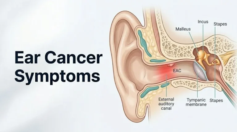
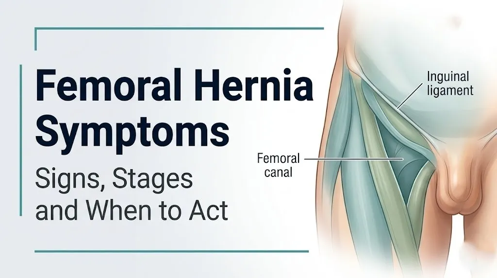
The femoral canal runs in the medial compartment of the femoral sheath, which is a downward extension of the transversalis fascia and iliac fascia below the inguinal ligament. In its normal state, the femoral canal is a small, nearly virtual space containing only loose connective tissue and a lymph node (the node of Cloquet). When intra-abdominal pressure forces omentum or bowel through the femoral ring above and into this canal, the previously empty space becomes occupied, and symptoms begin.
What makes femoral hernia symptoms particularly clinically important is the anatomy of the femoral ring itself. Three of its four boundaries are rigid, non-expandable structures: the inguinal (Poupart’s) ligament anteriorly, the lacunar (Gimbernat’s) ligament medially, and the pectineal (Cooper’s) ligament posteriorly. Only the femoral vein lies laterally, providing minimal flexibility. This rigid ring cannot accommodate an enlarging hernia sac, which is precisely why symptoms can escalate from mild and intermittent to an acute emergency without the gradual warning progression people often expect.
Key symptoms produced by femoral hernia:
- A small lump or bulge below and lateral to the pubic tubercle, in the upper inner thigh (location distinguishes it from inguinal hernia)
- Groin discomfort that worsens with prolonged standing, walking, or physical exertion
- Inner thigh aching or a sense of heaviness, produced by pressure on the anterior femoral cutaneous nerve branches
- Nausea with or without vomiting, indicating herniated bowel with early obstruction
- Acute, severe pain with a hard, non-reducible lump indicating incarceration or strangulation
People with obesity may have femoral hernia symptoms masked by adipose tissue in the groin region, making the bulge difficult to palpate and potentially delaying recognition of even a moderately advanced hernia.
What Is a Femoral Hernia and How Does It Form
A femoral hernia forms when increased intra-abdominal pressure forces a protrusion of peritoneal fat or a peritoneal sac through the femoral ring, allowing abdominal contents to descend into the femoral canal below the inguinal ligament.
The peritoneum is the thin membranous lining of the abdominal cavity. Under conditions of chronically or acutely elevated intra-abdominal pressure (heavy lifting, straining at stool, pregnancy, chronic cough, or obesity), the peritoneum is repeatedly pushed against the weakest points of the abdominal floor. The femoral ring, particularly in individuals with a wider or structurally weaker femoral canal, becomes a focal point for this pressure.
Think of the femoral ring like a narrow drain pipe covered by a rubber membrane. Normal abdominal pressure presses lightly on the membrane. Repeated or sustained high pressure gradually pushes the membrane down into the drain. Once the membrane is partially through the opening, each subsequent pressure event pushes more of it through, and the contents of the drain (bowel, omentum) begin following the membrane into the pipe.
The femoral hernia sac typically contains omentum (the fatty peritoneal apron that drapes over the intestines) in the early stages, which produces a soft, reducible lump with minimal symptoms. When small bowel loops herniate into the sac, the clinical picture changes substantially: the lump becomes larger, firmer, and more symptomatic.
According to the American College of Surgeons, risk factors for femoral hernia formation include female sex, older age, multiparity, chronic constipation, chronic cough from conditions such as COPD, heavy repetitive occupational lifting, and obesity. Connective tissue disorders including Marfan syndrome and Ehlers-Danlos syndrome impair the structural integrity of the fascial layers surrounding the femoral ring, increasing the risk in affected individuals.
Femoral Hernia Early Symptoms
Femoral hernia early symptoms are typically subtle: a small, soft, non-tender lump in the upper inner thigh or groin crease that may intermittently appear and disappear, accompanied by mild discomfort or a sense of pressure that worsens with physical activity.
In the earliest stage, many people notice the lump only after a sustained period of standing or physical exertion. It may partially or completely disappear when lying flat (the hallmark of a reducible hernia), because gravity allows the herniated omentum or peritoneal fat to fall back through the femoral ring when intra-abdominal pressure is reduced in the supine position.
The early discomfort is typically described as a dull ache, a dragging sensation, or a feeling of fullness in the groin rather than sharp pain. This mild quality is one reason femoral hernias are frequently dismissed as a muscle pull, lymph node, or groin strain for weeks to months before a correct evaluation is sought. The U.S. National Library of Medicine / MedlinePlus notes that many femoral hernias are found incidentally during physical examination or imaging for another complaint, reflecting how minimally symptomatic the early presentation can be.
Quick Tip:
- The lump of a femoral hernia appears below and slightly lateral to the pubic tubercle, placing it below the inguinal ligament. A lump above the inguinal ligament suggests an inguinal hernia, not a femoral hernia.
- Early discomfort often worsens after prolonged walking or standing and improves substantially after lying down for 20 to 30 minutes.
- Women who notice a groin lump during or after pregnancy should be evaluated by a primary care physician or board-certified general surgeon promptly, because relaxin-mediated ligamentous laxity during pregnancy significantly increases femoral hernia risk in that period.
Femoral Hernia Lump and Groin Bulge
The lump produced by a femoral hernia is typically a small, rounded, firm-to-soft swelling located in the crease of the upper inner thigh, just below the inguinal ligament and medial to the femoral vessels.
The precise anatomical location is directly below the mid-inguinal point (the midpoint between the anterior superior iliac spine and the pubic symphysis), slightly more medial. This location places it in the femoral triangle, the anatomical depression in the upper thigh bounded by the inguinal ligament superiorly, the sartorius muscle laterally, and the adductor longus muscle medially. The femoral artery and vein run through this triangle, which is why large femoral hernias can occasionally produce symptoms related to vascular compression.
The lump varies in consistency depending on its contents. When the hernia sac contains only omentum, the lump is soft, smooth, and partially compressible. When small bowel has entered the sac, the lump may feel firmer, sometimes with a slight gurgling sensation on palpation (borborygmi transmitted from gas-containing intestinal loops). A hard, stone-like lump that does not change with palpation or position is the physical hallmark of an incarcerated or strangulated hernia.
According to the Mayo Clinic, the femoral hernia lump often appears more pronounced after activities that raise intra-abdominal pressure: coughing, straining, lifting, or prolonged standing. In the clinical examination, a physician may ask the patient to cough or perform a Valsalva maneuver specifically to reproduce the bulge or its expansion, confirming the hernia’s communication with the abdominal cavity.
Overweight and obese individuals may not be able to see or feel the lump clearly due to overlying adipose tissue, meaning that groin or thigh discomfort without an obvious visible bulge does not rule out a femoral hernia. Ultrasound imaging is the appropriate next step in this scenario.
Key Takeaway: The femoral hernia lump sits below the inguinal ligament in the upper inner thigh, changes size with physical activity and position in early stages, and becomes hard and non-movable when incarcerated or strangulated. Hard and non-movable means go to the emergency room, not wait for a clinic appointment.
Femoral Hernia Pain Location and Character
Femoral hernia pain is typically located in the upper inner thigh or groin crease, with a character that changes from a dull, activity-related ache in early-stage hernias to severe, constant, and unrelenting pain when the hernia becomes incarcerated or strangulated.
In a reducible femoral hernia, pain is intermittent and positional. It worsens when standing, walking, coughing, or straining, and improves with rest or recumbency. The mechanism is mechanical: when the hernia contents are pushed through the ring by increased intra-abdominal pressure, they compress the surrounding tissues and stretch the peritoneal sac, activating nociceptors in the peritoneal lining and the surrounding connective tissue. When pressure is removed (lying flat), the contents partially reduce and the nociceptor activation subsides.
The character of pain shifts in an incarcerated hernia. Incarceration means the hernia contents can no longer be pushed back through the femoral ring. The contents remain trapped, venous outflow is obstructed, and the sac becomes progressively more congested and edematous. Pain at this stage is constant, moderately to severely intense, and not relieved by position change. The lump becomes tender to touch.
Research published in the British Journal of Surgery has noted that pain intensity in femoral hernia does not reliably correlate with the severity of the underlying pathology. A hernia with early strangulation may initially produce pain that seems manageable, particularly in older adults or in cases of Richter hernia (where only a portion of the bowel wall is trapped and full obstruction has not occurred). This makes clinical examination and imaging more reliable than pain self-assessment for determining urgency.
For people with diabetic peripheral neuropathy or other conditions reducing pain perception, even a strangulated femoral hernia may initially present with less pain than expected. These individuals require a lower threshold for emergency evaluation whenever a groin lump is noticed.
Femoral Hernia Inner Thigh and Hip Symptoms
Femoral hernia inner thigh and hip symptoms include aching, tingling, or a sense of pressure along the anterior and medial thigh, produced by the proximity of the herniated sac to the femoral nerve and its branches within the femoral triangle.
The femoral nerve enters the femoral triangle from beneath the inguinal ligament, running lateral to the femoral artery. As a femoral hernia enlarges within the femoral canal medial to the femoral vessels, the expanding mass can exert pressure on the femoral nerve sheath or its branches, particularly the anterior cutaneous branches of the femoral nerve that supply sensation to the anterior thigh. This compression produces the inner thigh aching and tingling that many patients describe.
One underrecognized clinical observation is the hip flexion posture. People with a symptomatic femoral hernia sometimes unconsciously adopt a slight hip flexion (bending forward at the hip) when standing or walking. This posture relaxes the iliopsoas muscle and reduces tension on the inguinal ligament, which in turn marginally reduces pressure on the herniated sac within the femoral canal. Clinicians familiar with this finding use it as a physical examination clue during gait assessment.
The inner thigh symptoms can be mistaken for a hip adductor muscle strain, hip joint pathology such as femoroacetabular impingement, or obturator nerve entrapment, because all three conditions produce medial thigh discomfort. The distinguishing feature of femoral hernia-related inner thigh pain is its association with the visible or palpable groin lump and its exacerbation by activities that raise intra-abdominal pressure (straining, coughing) rather than hip range-of-motion testing alone.
Older adults with pre-existing hip osteoarthritis may attribute inner thigh pain entirely to their hip condition, delaying recognition of a co-existing femoral hernia. A primary care physician who is evaluating groin or inner thigh pain in an older patient should include femoral hernia in the differential even when hip pathology is already established.
Femoral Hernia Symptoms in Females
Femoral hernia symptoms in females are produced by the same mechanical process as in males, but females are affected far more frequently because the female pelvis has a wider transverse diameter, which creates a structurally larger femoral canal relative to the surrounding supportive tissue.
The female pelvis is broader than the male pelvis to accommodate childbearing. This wider pelvic architecture increases the transverse dimension of the femoral ring, creating a larger potential entry point for abdominal contents. According to data published in the Annals of Surgery, femoral hernias occur approximately 4 to 5 times more frequently in females than in males, and females account for approximately 70% of all femoral hernia cases. In contrast, inguinal hernias are far more common in males because the male inguinal canal is widened by the descent of the testis during fetal development.
In females, the femoral hernia lump may be mistaken for a number of other conditions that present in the same anatomical region, including an enlarged inguinal lymph node, a lipoma, a Bartholin gland cyst (which sits more inferiorly and posteriorly), a saphenous varix (a varicosity of the great saphenous vein at the saphenofemoral junction), or an inguinal hernia. Clinical examination and ultrasound imaging are required to distinguish these reliably.
The symptom presentation in females does not fundamentally differ from the general description above, but there are two population-specific considerations. First, postmenopausal women have reduced estrogen, which is associated with decreased collagen density in fascial structures, increasing the structural vulnerability of the femoral canal. Second, females who have had multiple pregnancies (multiparity) have experienced repeated episodes of raised intra-abdominal pressure combined with relaxin-mediated ligamentous laxity, increasing cumulative mechanical stress on the femoral ring.
A female patient who notices a lump below the groin crease should be evaluated by a primary care physician who should arrange referral to a board-certified general surgeon for assessment, rather than assuming the lump is a lymph node without imaging confirmation.
Femoral Hernia Symptoms in Pregnancy
Femoral hernia symptoms in pregnancy can develop or worsen during the second and third trimesters as a result of two simultaneous physiological changes: rising intra-abdominal pressure from the expanding uterus and relaxin-mediated ligamentous laxity affecting the structural boundaries of the femoral ring.
Relaxin is a peptide hormone produced first by the corpus luteum of the ovary and later by the placenta during pregnancy. Its primary physiological role is to loosen the pelvic ligaments and cervix in preparation for labor. Relaxin acts on relaxin receptors in connective tissue cells, reducing collagen fiber cross-linking and increasing tissue compliance. The ligaments bounding the femoral ring, including the inguinal ligament and lacunar ligament, are subject to this effect. As they soften, the femoral ring widens slightly, increasing the anatomical vulnerability to hernia formation.
Simultaneously, the growing uterus displaces the abdominal contents upward and outward, increasing baseline intra-abdominal pressure. This pressure is particularly pronounced during activities like standing from a seated position, coughing, or carrying additional weight. The combination of an anatomically wider femoral ring and higher sustained intra-abdominal pressure creates the precise conditions for femoral hernia development.
Symptoms during pregnancy may be particularly difficult to attribute correctly. Groin pain and a sense of pelvic heaviness are frequent in pregnancy for unrelated reasons (round ligament pain, pubic symphysis dysfunction, varicose veins of the vulva and groin). A new, localized lump in the upper inner thigh during pregnancy warrants evaluation by an obstetrician-gynecologist, who should include femoral hernia in the differential and arrange ultrasound imaging to confirm or exclude it.
Surgical repair during pregnancy is generally reserved for cases of incarceration or strangulation rather than elective repair, making accurate symptom monitoring and prompt reporting to the obstetric care team particularly important for pregnant women with a known or suspected femoral hernia.
Key Takeaway: In females, femoral hernias occur 4 to 5 times more frequently than in males because of the wider female femoral canal, and pregnancy amplifies this risk through relaxin-mediated widening of the femoral ring. Any new groin lump in a pregnant woman deserves imaging evaluation, not watchful waiting.
Femoral Hernia vs. Inguinal Hernia Symptoms
Femoral and inguinal hernias both produce a groin lump with associated pain and discomfort, but they differ in the precise location of the lump, the anatomical pathway of herniation, and their relative complication rates.
The most reliable clinical distinguishing feature is the location of the lump in relation to the inguinal ligament. A femoral hernia lump sits below and lateral to the pubic tubercle, appearing below the inguinal ligament in the upper inner thigh. An inguinal hernia lump sits above the inguinal ligament, in the groin crease, and may extend into the scrotum in males. This distinction, while anatomically clear in principle, is often difficult for patients to self-assess and requires physical examination by a clinician or imaging to confirm.
| Feature | Femoral Hernia | Inguinal Hernia |
|---|---|---|
| Lump location | Below inguinal ligament; upper inner thigh | Above inguinal ligament; groin crease or scrotum |
| Sex distribution | 70% female | 90% male |
| Strangulation risk | High (approximately 15-20%) | Lower (approximately 3-5%) |
| Typical presentation | Often subtle; may be asymptomatic until strangulation | Usually palpable lump with activity-related discomfort |
| Pain character | Aching inner thigh; hip flexion posture may be present | Groin aching; scrotal heaviness in indirect type |
| Reducibility | Frequently irreducible even early on | More often reducible in the early phase |
| Primary patient group | Older women; multiparous women | Middle-aged to older men |
According to the British Journal of Surgery, the strangulation rate for femoral hernias is estimated at 15 to 20%, compared to approximately 3 to 5% for inguinal hernias. This difference exists because the femoral ring’s rigid three-sided boundary cannot accommodate growth of the hernia sac, making vascular compromise faster and more likely than in the more expandable inguinal canal.
For males with a groin lump, inguinal hernia is statistically far more likely than femoral hernia. For females with a groin lump below the inguinal ligament, femoral hernia is the priority differential. For both sexes, imaging (groin ultrasound as the first-line test) is the appropriate step when physical examination leaves the diagnosis uncertain.
Reducible vs. Incarcerated Femoral Hernia Symptoms
A reducible femoral hernia produces intermittent, mild symptoms that resolve with rest; an incarcerated femoral hernia produces constant, worsening pain with a firm, non-reducible lump, and represents a medical emergency that must be evaluated the same day.
Reducible means the hernia contents can be gently pushed back through the femoral ring, either spontaneously (when lying flat and intra-abdominal pressure drops) or manually with gentle physician-applied pressure. A reducible femoral hernia produces a soft, partially tender lump that changes in size with activity and position. Pain is positional and intermittent.
Incarcerated means the hernia contents are trapped in the femoral canal and cannot be returned to the abdominal cavity. The contents are typically swollen from venous congestion and edema within the sac, making them too large to fit back through the ring even with position change or manual pressure. An incarcerated femoral hernia produces constant pain, a firm and tender lump that does not change with position or palpation, and the beginning of systemic symptoms as bowel distention develops.
| Hernia Status | Lump Character | Pain Character | Reducibility | Urgency |
|---|---|---|---|---|
| Reducible | Soft to firm; changes with position | Intermittent; activity-related | Yes: reduces with recumbency | Elective surgical evaluation |
| Incarcerated | Firm, tender; does not change | Constant, moderate to severe | No | Same-day emergency evaluation |
| Strangulated | Hard, extremely tender; may be discolored | Severe, constant; systemic symptoms | No | Call 911; emergency surgery |
The clinical transition from incarceration to strangulation is time-dependent. Venous obstruction within the sac produces congestion and edema; if not relieved, arterial inflow is eventually compromised, producing ischemia. According to clinical data from JAMA Surgery, the window from incarceration to established intestinal necrosis can be as short as 4 to 6 hours in cases of complete vascular compromise, which is why incarceration is not a “wait and see” situation.
For people with a known femoral hernia who notice that the lump has become suddenly tender, hard, and non-reducible, the appropriate action is immediate medical evaluation, not trying to reduce it further at home.
Femoral Hernia Strangulation Symptoms
Femoral hernia strangulation symptoms are a surgical emergency: a hard, acutely tender, non-reducible lump in the upper inner thigh combined with severe constant pain, nausea, vomiting, and possible fever indicates that the blood supply to herniated bowel has been compromised and bowel necrosis may be developing.
Strangulation occurs when the rigid femoral ring compresses the blood supply to the herniated contents to the point of arterial occlusion. The sequence begins with venous obstruction (producing a congested, purple-red, edematous sac) and progresses to arterial occlusion (producing ischemia, then necrosis, then perforation). Bacterial translocation from the ischemic bowel wall into the peritoneal cavity produces peritonitis, which if unchecked can lead to sepsis and multi-organ failure.
The external hallmarks of strangulation visible to the patient:
- The lump becomes dramatically more tender than it has ever been before
- The lump feels hard, stone-like, and fixed in position
- The overlying skin may appear red, dusky, or bruised in color
- Touching the lump reproduces severe pain
Systemic symptoms that accompany strangulation:
- Nausea and vomiting (from bowel obstruction and peritoneal irritation)
- Fever and chills (from bacterial translocation and early sepsis)
- Rapid heart rate (tachycardia) from pain, volume loss, and early sepsis
- Abdominal distension and diffuse abdominal tenderness as peritonitis spreads
- Inability to pass gas or have a bowel movement
A special clinical entity called Richter hernia deserves specific mention. In a Richter hernia, only one wall of the bowel is pinched in the femoral ring rather than the full bowel lumen. Full bowel obstruction does not occur, so vomiting and inability to pass stool may be absent or delayed, making the presentation less obvious. The strangulated bowel wall segment still undergoes necrosis, however, and the absence of obstruction symptoms does not mean absence of the emergency.
People with diabetes and peripheral neuropathy may have reduced pain perception even with a strangulated femoral hernia, meaning that severe systemic signs (fever, vomiting, tachycardia) can appear while local pain remains deceptively mild. Any combination of a new or changed groin lump with fever and vomiting in a person with diabetes warrants immediate emergency evaluation.
Femoral Hernia Symptoms in Older Adults and Men
Femoral hernia symptoms in older adults of both sexes reflect a clinical picture complicated by reduced pain perception, decreased connective tissue tensile strength, and the higher likelihood of co-existing medical conditions that can mask or delay the recognition of serious complications.
In older adults, the nociceptive response to hernia-related tissue stress is often blunted. An elderly person may describe a strangulated femoral hernia as “a bit uncomfortable” when the same degree of bowel ischemia would produce severe pain in a younger person. This reduced pain signaling is a genuine physiological difference, not stoicism, reflecting age-related changes in peripheral nociceptor density and central pain processing. The clinical implication is that any groin lump in an older adult that has changed in character, size, or reducibility requires same-day medical assessment regardless of self-reported pain level.
Older adults are also more likely to be on medications including anticoagulants (warfarin, apixaban, rivaroxaban) or corticosteroids, both of which affect the appearance and behavior of the hernia. Anticoagulant use can produce a hematoma within the hernia sac that mimics strangulation. Chronic corticosteroid use weakens fascial and connective tissue throughout the abdominal wall, making hernia formation more likely and repair more technically complex.
In men, femoral hernia is considerably less common than inguinal hernia, occurring in approximately 1 in 20 groin hernias in males. The narrower male femoral canal (reflecting the narrower male pelvis) provides more structural resistance to femoral ring herniation. However, when femoral hernias do occur in men, they should be taken seriously, because male patients with femoral hernia are more likely to present with incarceration or strangulation at their first presentation than female patients are, according to published data in the Hernia journal. This higher complication rate at presentation in males may reflect delayed recognition because femoral hernia is not the first differential considered in male groin pathology.
Femoral Hernia and Bowel Obstruction Symptoms
Femoral hernia bowel obstruction symptoms occur when herniated loops of small intestine within the femoral canal become kinked, compressed, or edematous enough to prevent normal intestinal transit.
Bowel obstruction from a femoral hernia is a mechanical small bowel obstruction: the physical narrowing of the intestinal lumen within the hernia sac prevents the normal passage of intestinal contents downstream. As gas and fluid accumulate proximal to the obstruction, the intestine dilates and its peristaltic contractions intensify in an attempt to overcome the blockage. These intensified contractions produce the cramping, wave-like abdominal pain characteristic of bowel obstruction.
Observable symptoms of femoral hernia-related bowel obstruction:
- Intermittent cramping abdominal pain that comes in waves, typically centered around the navel and lower abdomen
- Abdominal distension visible as bloating or a visibly enlarged abdomen
- Nausea progressing to vomiting, initially of gastric contents, then bilious (green-yellow bile-stained vomit), and eventually feculent (foul-smelling, feces-like vomit indicating late obstruction)
- Inability to pass gas or have a bowel movement after a period during which this was normal
- High-pitched or tinkling bowel sounds, produced by gas moving through dilated, fluid-filled intestinal loops
According to the European Hernia Society, small bowel obstruction from a femoral hernia is one of the leading causes of emergency hernia surgery, precisely because the narrow femoral ring traps bowel with sufficient force to produce obstruction while the hernia itself may be small and easily overlooked on physical examination. A patient presenting with bowel obstruction symptoms and a small, painful groin lump is a surgical emergency until proven otherwise.
Plain abdominal X-ray shows dilated loops of small bowel with air-fluid levels in established obstruction. CT scan of the abdomen and pelvis with contrast is the definitive imaging study, demonstrating the herniated bowel loop within the femoral canal, the degree of bowel dilation, and any signs of ischemia such as wall thickening, pneumatosis intestinalis (gas within the bowel wall), or free peritoneal air.
Key Takeaway: A femoral hernia with vomiting, inability to pass gas, and cramping abdominal pain is producing bowel obstruction. This combination means the same-day emergency room is the only appropriate destination, not an urgent care clinic or a primary care office appointment.
Chronic Femoral Hernia Symptoms
Chronic femoral hernia symptoms develop in people who have had a reducible femoral hernia for months to years without surgical repair, and they differ from the acute or emergency presentations in their more persistent but lower-intensity character.
Chronic symptoms reflect the gradual enlargement of the hernia sac over time and the progressive mechanical effects on surrounding structures. As the sac enlarges, the hernia becomes harder to reduce, the lump becomes larger and more visible, and the periods of discomfort become longer in duration. What was previously an intermittent ache appearing only after several hours of standing may become a near-constant dull heaviness in the groin and upper thigh.
The chronic mechanical compression of surrounding structures produces secondary effects. The anterior cutaneous branches of the femoral nerve may become persistently sensitized, producing a chronic aching or tingling along the anterior and medial thigh that is present even at rest. The adductor muscle group medial to the femoral triangle may develop chronic tension from the altered biomechanics of the groin, producing an apparent adductor tightness or medial thigh stiffness that can be mistaken for a hip adductor strain.
Chronic femoral hernias are not stable entities in the way a stable medical condition might be. They carry an ongoing and increasing risk of incarceration as the hernia contents change over time. A hernia that has been easily reducible for two years can become incarcerated during a single episode of vigorous activity, prolonged coughing, or constipation-related straining. The EHS guidelines state that all femoral hernias, including those with mild or minimal symptoms, should be referred for surgical evaluation because the strangulation risk is not eliminated by symptom mildness.
A person who has been monitored with a known reducible femoral hernia and notices that the lump is becoming larger, harder to reduce, or more consistently painful should contact their primary care physician or their managing surgeon the same day rather than waiting for a scheduled follow-up.
How Femoral Hernia Symptoms Are Diagnosed
Femoral hernia symptoms are diagnosed through a combination of physical examination, groin ultrasound as the primary imaging modality, and CT scanning when incarceration or strangulation is suspected.
Physical examination is the starting point. The examining clinician (a primary care physician or a board-certified general surgeon) assesses the groin with the patient standing and supine. Standing allows the hernia to be gravity-dependent and maximally apparent. The Valsalva maneuver (bearing down as if to have a bowel movement) or a cough impulse test (asking the patient to cough while the examiner’s hand is placed over the lump) reproduces or accentuates the bulge by transiently raising intra-abdominal pressure. The location of the bulge relative to the pubic tubercle and inguinal ligament is the key finding for distinguishing femoral from inguinal hernia.
To describe femoral hernia symptoms accurately to a clinician:
- Note the precise location of the lump: is it in the groin crease above the inner thigh fold, or in the upper inner thigh itself?
- Describe whether it appears and disappears with activity versus position change
- State whether it is tender to touch and whether its tenderness has changed recently
- Report whether you have had nausea, vomiting, or changes in bowel habit since noticing the lump
- Tell the clinician whether you have tried to push the lump back and what happened when you did
Groin ultrasound is the first-line imaging test for femoral hernia diagnosis, according to clinical practice endorsed by the European Hernia Society. It visualizes the hernia sac in real time, allows dynamic imaging during a Valsalva maneuver, and can identify the contents of the sac (omentum vs. bowel). Ultrasound is safe in pregnancy, making it the modality of choice for pregnant patients with suspected femoral hernia.
CT scan of the abdomen and pelvis is indicated when strangulation, incarceration, or bowel obstruction is suspected. CT provides definitive anatomical detail including bowel wall viability, free peritoneal fluid (indicating perforation), and the relationship of the hernia to the femoral vessels. MRI is reserved for equivocal cases where ultrasound and CT have not produced a definitive diagnosis.
When to See a Doctor for Femoral Hernia Symptoms
Any person who notices a new lump in the upper inner thigh or groin below the inguinal ligament should be evaluated by a primary care physician within one week, because all confirmed femoral hernias require surgical assessment given their higher strangulation risk compared to other hernia types.
The appropriate provider for initial evaluation is a primary care physician, who will perform or arrange a physical examination and order groin ultrasound if the diagnosis is not clinically certain. Following confirmed diagnosis, referral to a board-certified general surgeon or hernia specialist is the appropriate next step for surgical planning. Surgical repair is the only definitive treatment; no truss, binder, or physical therapy protocol eliminates the anatomical vulnerability of the femoral ring.
Same-day evaluation by an emergency medicine physician is appropriate (not optional) in the following circumstances:
- The hernia lump has become suddenly more tender, harder, or larger within the past 12 to 24 hours
- The lump cannot be reduced when lying flat, having previously been reducible
- Nausea or vomiting has developed alongside groin pain
- Fever has appeared alongside groin discomfort
- Any abdominal distension, inability to pass gas, or cramping abdominal pain has developed
Bring the following information to any medical appointment for femoral hernia:
- When the lump was first noticed and whether it has changed in size, character, or tenderness
- Whether it disappears when lying flat and returns when standing
- Any prior groin surgery, hernia repair, or abdominal surgery that may have altered the anatomy
- Current medications including anticoagulants and corticosteroids
- Any history of connective tissue disorders
For people who are managing a known femoral hernia while awaiting surgical evaluation, the one daily monitoring step that matters most is checking whether the lump can still be gently reduced when lying flat. Loss of that reducibility is the signal that incarceration may be beginning and same-day evaluation is needed.
Emergency Symptoms: When to Call 911 or Go to the ER
Certain symptoms associated with a femoral hernia require immediate emergency evaluation. Do not wait to see if these resolve on their own.
Call 911 or go to the nearest emergency room immediately if you experience:
- A groin or inner thigh lump that has suddenly become extremely tender, rock-hard, and cannot be reduced in any position, indicating incarceration or early strangulation requiring emergency surgical assessment
- Nausea and vomiting combined with a painful, non-reducible groin lump, indicating bowel obstruction from incarcerated hernia contents
- Fever (temperature above 38°C / 100.4°F) combined with groin pain and a lump, indicating early peritonitis or bacterial translocation from ischemic bowel
- Abdominal distension with cramping abdominal pain and inability to pass gas or stool, indicating progressive mechanical small bowel obstruction
- Skin over the groin lump that has become red, dusky, purple, or bluish in color, indicating vascular compromise to the hernia sac and its contents
- Rapid heart rate, sweating, or feeling faint with groin pain, indicating systemic response to strangulation or early sepsis
- Severe constant abdominal pain that cannot be relieved by any position, indicating peritonitis from bowel perforation
These presentations can indicate strangulated femoral hernia, intestinal necrosis, bowel perforation, or peritonitis, all of which require emergency surgical assessment and intervention within hours, not an urgent care appointment or a phone call to a clinic.
Frequently Asked Questions About Femoral Hernia Symptoms
What does a femoral hernia feel like?
A femoral hernia typically feels like a small, soft-to-firm lump in the upper inner thigh or groin crease, just below the inguinal ligament, with a dull aching or sense of pressure that worsens after prolonged standing or physical activity.
In the early reducible stage, the lump may partially disappear when lying flat and return when standing or straining.
When incarcerated or strangulated, the lump becomes hard, extremely tender, and fixed in position, accompanied by constant pain, nausea, and vomiting.
Where exactly does a femoral hernia hurt?
Femoral hernia pain is located in the upper inner thigh and groin crease, specifically below and lateral to the pubic tubercle, in the area known as the femoral triangle.
Pain can radiate along the anterior and medial thigh due to compression of the anterior cutaneous branches of the femoral nerve by the enlarging hernia sac.
When bowel obstruction or strangulation develops, abdominal pain is added to the thigh and groin pain, typically as cramping around the navel or a diffuse lower abdominal ache.
Can a femoral hernia go away on its own?
A femoral hernia cannot resolve without surgical repair because the anatomical defect in the femoral ring does not close spontaneously.
Symptoms may fluctuate (improving when the hernia is reducible and less physically active) but the underlying structural vulnerability persists and typically worsens over time.
Surgical repair by a board-certified general surgeon is the only definitive treatment, and the European Hernia Society recommends surgical evaluation for all confirmed femoral hernias given their elevated strangulation risk.
How do I know if my femoral hernia is strangulated?
A strangulated femoral hernia produces a suddenly extremely tender, rock-hard, non-reducible lump in the upper inner thigh, accompanied by constant severe pain, nausea, vomiting, and in progressing cases, fever and abdominal distension.
The lump may develop skin discoloration (red, purple, or dusky) indicating vascular compromise to the hernia contents.
Call 911 or go to the nearest emergency room immediately if these symptoms develop, because strangulated bowel can become necrotic within 4 to 6 hours of complete vascular occlusion.
Why are femoral hernias more common in women?
Femoral hernias occur approximately 4 to 5 times more frequently in women because the female pelvis is wider in its transverse dimension, producing a structurally larger femoral canal with less surrounding supportive tissue.
During pregnancy, the hormone relaxin loosens the ligaments that form three of the four boundaries of the femoral ring, widening it further and increasing vulnerability to hernia formation.
Multiparity compounds this effect: multiple pregnancies produce repeated episodes of raised intra-abdominal pressure combined with relaxin-mediated ligamentous laxity, cumulatively stressing the femoral ring.
What is the difference between a femoral hernia and an inguinal hernia?
A femoral hernia protrudes through the femoral ring below the inguinal ligament, producing a lump in the upper inner thigh, while an inguinal hernia protrudes through the inguinal canal above the inguinal ligament, producing a lump in the groin crease or scrotum in males.
Femoral hernias are far more common in women and carry a substantially higher strangulation rate (approximately 15 to 20%) than inguinal hernias (approximately 3 to 5%) because the femoral ring’s rigid three-sided boundary cannot expand.
Distinguishing the two requires physical examination or groin ultrasound, as self-assessment of lump location relative to the inguinal ligament is unreliable.
A femoral hernia’s defining clinical danger is its capacity to deteriorate from a mild nuisance to a life-threatening surgical emergency without the gradual warning progression that other hernias sometimes provide. The rigid femoral ring can compress herniated bowel to the point of necrosis within hours of complete incarceration.
If you have a groin or inner thigh lump, the single most important action is getting a confirmed diagnosis from a primary care physician, who will arrange groin ultrasound and refer you to a board-certified general surgeon for surgical assessment. Do not wait to see if it improves; femoral hernias do not resolve on their own and their strangulation risk does not diminish with time.
The symptom that changes everything is a sudden increase in lump tenderness combined with hardness and non-reducibility. That change, whenever it occurs, is the signal to go to the emergency room immediately, not the next morning and not after calling the clinic.