Menopause After Hysterectomy With Ovaries Intact: Signs
The symptoms of menopause after hysterectomy with ovaries intact are real, they follow a recognizable pattern, and they are often more confusing to track than natural menopause because the most reliable early indicator of perimenopause, menstrual irregularity, is no longer available. Your ovaries still produce hormones, they still age, and when their estrogen output begins to decline, your body responds with the full spectrum of vasomotor, genitourinary, neurological, and musculoskeletal changes that define the menopause transition.
According to research published in Obstetrics and Gynecology, women who undergo hysterectomy while retaining their ovaries reach natural menopause an estimated 2 to 4 years earlier than women with intact uteri, likely because ligation of the uterine arteries during surgery reduces collateral blood supply to the ovarian cortex. This is not a minor timing difference. For a woman who had her hysterectomy at 38, it may mean facing menopause symptoms in her early to mid-40s without the menstrual roadmap that women without hysterectomy typically use to orient themselves.
This article explains every recognized symptom with the physiological mechanism behind it, clarifies how the post-hysterectomy hormonal timeline differs from standard natural menopause, and gives you the specific diagnostic and clinical information you need to have a productive conversation with a board-certified gynecologist or menopause specialist certified by The Menopause Society.
Symptoms of Menopause After Hysterectomy With Ovaries Intact
The symptoms of menopause after hysterectomy with ovaries intact are identical in type to those of natural menopause but occur without the menstrual cycle changes that normally signal their arrival, making them harder to attribute and easier to misidentify as stress, thyroid disease, or other conditions. The symptom set is driven by declining ovarian estradiol (E2) production as the primordial follicle pool in the ovarian cortex depletes with age.
The core symptom picture includes vasomotor symptoms (hot flashes and night sweats), genitourinary changes (vaginal dryness, recurrent urinary symptoms), neurological changes (brain fog, mood instability, sleep disruption), physical symptoms (joint pain, reduced skin collagen, hair thinning), and cardiovascular changes (palpitations, increased arterial stiffness). Each symptom has a distinct physiological driver, and understanding that driver is the most useful thing a woman in this situation can have.
The absence of periods makes this population unique. Without menstrual irregularity to track, many women in the early post-hysterectomy years reach perimenopause without recognizing it for months or longer. Hot flashes are often the first symptom to break through clearly enough to prompt evaluation.
Key symptoms of menopause after hysterectomy with ovaries intact, ordered by typical prominence at presentation:
- Hot flashes and sudden heat episodes, affecting an estimated 75% of menopausal women according to The Menopause Society
- Night sweats with sleep disruption, often preceding daytime hot flashes in onset
- Vaginal dryness and genitourinary discomfort, including dyspareunia (pain with intercourse)
- Mood changes: irritability, low mood, increased anxiety
- Cognitive changes: difficulty concentrating, word retrieval problems, memory lapses
- Joint pain and morning stiffness
- Fatigue disproportionate to activity
- Heart palpitations or awareness of heartbeat
- Skin, hair, and nail changes from collagen reduction
- Reduced libido
Why Menopause Happens Differently After Hysterectomy With Ovaries Intact
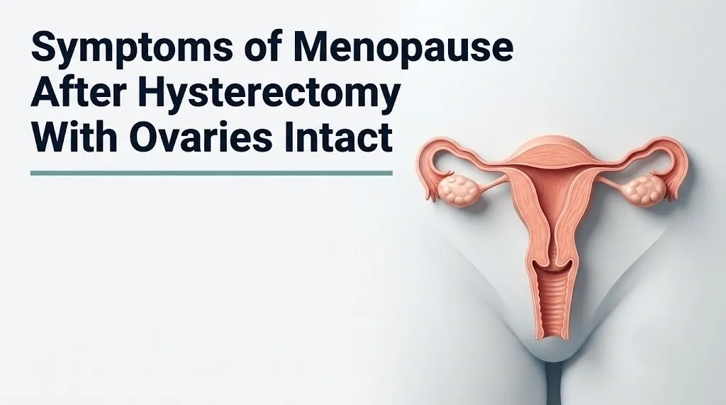
Menopause happens earlier and without menstrual warning signs after hysterectomy with ovaries intact because of a specific vascular mechanism: the surgical ligation of the uterine arteries during hysterectomy removes a secondary blood supply that normally reaches the ovarian cortex through utero-ovarian arterial anastomoses, the fine connecting vessel network between the uterine and ovarian arteries.
Think of the ovarian blood supply like a city with two water mains. The primary ovarian artery (from the aorta) stays intact after hysterectomy. But the secondary supply through the uterine artery branches is cut. The reduced perfusion pressure in the ovarian cortex does not stop ovarian function, but it does appear to accelerate the rate at which ovarian follicles undergo follicular atresia, the natural process of follicle death that depletes the primordial follicle pool over a woman’s reproductive lifetime.
Research published in Obstetrics and Gynecology has found that this vascular effect is most pronounced in women who had hysterectomy before age 45. The younger the woman at surgery, the longer the window during which reduced ovarian perfusion can accumulate its effect on follicular reserve before natural menopause age would otherwise occur.
From a hormonal axis perspective, the hypothalamic-pituitary-ovarian (HPO) axis continues to function after hysterectomy. The GnRH pulse generator in the arcuate nucleus of the mediobasal hypothalamus continues sending pulsatile GnRH signals to the anterior pituitary. FSH and LH continue to stimulate the ovaries. What changes is that the ovaries’ capacity to respond with estradiol production diminishes more quickly than it would have without surgery.
- Women who had their fallopian tubes removed (salpingectomy) alongside hysterectomy may experience additional reduction in ovarian blood supply, as the fallopian tube mesosalpinx carries ovarian vascular branches that contribute to ovarian perfusion.
- Women who had bilateral hysterectomy with ovarian cystectomy (removal of ovarian cysts at the time of hysterectomy) may have further reduced ovarian reserve due to removal of cortical tissue alongside the cyst.
When Does Menopause Start After Hysterectomy With Ovaries Intact?
Menopause after hysterectomy with ovaries intact typically begins 2 to 4 years earlier than it would have without surgery, based on published cohort data, with the average natural menopause age in the general population being approximately 51 years in the United States according to the National Institute on Aging. This means a woman who retains her ovaries after hysterectomy may begin experiencing perimenopause symptoms in her mid-to-late 40s rather than her early 50s.
The timing is not the same for every woman. Age at the time of hysterectomy is the strongest individual predictor. A woman who had her hysterectomy at 30 faces a longer interval of reduced ovarian perfusion before natural menopause age, potentially accumulating more follicular attrition, than a woman who had her hysterectomy at 48 when the follicle pool was already substantially depleted.
Symptom onset after hysterectomy with ovaries intact typically follows no obvious precipitating event, unlike surgical menopause (where oophorectomy produces immediate, steep estrogen withdrawal). Instead, symptoms appear gradually, as they would in natural perimenopause, but without menstrual irregularity to flag the transition.
| Hysterectomy Age | Estimated Menopause Onset | Perimenopause Duration (Approximate) |
|---|---|---|
| Under 35 | Mid to late 40s (estimated) | 5 to 8 years |
| 35 to 44 | Late 40s (estimated) | 4 to 6 years |
| 45 to 50 | Early to mid-50s (estimated) | 2 to 4 years |
| Over 50 | May already be in perimenopause at time of surgery | Variable |
- Women with a family history of early menopause (mother or sisters reaching menopause before 45) should expect their post-hysterectomy menopause timeline to be at the earlier end of the range, as genetic follicle pool size is heritable.
- Women who smoke reach menopause approximately 1 to 2 years earlier than non-smokers even without hysterectomy, according to data from the SWAN study; combined with the surgical effect, smoking significantly compresses the reproductive window post-hysterectomy.
Key Takeaway: Menopause after hysterectomy with ovaries intact typically arrives 2 to 4 years earlier than it would have without surgery, and without any menstrual irregularity to signal its approach, making symptom recognition the primary diagnostic tool.
Hot Flashes After Hysterectomy With Ovaries Intact
Hot flashes after hysterectomy with ovaries intact occur through the same physiological mechanism as in natural menopause: declining estradiol from the aging ovaries destabilizes the thermoregulatory center in the hypothalamic preoptic area, narrowing the thermoneutral zone (the temperature range within which the body maintains stable core temperature without triggering heat dissipation).
The specific mechanism involves norepinephrine (NE) and serotonin (5-HT). As estradiol declines, hypothalamic noradrenergic activity increases, which raises the threshold sensitivity of the preoptic thermoregulatory neurons. Small, normal fluctuations in core body temperature that would ordinarily be ignored now trigger full vasodilatory responses: sudden peripheral blood vessel dilation, flushing, sweating, and a rapid rise in skin temperature. The episode typically lasts 1 to 5 minutes and can be followed by chills as the body overcompensates.
According to The Menopause Society, vasomotor symptoms affect approximately 75% of women transitioning through menopause, making hot flashes the most consistently reported symptom in this population. In women who have had hysterectomy with ovaries intact, the experience is qualitatively identical to natural menopause hot flashes, but because there are no menstrual changes to contextualize them, they are more likely to be attributed to anxiety, thyroid disease, or other causes initially.
Hot flash characteristics that help identify them specifically in post-hysterectomy women:
- Sudden onset of intense warmth spreading from the chest upward to the neck and face
- Skin reddening and visible flushing
- Sweating, particularly on the upper body and face
- Heart rate increase during the episode (typically 10 to 15 beats per minute)
- Resolution within 1 to 5 minutes, often followed by a brief chill
- Occurring multiple times per day, sometimes waking the woman from sleep
- Women with higher body mass index (BMI) tend to experience more frequent hot flashes because adipose tissue generates heat during metabolic activity, and because the noradrenergic thermoregulatory sensitivity is not offset by peripheral estrogen (estrone from adipose aromatization) to the same degree that was previously assumed; research published in the Menopause journal has found that higher BMI is associated with greater, not lesser, vasomotor symptom frequency in the early transition.
Night Sweats and Sleep Problems After Hysterectomy Menopause
Night sweats after hysterectomy menopause are nocturnal hot flash events that occur during sleep and disrupt sleep architecture by forcing brief awakenings, sometimes without the woman fully recognizing the cause. The physiological trigger is identical to daytime hot flashes: hypothalamic preoptic area thermoregulatory instability from declining estradiol. Night sweats are often the first symptom that brings post-hysterectomy perimenopause to clinical attention.
Sleep disruption in the menopause transition after hysterectomy operates through multiple overlapping mechanisms beyond just night sweats. Declining estradiol directly affects the serotonergic system, which regulates REM sleep architecture and the transitions between non-REM and REM sleep stages. Research published in JAMA Internal Medicine has found that perimenopausal women report more awakenings, longer sleep latency (time to fall asleep), and less restorative slow-wave (N3) sleep than premenopausal women matched for age and health status.
The sleep deprivation produced by night sweats and hormonal sleep disruption is not simply fatigue. Fragmented sleep drives elevated cortisol output the following day via HPA axis activation, which compounds mood instability, cognitive difficulty, and daytime fatigue into a self-reinforcing cycle that worsens throughout perimenopause without intervention.
Quick Tip:
- Keeping the sleep environment below 67°F (19.4°C) reduces the frequency of night sweat awakenings by narrowing the gap between ambient and skin temperature, reducing the trigger threshold for nocturnal vasomotor events.
- Alcohol and spicy food in the evening hours lower the thermoregulatory threshold, increasing night sweat frequency; avoiding both within 3 hours of sleep reduces nocturnal episodes in many women.
- Women with pre-existing sleep apnea should inform their sleep medicine physician about hormone status, as estrogen withdrawal is associated with worsening upper airway tone during sleep, and post-hysterectomy perimenopause may accelerate sleep apnea severity.
Vaginal Changes and Genitourinary Symptoms After Hysterectomy Menopause
Genitourinary syndrome of menopause (GSM), the clinical term that replaced the older term “vaginal atrophy,” encompasses the range of vulvovaginal and lower urinary tract changes produced by estrogen withdrawal acting on estrogen receptor alpha (ERalpha) in the epithelial cells of the vaginal wall, vulvar tissue, and urethral mucosa. These changes occur after hysterectomy with ovaries intact just as in natural menopause, though the absence of the cervix (in total hysterectomy) alters the anatomy of the vaginal apex.
The vaginal epithelium is exquisitely estrogen-dependent. Under adequate estrogen, vaginal epithelial cells are thick, glycogen-rich, and maintain a lactobacillus-dominant microbiome that keeps vaginal pH between 3.8 and 4.5. As estradiol declines, epithelial thickness decreases, glycogen content falls, lactobacillus colonies reduce, and vaginal pH rises above 5.0. The result is dryness, reduced lubrication during arousal, microscopic tearing with friction, dyspareunia (pain with intercourse), and increased susceptibility to urinary tract infections and yeast infections.
Urinary symptoms of GSM include urinary urgency, increased frequency, urge incontinence, and recurrent lower urinary tract infections, all produced by urethral mucosal thinning and reduced periurethral collagen support. According to the American College of Obstetricians and Gynecologists, GSM affects approximately 45% of postmenopausal women but is underreported because many women do not connect urinary symptoms to estrogen status.
- Women who underwent total hysterectomy (uterus and cervix removed) will notice that the vaginal canal terminates at a surgical scar rather than a cervix; this does not change the symptom experience of GSM but means that some over-the-counter vaginal moisturizers and lubricants that were designed around cervical anatomy may need to be positioned differently for comfort.
- Women who have not been sexually active since their hysterectomy may not notice dyspareunia but should still be evaluated for GSM because the urinary symptoms (frequency, urgency, recurrent infections) occur independently of sexual activity and significantly affect quality of life.
Key Takeaway: Genitourinary syndrome of menopause after hysterectomy is driven by estrogen withdrawal at ERalpha receptors in vaginal and urethral epithelium, produces dryness, pain with intercourse, and urinary urgency, and is underreported because women do not connect urinary symptoms to hormonal change.
Mood Changes and Brain Fog After Hysterectomy Menopause
Mood changes and cognitive symptoms during menopause after hysterectomy with ovaries intact are among the most disorienting symptoms in this population, partly because they appear without the menstrual context that would normally prompt a woman to attribute them to hormonal change, and partly because they overlap with anxiety disorders, depression, and thyroid disease in ways that require hormonal testing to clarify.
Estradiol acts as a neuroactive steroid throughout the central nervous system. It modulates serotonin (5-HT) transporter expression, regulates dopamine synthesis in the nigrostriatal and mesolimbic pathways, and maintains BDNF (brain-derived neurotrophic factor) levels in the hippocampus, the primary brain structure for memory formation and encoding. As estradiol declines during perimenopause, all three of these systems are disrupted simultaneously.
The serotonin disruption accounts for the low mood and increased irritability. The dopamine reduction affects motivation and reward sensitivity. The BDNF decline is the mechanism most directly linked to the memory lapses and word-retrieval difficulties that women describe as brain fog, clinically termed subjective cognitive decline. Research published in the Menopause journal has found that perimenopausal women perform measurably lower on verbal memory and processing speed tasks during the early transition compared to their premenopausal baseline, and that performance partially recovers in the postmenopausal phase as the hormonal environment stabilizes.
If you are in crisis or experiencing thoughts of self-harm or suicide, contact the 988 Suicide and Crisis Lifeline by calling or texting 988 at any time. This service is free, confidential, and available 24 hours a day, 7 days a week.
- Women with a prior history of major depressive disorder or premenstrual dysphoric disorder (PMDD) are at elevated risk for more severe mood symptoms during the perimenopause transition after hysterectomy, as their serotonergic systems have demonstrated previous sensitivity to estrogen fluctuation. A board-certified gynecologist or menopause specialist should coordinate care with a psychiatrist for these women.
- Women taking SSRIs or SNRIs for depression or anxiety should know that these medications also have modest evidence for reducing vasomotor symptom frequency through serotonergic and noradrenergic pathways; their prescribing psychiatrist and gynecologist should be aware of hormone status when evaluating medication efficacy.
Joint Pain and Physical Symptoms of Menopause After Hysterectomy
Joint pain and musculoskeletal symptoms during menopause after hysterectomy with ovaries intact arise from estrogen’s widespread regulatory role in connective tissue maintenance, cartilage health, and inflammatory modulation. Estrogen receptors are expressed in chondrocytes (cartilage cells), synovial tissue, tendons, and ligaments, and estrogen withdrawal upregulates local inflammatory cytokines including IL-1beta and TNF-alpha in joint tissues, producing morning stiffness, aching, and increased joint sensitivity that closely mimics early inflammatory arthritis.
The joints most commonly affected in menopausal joint syndrome are the small joints of the hands, the knees, the hips, and the shoulders. The symptoms are typically bilateral, symmetric, and accompanied by morning stiffness lasting less than 30 minutes, distinguishing the pattern from rheumatoid arthritis (which produces stiffness lasting longer than 60 minutes and is confirmed by specific antibody testing). A board-certified rheumatologist should evaluate any joint symptoms lasting more than 6 weeks, particularly with elevated inflammatory markers, to rule out autoimmune arthritis.
Bone loss is a longer-term physical consequence that begins during perimenopause and accelerates in the first 3 to 5 years after final menstrual period (or after confirmed ovarian failure in post-hysterectomy women). Estrogen suppresses RANKL (receptor activator of nuclear factor kappa-B ligand) on osteoblasts, which drives osteoclast maturation and bone resorption. Without estrogen’s suppression, osteoclast activity increases, bone mineral density falls, and risk for osteoporosis rises substantially.
According to the National Institute on Aging, women can lose up to 20% of bone density in the 5 to 7 years following menopause. For women post-hysterectomy who may reach menopause earlier, this bone loss window begins sooner.
- Women who had hysterectomy before age 45 should discuss dual-energy X-ray absorptiometry (DEXA) scan scheduling with their gynecologist earlier than the standard recommendation (which typically begins at age 65 for low-risk women or age 60 for women with risk factors), because their earlier estrogen withdrawal represents a longer cumulative bone exposure risk.
Heart Palpitations and Cardiovascular Symptoms After Hysterectomy Menopause
Heart palpitations during menopause after hysterectomy with ovaries intact are produced by two concurrent mechanisms: estrogen withdrawal reduces endothelial nitric oxide (NO) synthase (eNOS) activity in cardiac and vascular endothelium, altering vascular tone and cardiac electrical stability; and the noradrenergic hyperactivity that drives hot flashes also increases sympathetic nervous system input to the heart, raising the rate of atrial ectopic beats that produce the fluttering, skipped-beat, or pounding sensations women describe as palpitations.
These palpitations are typically benign in women without pre-existing cardiac disease. They tend to occur alongside or immediately after hot flash episodes, when sympathetic activation is highest. They are self-limiting, lasting seconds to minutes, and resolve as the hot flash episode resolves.
The cardiovascular significance of menopause after hysterectomy is not limited to palpitations. Estrogen is a direct protective factor for endothelial function. Before menopause, estrogen maintains eNOS activity, which keeps blood vessels flexible and reduces arterial stiffness. After menopause, eNOS activity falls, LDL cholesterol rises (due to reduced hepatic LDL receptor expression), HDL cholesterol decreases modestly, and systemic arterial stiffness increases. The Women’s Health Initiative study established that these cardiovascular risk changes are real and measurable in the postmenopausal period.
Women who reach menopause before age 45, as many post-hysterectomy women do, have a longer duration of exposure to the post-menopausal cardiovascular environment, which is associated with higher cardiovascular event rates in long-term observational studies.
- Women with pre-existing atrial fibrillation or supraventricular tachycardia should inform their cardiologist when perimenopause begins post-hysterectomy, as the sympathetic and estrogen-withdrawal-related changes can increase the frequency of arrhythmic episodes and may alter their rhythm medication requirements.
- Women with hypertension should monitor blood pressure more frequently during the perimenopause transition, as declining eNOS activity and rising arterial stiffness can elevate blood pressure readings that were previously well-controlled.
Key Takeaway: Palpitations during post-hysterectomy menopause are typically caused by sympathetic nervous system activation during hot flashes and declining eNOS cardiac endothelial activity, not primary cardiac disease, but new or persistent palpitations always warrant evaluation by a primary care physician or cardiologist.
Menopause Symptoms After Partial Hysterectomy
Menopause symptoms after partial hysterectomy (subtotal hysterectomy, where the uterine body is removed but the cervix is retained) are identical in mechanism and pattern to those after total hysterectomy with ovaries intact, because the determining factor for menopause symptom onset is ovarian function, not whether the cervix is present or absent. The ovaries age, follicle pools deplete, and estradiol declines on the same accelerated post-hysterectomy trajectory regardless of cervical status.
One meaningful anatomical difference in partial hysterectomy is that the uterine cervix remains, which means the utero-ovarian vascular relationship may be partially preserved if the cervical stroma retains anastomotic connections to the ovarian vascular supply. There is some evidence, based on small observational studies, that cervical preservation may modestly protect ovarian blood flow compared to total hysterectomy, though the clinical effect on menopause timing is not clearly established in large trials.
Women who had partial hysterectomy retain the cervix and therefore require continued cervical cancer screening (Pap tests and HPV testing) on a schedule recommended by their gynecologist, unlike women who had total hysterectomy for non-cervical indications, who generally do not require Pap testing. This distinction is clinically important and is sometimes overlooked when menopause symptoms become the focus of follow-up care.
What to tell your gynecologist at a post-hysterectomy menopause evaluation appointment:
- The type of hysterectomy you had: partial (subtotal) or total, and whether the cervix was retained.
- The age at which you had the surgery and the reason for it.
- Whether both ovaries and both fallopian tubes were left intact, or whether one or both tubes were removed.
- The approximate time at which symptoms began and how they have changed over time.
- Your family history of menopause age in first-degree female relatives.
- Current medications, including any hormonal contraceptives or antidepressants.
- Women who had partial hysterectomy for uterine fibroids (leiomyomas) should be aware that fibroid development is estrogen-driven, and the perimenopausal estrogen decline typically causes residual small fibroids in the cervical stump to shrink; sudden return of pelvic pressure or rectal pressure warrants evaluation, as cervical fibroid growth after menopause is atypical.
Perimenopause Symptoms After Partial Hysterectomy: How to Recognize Them Without Periods
Perimenopause symptoms after partial hysterectomy are particularly challenging to recognize because perimenopause is defined clinically by menstrual cycle irregularity in women with an intact uterus, and that marker is absent post-hysterectomy regardless of whether the cervix was retained. The physiological perimenopause transition is happening in the ovaries, but without its primary clinical signal.
The SWAN study (Study of Women’s Health Across the Nation), one of the most comprehensive longitudinal cohort studies of the menopause transition, characterizes the perimenopause transition as beginning when ovarian estradiol and inhibin B production starts to fluctuate significantly, FSH begins rising intermittently, and vasomotor or mood symptoms emerge. In women with an intact uterus, this corresponds to menstrual cycle shortening, followed by irregular cycles, followed by cycles longer than 60 days. Post-hysterectomy, only the hormonal and symptomatic events are present.
The earliest symptoms of perimenopause that appear without menstrual changes in this population tend to be subtle: sleep becomes lighter, mood becomes more reactive to minor stressors, concentration feels slightly harder, and body temperature regulation feels different during sleep. Hot flashes, the most unmistakable symptom, often appear 1 to 2 years after these subtler symptoms, by which time the woman has been in the perimenopausal transition without knowing it.
| Perimenopause Stage | Women With Intact Uterus | Women Post-Hysterectomy (Ovaries Intact) |
|---|---|---|
| Early perimenopause | Cycle shortening, irregular cycles | Subtle sleep, mood, and concentration changes only |
| Mid perimenopause | Cycles 60+ days apart | Hot flashes begin, night sweats emerge |
| Late perimenopause | Final menstrual period | Vasomotor, genitourinary, and mood symptoms peak |
| Early postmenopause | 12 months after final period | Symptoms may continue; diagnosed by FSH and estradiol |
- Women who used hormonal contraception (oral contraceptives, hormonal IUD) after their hysterectomy will not notice perimenopause onset while using these methods, as exogenous hormones suppress the HPO axis and mask the hormonal changes of the transition. Perimenopause symptoms may emerge abruptly when contraception is discontinued.
Premature Ovarian Insufficiency vs Menopause After Hysterectomy
Premature ovarian insufficiency (POI) is the clinical condition defined by loss of normal ovarian function before age 40, characterized by elevated FSH levels (greater than 25 to 40 IU/L on two measurements at least 4 weeks apart) and low estradiol in a woman under 40. It is distinct from natural menopause after hysterectomy with ovaries intact, which follows the expected age-related trajectory (even if earlier than average), but the two conditions share overlapping symptoms and the distinction requires laboratory confirmation.
POI occurs in approximately 1% of women under 40 in the general population, according to the American College of Obstetricians and Gynecologists. In post-hysterectomy women, the risk of POI may be modestly elevated because surgical reduction of ovarian blood supply can, in susceptible individuals, accelerate follicular loss to the degree that ovarian failure occurs before age 40 rather than simply earlier than average.
The clinical distinction matters for two specific reasons. First, POI is associated with a higher long-term cardiovascular and bone risk than natural early menopause because estrogen deficiency begins at a younger age with more years of exposure ahead. Second, POI may have associated causes (autoimmune conditions including autoimmune thyroid disease, fragile X premutation, or chromosomal abnormalities) that require specific investigation and monitoring beyond routine menopause management.
A woman who had hysterectomy before age 40 and develops hot flashes, sleep disruption, and vaginal dryness should be evaluated by a reproductive endocrinologist for POI, not simply reassured that these are expected post-surgical changes.
- Women diagnosed with POI after hysterectomy should also be tested for thyroid peroxidase antibodies (TPO antibodies) and antinuclear antibodies (ANA), as autoimmune conditions co-occur with POI at rates higher than in the general population, according to research published in the Journal of Clinical Endocrinology and Metabolism.
Key Takeaway: Premature ovarian insufficiency in a woman under 40 who has had hysterectomy requires evaluation by a reproductive endocrinologist, not routine menopause counseling, because the causes, long-term risks, and management differ from standard post-hysterectomy menopause.
How Menopause Is Diagnosed After Hysterectomy With Ovaries Intact
Menopause after hysterectomy with ovaries intact is diagnosed through hormonal laboratory testing rather than the standard clinical criterion (12 consecutive months without a menstrual period), because menstrual periods are absent by definition post-hysterectomy. The primary diagnostic tools are serum FSH, serum estradiol (E2), and anti-Mullerian hormone (AMH).
FSH rises progressively as ovarian follicular reserve depletes and inhibin B falls. A single elevated FSH reading does not confirm menopause, because FSH fluctuates substantially during perimenopause, sometimes returning to normal ranges between episodes of follicular failure. The Menopause Society recommends that FSH above 25 IU/L on two measurements at least 4 to 6 weeks apart, in the clinical context of menopause symptoms and appropriate age, supports a diagnosis of menopause in post-hysterectomy women.
Serum estradiol below 20 pg/mL (some guidelines use 30 pg/mL) in the context of elevated FSH and menopause symptoms is consistent with the postmenopausal hormonal profile. AMH, produced by granulosa cells of pre-antral and small antral follicles, reflects the remaining primordial follicle pool. Low or undetectable AMH in a symptomatic woman post-hysterectomy confirms substantially diminished ovarian reserve even when FSH has not yet reached the menopausal threshold.
To prepare for a hormonal evaluation appointment for post-hysterectomy menopause:
- Record all symptoms, including onset, frequency, and severity, using a validated tool such as the Menopause Rating Scale (MRS) or Greene Climacteric Scale, available for download from menopause society websites.
- Bring a list of all current medications, supplements, and any recent hormonal treatments, as these affect FSH and estradiol measurements.
- Request that FSH and estradiol be drawn in the early morning (8 to 10 AM), when LH and FSH are at their daily peak, for the most reliable reading.
- Discuss whether AMH testing is appropriate given your age and symptom timeline.
- Ask specifically about DEXA scan scheduling to establish a baseline bone mineral density, which should be done before initiating any hormone therapy.
- Women who are taking oral contraceptive pills (even low-dose formulations) should stop the pills for at least 2 full weeks before FSH and estradiol testing, as exogenous estrogen suppresses pituitary FSH secretion and will falsely normalize the result.
Who Experiences Worse Menopause Symptoms After Hysterectomy: Individual Variation
The severity of menopause symptoms after hysterectomy with ovaries intact varies substantially between individuals and is predicted by identifiable biological, genetic, and lifestyle factors that determine how steeply estradiol falls, how sensitively the hypothalamic thermoregulatory system responds to withdrawal, and how efficiently the body maintains alternative estrogen sources through peripheral aromatization.
The SWAN study provides the most robust population-level data on menopause symptom variation. Black women experience more frequent and more intense vasomotor symptoms than white women at equivalent hormonal status. Hispanic and Asian women on average report fewer vasomotor symptoms, though all populations experience genitourinary and musculoskeletal changes. These differences are not fully explained by BMI or socioeconomic factors alone and likely reflect genetic variation in noradrenergic receptor sensitivity and estrogen receptor density.
| Risk Factor | Effect on Symptom Severity | Evidence Basis |
|---|---|---|
| Black race/ethnicity | More frequent, intense vasomotor symptoms | SWAN study cohort data |
| Higher BMI | More frequent vasomotor symptoms (counterintuitively) | Menopause journal research |
| Smoking history | Earlier menopause onset, more severe bone loss | SWAN and multiple cohort studies |
| History of PMDD | More severe mood symptoms in transition | Clinical and epidemiological data |
| Age under 45 at hysterectomy | Longer estrogen deficiency window, greater bone/CV risk | Obstetrics and Gynecology cohort data |
| Autoimmune thyroid disease | Symptom overlap complicates picture | Clinical observation |
| Prior anxiety or depression | More intense mood and sleep symptoms | Clinical and observational data |
- Women with type 2 diabetes and post-hysterectomy menopause face compounded cardiovascular risk because both insulin resistance and estrogen deficiency independently impair endothelial function; their primary care physician and gynecologist should coordinate cardiovascular risk management actively.
- Women who had hysterectomy for endometriosis may continue to experience pelvic pain during the post-hysterectomy transition if endometrial implants on the ovaries or peritoneum remain, as these implants are estrogen-sensitive and fluctuating perimenopausal estrogen can reactivate them even without a uterus; a board-certified gynecologist with endometriosis expertise should manage this combination.
Hormone Therapy After Hysterectomy With Ovaries Intact: What to Know
Hormone therapy after hysterectomy with ovaries intact differs in one clinically important way from hormone therapy for women with an intact uterus: women without a uterus do not need progestogen (a synthetic progesterone) to protect the uterine endometrium from estrogen-driven endometrial hyperplasia, because the endometrium was removed with the uterus. This means unopposed estrogen therapy (estrogen alone, without progestogen) is the appropriate formulation for eligible women post-hysterectomy.
This distinction matters because the progestogen component of combined hormone therapy carries the breast cancer risk signal that was amplified in the Women’s Health Initiative study. The WHI’s estrogen-only arm (enrolled women who had prior hysterectomy) found that unopposed estrogen was not associated with increased breast cancer risk and was actually associated with a reduced breast cancer incidence compared to placebo in that specific population, a finding published in JAMA. Women post-hysterectomy should be aware of this distinction when discussing hormone therapy with their gynecologist.
According to The Menopause Society, hormone therapy remains the most effective treatment for vasomotor symptoms, genitourinary syndrome of menopause, and prevention of bone loss in the menopause transition. For women under age 60 or within 10 years of menopause onset who do not have contraindications, the benefit-risk profile of hormone therapy is generally favorable based on current clinical evidence.
- Women who have a history of estrogen receptor-positive breast cancer are not appropriate candidates for systemic hormone therapy regardless of hysterectomy status; this decision must be made in consultation with a board-certified oncologist and gynecologist.
- Women with prior deep vein thrombosis or pulmonary embolism should discuss the route of hormone therapy administration specifically with their gynecologist; transdermal estrogen (patch or gel) does not carry the same first-pass hepatic effect on coagulation factors as oral estrogen and may have a more favorable venous thromboembolism risk profile, according to observational data published in the BMJ.
When to See a Gynecologist for Menopause Symptoms After Hysterectomy
Any woman who has had a hysterectomy with ovaries intact and is experiencing symptoms consistent with perimenopause or menopause should establish care with a board-certified gynecologist or a menopause specialist certified by The Menopause Society specifically for hormonal evaluation, rather than relying on a general practitioner who may not be familiar with the diagnostic nuances of the post-hysterectomy hormonal picture.
The appropriate timing for the first evaluation is when symptoms first appear and are affecting quality of life, not when they become unbearable. Early hormonal assessment establishes a baseline FSH, estradiol, and AMH that allows tracking of the trajectory over time and enables proactive discussion of bone density monitoring and cardiovascular risk management before the estrogen deficiency has accumulated its effects.
Specific circumstances that warrant prompt, not routine, gynecological evaluation:
- Symptoms beginning before age 40 (requires evaluation for premature ovarian insufficiency, not standard menopause counseling)
- Severe depression or thoughts of self-harm during the transition (requires coordination between gynecologist and psychiatrist)
- Pelvic pain recurring after hysterectomy (requires evaluation for residual endometriosis or other causes)
- Urinary symptoms including recurrent infections or urge incontinence substantially affecting daily function
- New-onset palpitations or chest discomfort (requires cardiac evaluation before attribution to menopause)
- Any symptom the woman cannot manage with lifestyle adjustments and that is affecting work, relationships, or mental health
Emergency Symptoms: When to Call 911 or Go to the ER
Certain symptoms that can occur during the menopause transition after hysterectomy require immediate emergency evaluation. Do not wait to see if these resolve on their own.
Call 911 or go to the nearest emergency room immediately if you experience:
- Chest pain with or without palpitations, shortness of breath, or left arm pain (possible cardiac event, the risk of which increases with postmenopausal estrogen deficiency)
- Sudden severe headache unlike any previous headache (possible hemorrhagic stroke, requiring immediate neurological evaluation)
- Sudden one-sided weakness, facial drooping, or speech difficulty (acute stroke requiring emergency intervention within a narrow treatment window)
- Chest pain with shortness of breath and leg swelling or leg pain (possible pulmonary embolism, risk elevated in the post-surgical period)
- Sudden severe pelvic or abdominal pain that is different in character from any prior menopause-related cramping (possible ovarian torsion, bowel obstruction, or internal hemorrhage)
- Fainting or near-fainting with palpitations and chest discomfort (possible serious cardiac arrhythmia)
These presentations can indicate myocardial infarction, hemorrhagic or ischemic stroke, pulmonary embolism, or ovarian torsion, and require emergency medical assessment, not an urgent care appointment or a phone call.
Frequently Asked Questions About Menopause After Hysterectomy With Ovaries Intact
Can you go through menopause if you had a hysterectomy but kept your ovaries?
Yes, women who had a hysterectomy with ovaries intact go through natural menopause when their ovaries’ follicle pool depletes and estradiol production falls, exactly as in women who did not have a hysterectomy.
The difference is that this menopause typically occurs 2 to 4 years earlier than it would have without surgery, and without any menstrual irregularity to signal its approach.
Symptoms are the primary clinical indicator, and hormonal testing (FSH, estradiol, AMH) is required for diagnosis in the absence of menstrual tracking.
How do you know you’re in menopause if you don’t have periods after hysterectomy?
Without periods, menopause after hysterectomy with ovaries intact is recognized through its symptoms (hot flashes, night sweats, vaginal dryness, mood changes) and confirmed through blood tests showing elevated FSH above 25 IU/L on two measurements at least 4 weeks apart alongside low estradiol.
A board-certified gynecologist or menopause specialist should order these tests and interpret them in the context of symptom onset and age.
AMH testing can also indicate remaining ovarian reserve and help anticipate the trajectory of the transition.
Does having a hysterectomy with ovaries intact make menopause worse?
Having a hysterectomy with ovaries intact does not necessarily make menopause symptoms more severe, but it does cause menopause to arrive earlier, which means a longer total duration of exposure to estrogen deficiency and its health consequences.
The quality and intensity of individual symptoms are driven by the same factors as in natural menopause: genetics, BMI, smoking history, race, and pre-existing conditions.
The absence of menstrual tracking does make the transition harder to recognize early, which can delay evaluation and support.
What are the first signs of menopause after hysterectomy with ovaries intact?
The earliest signs of menopause after hysterectomy with ovaries intact are often subtle: lighter sleep with more frequent awakenings, slightly increased mood reactivity, and mild difficulty with concentration, all appearing before hot flashes begin.
Hot flashes and night sweats, when they appear, are typically the first symptoms unmistakable enough to prompt a woman to seek evaluation.
According to The Menopause Society, vasomotor symptoms are the most consistently reported early menopause indicator across populations, appearing in approximately 75% of women transitioning through menopause.
How long does menopause last after hysterectomy with ovaries intact?
The total menopause transition after hysterectomy with ovaries intact spans the same duration as natural menopause, typically 4 to 8 years of perimenopause followed by an early postmenopause phase during which symptoms gradually stabilize.
Vasomotor symptoms, specifically hot flashes and night sweats, persist for a median of 7 to 9 years in many women, according to research from the SWAN study, and some women continue experiencing hot flashes into their 60s.
Genitourinary symptoms tend to be progressive rather than self-resolving and require ongoing management beyond the acute transition period.
Do you need hormone therapy if you still have your ovaries after hysterectomy?
Hormone therapy is not automatically required after hysterectomy with ovaries intact, as the ovaries continue producing estrogen until natural menopause occurs.
Once ovarian estrogen production declines sufficiently to produce significant symptoms or measurable bone loss, hormone therapy may be appropriate, and because the uterus has been removed, the appropriate formulation is unopposed estrogen rather than combined estrogen-progestogen therapy.
The decision to begin hormone therapy should be made in consultation with a board-certified gynecologist or menopause specialist who can weigh individual symptom burden, bone density, cardiovascular risk factors, and personal medical history.
Closing
The most practically important clinical fact for any woman in this situation: your menopause experience after hysterectomy with ovaries intact follows a real, documented physiological trajectory, it arrives earlier than you would expect based on general menopause information, and it happens without the menstrual roadmap that most menopause guidance assumes you have. Hot flashes are usually the symptom that finally makes the picture clear. By that point, the transition has typically been underway for some time.
What to do next: schedule an appointment with a board-certified gynecologist or a menopause specialist certified by The Menopause Society. Ask specifically for serum FSH, estradiol, and AMH testing. Ask when your DEXA bone density baseline should be established given your age at hysterectomy and current symptom status. If symptoms began before age 40, request referral to a reproductive endocrinologist rather than a general gynecologist.
You now have the physiological and clinical foundation to understand what is happening in your body, what the evidence says about it, and what your specific situation calls for.