What Causes Yeast Infections in Dogs? Full 2026 Guide
Yeast infections in dogs are caused by the overgrowth of Malassezia pachydermatis, a yeast organism that normally lives on healthy canine skin in small numbers but proliferates rapidly when the skin’s immune defenses or physical barrier are compromised. The question of what causes yeast infections in dogs almost always leads to a second, more important question: what is the underlying condition disrupting that defense?
According to the Merck Veterinary Manual, Malassezia dermatitis is one of the most frequently diagnosed skin conditions in dogs across all breeds and ages, and in the majority of cases it is a secondary condition, meaning something else has changed the skin environment first. Identifying that underlying cause is the difference between treating a single infection and stopping the cycle of recurrence.
This article covers every documented cause of canine yeast infections, from the most common (allergic skin disease) to the systemic endocrine causes that competitors consistently miss, with the species-specific physiological explanation for each. It also covers which breeds are most anatomically vulnerable, when a yeast infection signals an emergency, and exactly what a veterinarian does to find the root cause.
What Causes Yeast Infections in Dogs: The Core Answer
Yeast infections in dogs are caused when Malassezia pachydermatis, a commensal yeast normally present on canine skin, overgrows because of a disruption in the skin’s physical barrier, immune defenses, or microbial balance.
The causes fall into two categories. Primary or predisposing causes change the skin’s microenvironment directly: allergic skin disease, moisture accumulation in skin folds, and anatomy of the ear canal are the most frequent. Secondary or systemic causes impair the immune system’s ability to control yeast populations: hypothyroidism, hyperadrenocorticism (Cushing’s disease), and immunosuppressive medications fall into this group.
Most yeast infections in dogs have at least one identifiable underlying cause. Finding and addressing that cause is what separates effective treatment from a revolving door of recurrent infections. A dog that clears a yeast infection with antifungal treatment but whose atopic dermatitis or Cushing’s disease remains unmanaged will develop another infection within weeks to months.
Key causes of yeast infections in dogs:
- Canine atopic dermatitis (environmental allergies): the most frequent underlying cause in clinical practice
- Cutaneous adverse food reaction (food allergy): a subset of allergic skin disease
- Antibiotic therapy: disrupts commensal bacterial competition with Malassezia
- Moisture retention in skin folds, ear canal, and paw interdigital spaces
- Hypothyroidism: alters skin sebum and cell turnover rate
- Hyperadrenocorticism (Cushing’s disease): suppresses cell-mediated immunity
- Corticosteroid or immunosuppressive medication use
- Seborrhea (primary or secondary): increases lipid substrate for Malassezia
- Young age (immature immune system) or advanced age (declining immune function)
According to the World Association for Veterinary Dermatology (WAVD) consensus guidelines, identifying and treating the primary predisposing condition is the essential step in managing Malassezia dermatitis long-term. Without addressing the underlying cause, antifungal treatment alone produces only temporary improvement.
Malassezia Pachydermatis: The Organism Behind Dog Yeast Infections
Malassezia pachydermatis is the specific yeast species responsible for virtually all canine yeast dermatitis, and it has a unique biological characteristic that explains its prevalence in dogs: unlike most Malassezia species, it does not require external lipid supplementation to survive.
Most Malassezia species (such as Malassezia globosa, found on human skin) are strictly lipid-dependent and require external lipid sources in their growth medium. M. pachydermatis is classified as non-lipid-dependent, which means it can grow on standard laboratory culture media and, more importantly, can thrive on canine skin surfaces with variable lipid content. It still strongly prefers lipid-rich environments, but its ability to survive without exogenous lipids makes it better adapted to the canine skin surface than other species.
This yeast produces several enzymes that actively damage the host skin. Its phospholipase and lipase enzymes hydrolyze sebum triglycerides, breaking them into free fatty acids. Some of these fatty acids (particularly oleic acid and arachidonic acid) are directly irritating to the stratum corneum and trigger local inflammatory responses. Others serve as growth substrates for additional yeast proliferation, creating a self-reinforcing cycle of skin irritation and yeast expansion.
The Veterinary Dermatology journal has published research demonstrating that M. pachydermatis activates Toll-like receptors (TLRs) on keratinocytes and dendritic cells, initiating innate immune responses that amplify the local inflammatory state. In dogs with atopic dermatitis, this already-inflamed immune baseline means the yeast triggers an even more pronounced inflammatory reaction, which is why atopic dogs with secondary Malassezia overgrowth are dramatically more itchy than either condition alone would produce.
Intact male dogs and females in estrus may have transient alterations in skin surface lipid composition from hormonal fluctuations, though the clinical significance of sex hormones on Malassezia growth rate is considered less important than allergic or endocrine causes in clinical practice.
Why Dogs Develop Yeast Overgrowth From a Commensal Organism
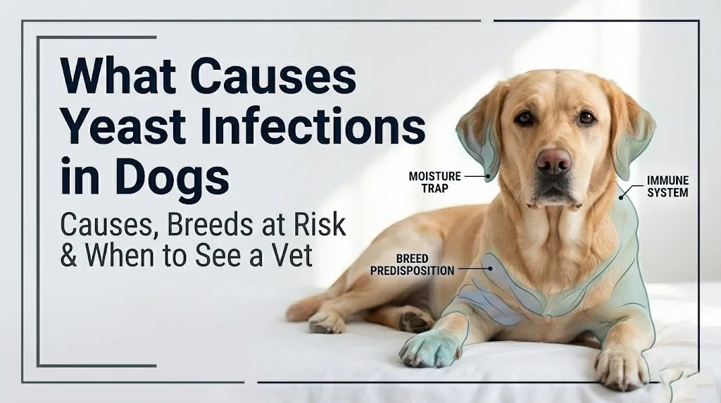
Dogs develop Malassezia overgrowth when changes in the skin’s physical microenvironment, immune competence, or microbial community tip the balance from commensal coexistence to pathogenic proliferation.
Think of the skin surface as a finely tuned ecosystem. In a healthy dog, Malassezia lives in the ecosystem as one member of a balanced community. The stratum corneum (the outermost layer of the epidermis) provides a physical barrier that limits yeast attachment. Commensal bacteria, principally Staphylococcus pseudintermedius on canine skin, compete with Malassezia for lipid substrates and attachment sites. T-lymphocytes in the local skin immune surveillance system keep yeast populations from expanding. Sebum production is relatively controlled, limiting the available lipid food source.
Disrupt any of these three elements and the ecosystem tilts toward Malassezia. A disrupted stratum corneum (from allergy-driven inflammation) gives the yeast better adhesion and deeper access to the skin surface. Depleted commensal bacteria (from antibiotics) remove the competitive check. Suppressed T-lymphocyte function (from corticosteroids or Cushing’s disease) removes the immune check. Excess sebum production (from seborrhea or hypothyroidism) floods the ecosystem with yeast food.
According to the VCA Animal Hospitals clinical reference on yeast dermatitis, Malassezia is an opportunistic pathogen, meaning its ability to cause disease is almost entirely dependent on changes in the host rather than changes in the organism itself. M. pachydermatis does not become more virulent; the dog becomes less able to control it.
Puppies and senior dogs represent the two age extremes most vulnerable for different reasons. Puppies have an immature adaptive immune system with less developed Th1/Th2 cytokine regulation, making immune surveillance less effective. Senior dogs have declining T-lymphocyte populations and slower skin cell turnover, reducing barrier renewal. Both groups warrant closer monitoring for yeast overgrowth after antibiotic courses or during management of other skin conditions.
Key Takeaway: Malassezia pachydermatis does not become more aggressive; your dog’s skin defenses become less effective at controlling it. Finding and fixing what compromised those defenses is the only way to prevent recurrence.
Canine Allergic Skin Disease: The Primary Cause of Dog Yeast Infections
Canine atopic dermatitis (CAD) is the most frequently identified underlying cause of Malassezia overgrowth in dogs, producing a specific immunological chain reaction that transforms healthy skin into an ideal growth environment for yeast.
CAD is an IgE-mediated hypersensitivity disorder. When a dog with atopic dermatitis is exposed to environmental allergens (dust mites, pollen, mold spores, or storage mites), allergen-specific IgE antibodies on mast cells in the skin’s dermis become cross-linked. This cross-linking triggers mast cell degranulation, releasing histamine and activating the Th2 cytokine pathway. The Th2 cytokines IL-4 and IL-13 are the key drivers of skin barrier dysfunction.
These cytokines directly suppress ceramide production in keratinocytes (the primary structural cells of the epidermis). Ceramides are the primary lipid component of the stratum corneum, forming the “mortar” between cornified skin cells. When ceramide production falls, the stratum corneum becomes permeable. Trans-epidermal water loss increases. The skin surface becomes warmer and more humid. The pH rises from its normal mildly acidic value. And the lipid composition of the skin surface shifts, accumulating the exact types of fatty acids that Malassezia uses as a food source.
The result is a skin surface that is physically compromised, immunologically inflamed, and nutritionally enriched for yeast. Malassezia’s lipase enzymes exploit this environment immediately, and the yeast’s own inflammatory products add to the itch-scratch cycle, creating a worsening spiral.
Research published in Veterinary Dermatology has found that a substantial proportion of dogs with Malassezia dermatitis have underlying atopic dermatitis as the primary diagnosis. Treatment of the yeast infection without managing the atopic dermatitis produces temporary improvement, with rapid recurrence in subsequent weeks.
West Highland White Terriers are a breed with documented hypersensitivity to Malassezia itself: their immune system mounts an exaggerated IgE response to M. pachydermatis antigens, making yeast infections both more likely and more inflammatory in this breed than the general atopic population.
How Antibiotics Cause Yeast Infections in Dogs
Antibiotic therapy causes yeast infections in dogs by eliminating the commensal bacterial populations on the skin and in the ear canal that normally compete with Malassezia for nutrients and colonization sites.
The canine skin surface and ear canal harbor a diverse community of commensal bacteria. Staphylococcus pseudintermedius is the dominant commensal bacterium on canine skin and plays a direct ecological role in limiting Malassezia expansion. It competes for the same lipid substrates that yeast requires, produces bacteriocins (antimicrobial peptides) that inhibit other organisms, and occupies adhesion sites on the stratum corneum that yeast would otherwise colonize.
When a dog is treated with antibiotics for any reason (a skin infection, a wound, a urinary tract infection, a respiratory infection), broad-spectrum antibiotics deplete Staphylococcus pseudintermedius and other commensal bacteria across the skin surface and ear canal simultaneously. Malassezia, being a yeast and not a bacterium, is completely unaffected by antibacterial antibiotics. With its bacterial competition eliminated, it expands into the vacated ecological space.
This is the same mechanism by which human patients develop oral or vaginal candidiasis during antibiotic courses, though the species differ. In dogs, the post-antibiotic yeast overgrowth presents most commonly in the ears (because the ear canal is an enclosed, warm, humid environment where yeast proliferates fastest without bacterial competition) and between the toes.
Quick Tip:
- Dogs receiving antibiotics for more than 5 to 7 days should have their ears checked weekly by the owner for increased wax production, odor, or head shaking
- Dogs with a history of recurrent yeast infections after antibiotic courses should have ear cytology performed by a licensed veterinarian at the start and end of any antibiotic treatment
- Dogs on immunosuppressive antibiotics (not standard antibacterials) are at risk for a different, more severe immune-driven pattern of yeast overgrowth
Dog Ear Canal Anatomy: Why Yeast Infections Develop There
The dog’s external ear canal is uniquely prone to yeast infection because of its L-shaped anatomical structure, which traps heat and moisture more effectively than the relatively straight human ear canal.
The canine external ear canal consists of a vertical portion (running downward from the ear opening) and a horizontal portion (running inward toward the tympanic membrane). These two portions meet at approximately a right angle, forming the characteristic L-shape. This architecture means that debris, moisture, and cerumen (ear wax) that enter the vertical canal must negotiate a 90-degree turn to exit, and in practice they frequently do not exit at all without mechanical cleaning or ear movement.
The ear canal is also lined with both sebaceous glands (producing lipid-rich sebum) and ceruminous glands (modified apocrine glands producing cerumen). Both secretion types provide the lipid-rich substrate that Malassezia requires. The combination of a moisture-trapping anatomy and a lipid-rich secretory lining makes the canine ear canal one of the most favorable microenvironments for yeast growth in the entire body.
Dogs with heavy, pendulous ear flaps (pinnae), such as Basset Hounds, Cocker Spaniels, and Bloodhounds, have reduced air circulation in the ear canal because the ear flap covers the opening. This creates a warmer, more humid internal environment than dogs with erect or open pinnae. According to the Merck Veterinary Manual, breeds with pendulous pinnae are consistently overrepresented in canine otitis externa case populations in veterinary practice.
Dogs that swim regularly, are bathed frequently, or live in humid climates are at elevated risk for moisture-related ear yeast overgrowth because water that enters the vertical ear canal is difficult to drain completely, providing the warm, wet conditions yeast requires.
Skin Folds and Moisture: Environmental Causes of Dog Yeast Infections
Skin fold yeast infections develop when the naturally warm, humid microenvironment of overlapping skin surfaces creates ideal conditions for Malassezia proliferation, independently of any systemic or immune-related cause.
Intertriginous zones are areas where two skin surfaces contact each other: facial folds, lip folds, neck folds, axillary (armpit) areas, inguinal (groin) areas, vulvar folds, and tail folds. In the skin folds, the opposing surfaces trap heat and moisture between them. Friction from movement abrades the stratum corneum in these zones, compromising the physical barrier. Moisture elevates local skin pH above the normal mildly acidic range, reducing the skin’s natural antimicrobial properties. Sebum and dead skin cells accumulate in the fold, providing nutrient substrate.
The result is a warm, moist, pH-elevated, lipid-rich microenvironment that is essentially a culture medium for Malassezia. No immune defect is needed for yeast to overgrow in a deep, consistently moist skin fold. The anatomy alone is sufficient.
The American Kennel Club notes that breeds with heavy wrinkling and skin folds (English Bulldogs, Shar-Peis, Neapolitan Mastiffs, French Bulldogs) are at substantially elevated baseline risk for skin fold dermatitis, which is frequently complicated by Malassezia overgrowth in addition to bacterial overgrowth.
Obese dogs develop deeper and more numerous intertriginous zones as excess adipose tissue creates new areas of skin contact that would not exist at a healthy body weight. According to clinical observation reported in Veterinary Clinics of North America: Small Animal Practice, weight reduction in obese dogs reduces the frequency and severity of recurrent skin fold infections by physically reducing the depth and moisture retention of intertriginous areas.
Dogs wearing medical bandages, casts, or protective collars for extended periods may develop moisture-related yeast overgrowth under the bandage or collar because these devices trap humidity against the skin surface.
Key Takeaway: Skin fold yeast infections in dogs do not require an immune defect to develop. The anatomy of deep skin folds creates a self-sufficient incubator for Malassezia, which is why weight management and regular fold cleaning are the practical interventions for fold-prone breeds.
Hypothyroidism as a Cause of Recurring Yeast Infections in Dogs
Canine hypothyroidism, the reduction of thyroid hormone (T3 and T4) production by the thyroid gland, causes recurring yeast infections by altering the skin’s sebum composition, reducing the rate of skin cell turnover, and impairing the immune cells that monitor the skin surface for pathogen overgrowth.
Thyroid hormones (T3: triiodothyronine, and T4: thyroxine, produced by the thyroid gland under the regulation of TSH from the pituitary gland) are required for normal epidermal cell turnover. They regulate the rate at which keratinocytes in the basal layer of the epidermis divide, mature, and eventually shed from the stratum corneum. When thyroid hormone levels fall, skin cell turnover slows. Dead keratinocytes accumulate on the skin surface longer than normal, producing the scaling, thickening, and dull coat quality that are classic signs of hypothyroidism in dogs.
Slower keratinocyte turnover creates an accumulation of the lipid-rich debris that Malassezia metabolizes. The sebum composition changes: hypothyroid dogs often develop seborrhea (excess oily skin secretion) or seborrhea sicca (excessive dryness, depending on the type of hypothyroidism), both of which alter the skin’s normal lipid balance in ways that favor yeast over bacteria.
According to data published in the Journal of Veterinary Internal Medicine, hypothyroidism is one of the most frequently identified systemic causes of recurrent Malassezia dermatitis in middle-aged to older large-breed dogs. Golden Retrievers, Labrador Retrievers, Doberman Pinschers, and Boxers are among the breeds with documented elevated hypothyroidism prevalence.
A dog that has repeated yeast infections despite adequate antifungal treatment, or whose yeast infections worsen over time without a clear allergic trigger, should have a thyroid panel (total T4, free T4 by equilibrium dialysis, and canine TSH) ordered by a licensed veterinarian to evaluate for hypothyroidism.
Cushing’s Disease and Yeast Infections in Dogs
Hyperadrenocorticism, commonly called Cushing’s disease, causes yeast infections in dogs by producing chronic excess cortisol that suppresses the T-lymphocyte-mediated immune surveillance responsible for controlling Malassezia at the skin surface.
Cushing’s disease in dogs most commonly results from a functional pituitary tumor (pituitary-dependent hyperadrenocorticism, PDH) that produces excess ACTH (adrenocorticotropic hormone), which in turn drives the adrenal glands to produce excess cortisol. Less commonly, it arises from a functional adrenal tumor producing cortisol autonomously. In both cases, the result is chronically elevated circulating cortisol.
Cortisol at normal levels is an essential immune modulator. At chronically elevated levels, it suppresses the cell-mediated immune arm: specifically, it reduces the proliferation and activity of CD4+ T-helper cells and CD8+ cytotoxic T-cells, which are the primary lymphocyte populations that patrol the skin surface and limit Malassezia expansion. With T-lymphocyte function impaired, Malassezia encounters almost no adaptive immune resistance.
Think of the skin’s immune surveillance like a security team patrolling a building. Normal cortisol keeps the team alert and appropriately regulated. Cushing’s disease is like flooding the building with a chemical that makes all the guards take a nap. Malassezia, given free access to the hallways, multiplies unchallenged.
Dogs with Cushing’s disease also develop skin thinning (due to cortisol’s catabolic effect on collagen), poor wound healing, pot-bellied appearance from abdominal muscle weakness, polyuria (excessive urination), polydipsia (excessive drinking), and bilateral symmetric hair loss. The combination of skin thinning and immune suppression makes their skin particularly vulnerable to secondary yeast and bacterial infections.
A licensed veterinarian evaluating a middle-aged to older dog with recurrent yeast infections alongside the signs described above should consider an ACTH stimulation test or low-dose dexamethasone suppression test to evaluate for Cushing’s disease. Referral to a board-certified veterinary internist is appropriate for complex or diagnostically uncertain presentations.
Immune Suppression and Steroid Use Causing Yeast Infections in Dogs
Immunosuppressive medications prescribed for canine atopic dermatitis and other inflammatory conditions cause yeast infections by directly reducing the dog’s immune-mediated control over Malassezia at the skin and ear surfaces.
Corticosteroids (prednisolone, prednisone, dexamethasone, methylprednisolone) are among the most frequently used anti-inflammatory medications in veterinary medicine. They work by suppressing the release of inflammatory cytokines, reducing mast cell activity, and broadly dampening the immune response. This immune dampening is therapeutically useful for controlling the itch and inflammation of atopic dermatitis, but it simultaneously reduces the T-lymphocyte immune surveillance that keeps Malassezia in check. Dogs receiving long-term or high-dose corticosteroids have a significantly elevated risk of secondary yeast overgrowth.
Oclacitinib (commercially known as Apoquel), a JAK1/JAK3 inhibitor used for canine atopic dermatitis, reduces the Th2 cytokine signaling that drives allergic inflammation but also partially impairs Th1-mediated responses that contribute to fungal control. Clinical observation consistently shows that a proportion of dogs on oclacitinib long-term develop recurrent ear or skin yeast infections as a secondary effect.
Ciclosporin (cyclosporine), a calcineurin inhibitor also used for atopic dermatitis, works by blocking IL-2-mediated T-lymphocyte activation. By reducing T-cell activity, it reduces allergic inflammation but simultaneously reduces the T-cell-dependent immune control of Malassezia and other opportunistic organisms.
According to the WAVD consensus guidance on managing Malassezia dermatitis, dogs receiving long-term immunosuppressive therapy for atopic dermatitis should have periodic ear and skin cytology monitoring, regardless of whether they are showing clinical signs of yeast infection, because subclinical Malassezia overgrowth is common in this population.
Senior dogs receiving immunosuppressive therapy are at the highest risk of this complication, because age-related decline in T-lymphocyte populations is compounded by the drug-induced immune reduction.
Key Takeaway: Corticosteroids, oclacitinib, and ciclosporin all reduce the immune control over Malassezia as a direct consequence of their mechanism. Dogs on any of these medications long-term need regular ear and skin cytology checks, not just when symptoms appear.
Food Allergies and Yeast Infections in Dogs
Food allergies cause yeast infections in dogs through the same IgE-mediated skin barrier disruption pathway as environmental atopic dermatitis, but the triggering antigen comes from dietary proteins rather than environmental allergens.
Cutaneous adverse food reaction (CAFR), the veterinary diagnostic term for food allergy affecting the skin, occurs when a dog’s immune system develops IgE-mediated hypersensitivity to one or more dietary proteins. The most frequently implicated protein sources are beef, dairy products, chicken, wheat, egg, lamb, and soy, though any dietary protein is theoretically capable of provoking hypersensitivity given sufficient individual immune variation.
When the sensitized dog ingests the offending protein, allergen-specific IgE on dermal mast cells is cross-linked, triggering the same Th2 cytokine cascade (IL-4, IL-13), ceramide depletion, and skin barrier disruption described for environmental atopic dermatitis. The skin surface changes in the same way: warmer, more humid, higher pH, lipid-shifted. Malassezia responds to the changed microenvironment in the same way: proliferating rapidly.
The practical distinction between food-driven and environmentally driven yeast infections is important for management. Food-driven skin disease does not respond to environmental allergy management (antihistamines, avoidance of dust mites, or immunotherapy). The only reliable way to confirm a food allergy as the underlying cause is a strict dietary elimination trial: a minimum of 8 weeks on a hydrolyzed protein or novel protein diet that the dog has never been exposed to before, with no other food sources of any kind.
According to the Merck Veterinary Manual, food allergy in dogs does not reliably respond to serum allergen-specific IgE testing or intradermal testing with food antigens in the way that environmental allergy does. The elimination trial is the current diagnostic gold standard.
Young adult dogs presenting with yeast infections between 6 months and 3 years of age, particularly with gastrointestinal signs (soft stool, increased frequency) alongside skin changes, should prompt consideration of CAFR as the primary underlying cause.
Dog Paw and Interdigital Yeast Infections: Why They Happen
Yeast infections in the paws and between the toes develop when the interdigital spaces (the skin between the metatarsal and metacarpal pads) provide a consistently warm, moist, and lipid-rich environment that is repeatedly exposed to environmental allergens and is difficult to keep dry.
The interdigital spaces in a dog’s paw are anatomically enclosed. The skin surfaces on either side of each interdigital space contact each other with every step. Moisture from ground contact, water, dew, or saliva from the dog’s own licking accumulates in these spaces and does not evaporate quickly because of the enclosed anatomy. The skin surface here also has a relatively high sebaceous gland density, providing the lipid substrate Malassezia requires.
Paw licking amplifies the problem. Dogs with any cause of allergic skin disease often lick their paws intensively because the paws are contact sites for environmental allergens (particularly grass pollen and storage mites in grass or soil). The saliva from licking raises local skin moisture, elevates surface pH, and mechanically abrades the stratum corneum, all of which favor Malassezia proliferation. The reddish-brown discoloration of the fur between the toes in dogs that lick their paws excessively is produced by the oxidation of porphyrins in dog saliva, and it is a reliable visual indicator of chronic licking from an underlying allergic or yeast-driven itch cycle.
Interdigital Malassezia pododermatitis (yeast infection of the paws) is consistently associated with canine atopic dermatitis and food allergy as the primary underlying causes in veterinary dermatology practice. It is less commonly caused by primary moisture exposure alone (though dogs that walk in wet conditions regularly without paw drying are at elevated risk).
Intact male dogs and female dogs in estrus are not specifically at elevated paw yeast infection risk from hormonal causes; however, dogs of any sex that are obese have wider, flatter paws with deeper interdigital spaces and more surface area for moisture accumulation.
Breeds Most Prone to Yeast Infections and Why
Certain dog breeds are dramatically overrepresented in canine yeast infection case populations because of specific anatomical features, documented genetic predispositions to atopic dermatitis, or both.
| Breed | Primary Anatomical or Genetic Risk Factor | Most Common Yeast Infection Site |
|---|---|---|
| West Highland White Terrier | Documented IgE hypersensitivity to Malassezia antigens; breed-associated atopic dermatitis | Skin (generalized), ears, paws |
| Basset Hound | Pendulous ear flaps; very deep ear canal; reduced air circulation; skin folds | Ears (otitis externa), facial folds |
| Cocker Spaniel | Heavy, closed pinnae; ceruminous otitis predisposition; seborrhea oleosa | Ears; skin |
| English Bulldog | Extensive facial, body, and tail folds; intertriginous moisture accumulation | Facial folds, body folds, tail fold |
| Shar-Pei | Extreme skin wrinkling; generalized skin fold anatomy; known atopic predisposition | Skin folds (face, neck, body) |
| Labrador Retriever | Atopic dermatitis predisposition; water exposure (swimming) | Ears, paws |
| Golden Retriever | Atopic dermatitis predisposition; hypothyroidism predisposition | Ears, paws, skin |
| Dachshund | Pendulous ears; seborrhea predisposition | Ears, skin |
| Shih Tzu | Facial fold anatomy; atopic predisposition | Facial folds, skin |
| German Shepherd | Atopic dermatitis predisposition; deep ear conformation | Ears, paws |
According to research summarized in Veterinary Clinics of North America: Small Animal Practice, the West Highland White Terrier has the highest documented breed-specific Malassezia hypersensitivity: these dogs mount an exaggerated IgE response specifically directed against M. pachydermatis antigens, meaning the yeast itself acts as an allergen in this breed in addition to the yeast being an opportunistic pathogen.
Mixed-breed dogs are not immune to yeast infections, particularly if they have physical characteristics inherited from predisposed breeds (floppy ears, skin folds, or water-retrieving behavioral tendencies that keep ears moist).
Chronic and Recurring Yeast Infections in Dogs
Recurring yeast infections in dogs almost always indicate that an underlying cause has not been identified or adequately treated, and each recurrence causes progressive skin changes that make subsequent infections more frequent.
A dog that clears a yeast infection with a standard antifungal course but redevelops it within 4 to 12 weeks is exhibiting a pattern that demands investigation of the primary cause, not repeated rounds of the same antifungal treatment. Each cycle of Malassezia overgrowth produces progressive skin changes: the stratum corneum becomes more permeable from repeated inflammation, skin thickening (lichenification) develops from chronic scratching and inflammation, and the skin surface’s microbial community becomes increasingly imbalanced, making future infections more likely.
Chronic yeast otitis externa (ear infection) is particularly consequential. Repeated ear infections cause:
- Progressive thickening and fibrosis of the ear canal walls, narrowing the canal lumen
- Calcification of the ear canal cartilage in severe long-term cases
- Increased cerumen production as the ceruminous glands respond to chronic inflammation
- Potential rupture of the tympanic membrane (eardrum), allowing infection to spread to the middle ear (otitis media)
- Progression from otitis media to otitis interna, producing vestibular dysfunction (head tilt, loss of balance, nystagmus) and in severe cases, facial nerve paralysis
A dog with its third or fourth yeast infection in one year, or whose ear infections have become difficult to resolve even with appropriate antifungal treatment, should be referred to a board-certified veterinary dermatologist (Diplomate ACVD) for comprehensive allergy evaluation, systemic cause investigation, and long-term management planning.
Middle-aged to senior dogs with recurrent infections who have not had thyroid or adrenal function evaluated should have that workup completed by a licensed veterinarian before further rounds of antifungal treatment are prescribed.
How Veterinarians Diagnose the Cause of Yeast Infections in Dogs
Veterinarians diagnose yeast infections in dogs and identify their underlying cause through a combination of physical examination, cytological testing, and targeted laboratory investigation based on the clinical history.
The first step is confirming that Malassezia is actually responsible for the dog’s symptoms, because bacterial overgrowth, mites (Demodex or Otodectes), and dermatophytosis (ringworm from Microsporum or Trichophyton species) can produce similar signs. Cytology is the primary confirmatory test.
To prepare for a veterinary appointment for a dog with suspected yeast infection:
- Record how long the symptoms have been present and whether they are getting worse, staying the same, or improving
- Note all areas of the body affected (ears, paws, skin folds, groin, armpits) so the veterinarian can perform cytology from the most affected sites
- Bring a list of all medications the dog is currently receiving, including flea and tick preventives, allergy medications, supplements, and any antibiotics given in the past 6 months
- Note any recent dietary changes, antibiotic courses, or new environmental exposures (new carpet, new yard, swimming)
- Describe the recurrence pattern: is this the first infection, or has the dog had recurrent infections and at what frequency?
| Diagnostic Test | What It Identifies | When It Is Ordered |
|---|---|---|
| Ear cytology (cotton swab, Diff-Quik stain) | Malassezia organisms, bacteria, white blood cells | Any ear infection; first-line test |
| Skin cytology (tape strip or impression smear) | Malassezia organisms, bacteria, acantholytic cells | Any skin infection; first-line test |
| Skin scraping | Demodex mites (rule out) | When hair loss and skin thickening are present |
| Wood’s lamp | Dermatophytes (rule out ringworm) | When circular lesions or patchy hair loss present |
| Thyroid panel (total T4, free T4, TSH) | Hypothyroidism | Recurrent infections; middle-aged to older dogs |
| ACTH stimulation test | Hyperadrenocorticism (Cushing’s) | Pot-belly, excessive drinking, hair loss alongside skin infection |
| Food elimination trial (8 weeks minimum) | Cutaneous adverse food reaction | Recurrent or year-round allergic skin disease |
| Intradermal or serum IgE allergy test | Environmental allergen sensitivity (CAD) | After food allergy ruled out; for immunotherapy planning |
According to the World Association for Veterinary Dermatology, cytology is the most cost-effective first-line diagnostic test for Malassezia, and it should be performed at every recurrence rather than treating empirically based on clinical signs alone, because bacterial infections frequently co-exist with yeast infections and require different treatment.
Emergency Signs: When to Take Your Dog to the Vet Immediately
Certain signs associated with canine yeast infections, particularly ear infections, require immediate veterinary evaluation. Do not wait to see if these resolve on their own.
Take your dog to a licensed veterinarian or a board-certified veterinary emergency and critical care specialist immediately if you observe:
- Head tilt that is sudden in onset, severe, or causing the dog to fall or circle, indicating possible otitis interna (inner ear infection) with vestibular nerve involvement
- Rapid, involuntary eye movement (nystagmus), in which the eyes flick rapidly from side to side or up and down, indicating vestibular system dysfunction from middle or inner ear disease
- Loss of balance, falling, rolling, or inability to walk normally following an ear infection, indicating that the infection has progressed to the inner ear and is affecting balance center function
- Drooping eyelid, asymmetric pupils, or protrusion of the third eyelid (nictitating membrane) on one side of the face, indicating facial nerve or sympathetic nerve involvement from otitis media
- A large, rapidly developing fluid-filled swelling of the ear flap (pinna) that is warm and painful to touch, indicating an aural hematoma from blood vessel rupture due to violent head shaking or ear scratching
- Any open, bleeding wound on or around the ears, face, or paws from self-trauma (scratching or rubbing) that is actively bleeding or appears infected
- Sudden complete loss of hearing on one side, or absence of response to sounds in a dog that previously had normal hearing
These presentations indicate that a localized ear yeast infection has progressed to involve deeper structures (middle ear, inner ear, or facial nerve canal) or that secondary trauma from self-scratching requires urgent wound management, and they require emergency veterinary assessment, not a wait-and-see approach or a scheduled clinic appointment.
When to Take a Dog With a Yeast Infection to the Vet
Any dog with suspected yeast infection symptoms should be evaluated by a licensed veterinarian before any home management is attempted, because the cause must be confirmed with cytology and the underlying predisposing condition must be identified.
A yeast infection in a dog is not the same as a human yeast infection that a person might recognize and treat with an over-the-counter product. In dogs, the yeast infection is almost always a secondary condition. Treating the yeast without finding and managing the primary cause (atopic dermatitis, food allergy, hypothyroidism, Cushing’s disease, or antibiotic disruption) leads to recurrence within weeks.
Contact a licensed veterinarian within 24 to 48 hours for the following:
- First-time ear scratching, head shaking, or ear odor in a dog that has not been evaluated for ear infection
- Paw licking that has been present for more than 2 weeks and has not responded to simple cleaning and drying
- Skin odor or skin thickening in any area of the body alongside itching
- Any skin infection in a dog that has already completed a course of antifungal treatment within the past 3 months
For a dog with a confirmed yeast infection that is not responding to treatment within 3 to 4 weeks, or that recurs more than twice per year, ask the treating veterinarian specifically about referral to a board-certified veterinary dermatologist for comprehensive allergy testing and long-term management planning.
For any suspected systemic cause (hypothyroidism or Cushing’s disease), a licensed veterinarian can perform or order the appropriate blood panels. If the panels indicate an endocrine disorder, referral to a board-certified veterinary internist is the appropriate next step for management guidance.
Frequently Asked Questions About What Causes Yeast Infections in Dogs
What is the main cause of yeast infections in dogs?
The most frequently identified underlying cause of yeast infections in dogs is canine atopic dermatitis (environmental allergies), which disrupts the skin barrier through IgE-mediated inflammation, creating warm, lipid-rich skin conditions that Malassezia pachydermatis thrives in.
Food allergy, antibiotic use, and anatomical factors like deep ear canals and skin folds are the other primary causes.
A licensed veterinarian must identify which cause applies to your dog before effective long-term management can begin.
Why does my dog keep getting yeast infections?
A dog that keeps getting yeast infections almost always has an underlying condition that has not been diagnosed or adequately managed.
The most common underlying conditions driving recurrent infections are canine atopic dermatitis, food allergy, hypothyroidism, and hyperadrenocorticism (Cushing’s disease).
A board-certified veterinary dermatologist can perform comprehensive allergy testing and systemic evaluation to identify the specific driver in dogs with recurrent Malassezia dermatitis.
Can dog food cause yeast infections?
Dog food can cause yeast infections when a dog has a cutaneous adverse food reaction (food allergy) that triggers the same skin barrier disruption as environmental atopic dermatitis.
The most frequently implicated dietary proteins are beef, dairy, chicken, wheat, egg, lamb, and soy, though any protein can theoretically trigger hypersensitivity in individual dogs.
The only reliable diagnostic test for food allergy in dogs is a strict 8-week dietary elimination trial using a hydrolyzed or novel protein diet, supervised by a licensed veterinarian.
What does a dog yeast infection smell like?
A dog yeast infection produces a distinctive musty, sour, or corn-chip-like odor from the skin or ears, produced by the metabolic byproducts (particularly short-chain fatty acids) that Malassezia pachydermatis releases when metabolizing sebum and skin lipids.
Ear yeast infections often have a darker brown to black waxy discharge with a similar musty odor distinct from the more pungent odor of a bacterial ear infection.
The presence of a musty skin or ear odor in a dog is a reliable owner-observable indicator that warrants cytological confirmation by a licensed veterinarian.
Can a dog yeast infection go away on its own?
A dog yeast infection does not reliably resolve without treatment, and allowing it to persist without veterinary evaluation carries the risk of progressive skin damage and spread to deeper structures, particularly in the ear canal.
Mild skin involvement in a dog whose underlying predisposing cause has been removed (for example, after completing an antibiotic course that caused bacterial competition disruption) may show some improvement, but this is not predictable.
A licensed veterinarian should evaluate any suspected yeast infection to confirm the diagnosis with cytology and prescribe appropriate antifungal treatment while investigating the underlying cause.
What breeds of dogs are most prone to yeast infections?
The breeds most prone to yeast infections are West Highland White Terriers (which have documented IgE hypersensitivity specifically to Malassezia antigens), Basset Hounds, Cocker Spaniels, English Bulldogs, Shar-Peis, Labrador Retrievers, and Golden Retrievers.
West Highland White Terriers are specifically overrepresented because their immune systems mount an exaggerated allergic response to Malassezia itself, making infections both more likely and more inflammatory than in other breeds.
Breed predisposition does not mean yeast infection is inevitable: identifying and managing the underlying anatomical or allergic driver significantly reduces infection frequency in at-risk breeds.
A dog that keeps developing yeast infections is telling you that something in its immune system, its skin environment, or its anatomy is giving Malassezia an opening it would not normally have. The infection itself is the symptom. The cause is what a licensed veterinarian needs to find.
If your dog has had more than one yeast infection in a year, ask your veterinarian directly: “What is the underlying cause, and what diagnostic tests do we need to identify it?” That question is the right starting point. A thyroid panel, an elimination diet trial, or an allergy workup may be the next appropriate step, and a board-certified veterinary dermatologist can guide that investigation when the primary veterinarian’s workup has not identified the driver.
The specific information in this article gives you the clinical vocabulary to have that conversation with your dog’s veterinarian. Use it.