Causes of Sunken Eyes: Why Your Eyes Look Hollow 2026
Sunken eyes causes span from everyday lifestyle factors like dehydration and poor sleep to age-related changes in orbital fat volume and, in some cases, underlying medical conditions that require prompt evaluation. The appearance people describe as “hollow eyes,” “sunken eyes,” or “eyes sinking in” results from a reduction in the soft tissue volume that normally fills the space beneath the brow and above the cheekbone.
According to the American Academy of Ophthalmology, the periorbital region is one of the earliest and most visually prominent areas to reflect both systemic health changes and the natural aging process. Research published in the Journal of Investigative Dermatology has established that dermal collagen production declines at a rate of approximately 1% per year after age 20, with the thin periorbital skin showing this loss earlier and more visibly than other facial regions.
This article covers every named cause of sunken eyes with specific physiological mechanisms, explains which causes require medical evaluation versus lifestyle adjustment, and gives clear guidance on when a sunken-eye presentation is an emergency that cannot wait.
Sunken Eyes Causes: An Overview of What’s Really Happening
Sunken eyes are caused by a reduction in the volume of soft tissue, orbital fat, or skin thickness in the periorbital region, which causes the area beneath the eye socket to appear recessed or hollow. This can happen gradually over years or appear relatively quickly depending on the underlying cause.
The medical term most clinicians use is periorbital hollowing. A different condition called true enophthalmos refers specifically to the actual physical recession of the eyeball inside the bony orbit, a finding with a distinct set of causes including orbital trauma and severe dehydration-related volume loss. Most people describing “sunken eyes” are experiencing periorbital hollowing rather than true enophthalmos, but the distinction matters because the causes and urgency differ significantly.
The periorbital area is structurally complex. It contains fat, muscle, skin, lymphatics, and a network of retaining ligaments, all of which contribute to the three-dimensional support that keeps the under-eye region appearing full and smooth. When any of these components loses volume, strength, or integrity, the visual result is the shadowed, hollow appearance most people associate with sunken eyes.
Key factors that determine the severity of periorbital hollowing include:
- The rate of fat volume loss in the orbital compartments
- The degree of skin collagen and elastin reduction
- The strength and depth of the facial retaining ligaments
- The amount of overlying periorbital edema masking or accentuating the hollow
- The individual’s baseline bony orbital anatomy
Adults over 40 are most commonly affected by gradual causes, while sudden changes at any age warrant clinical evaluation. People with very low body fat, dark skin tones (where shadowing is more visible), or deep orbital anatomy may notice hollowing earlier or more prominently than others, even when the underlying tissue volume is the same.
What Causes Sunken Eyes: The Anatomy Behind the Hollow Look
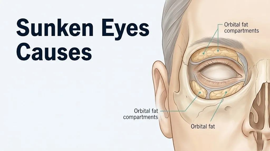
The orbital fat compartments are the primary structures whose loss produces the sunken appearance most people experience. The lower orbital region contains three main fat pads: the medial orbital fat pad, the central orbital fat pad, and the lateral orbital fat pad. These are separated by the lower eyelid retractors and the orbital septum, a fibrous membrane that acts like a curtain holding the fat in place anterior to the bony orbit.
In front of and below these orbital fat pads sits a separate fat layer called the sub-orbicularis oculi fat (SOOF). The SOOF lies directly beneath the orbicularis oculi muscle, the ring-shaped muscle responsible for closing the eyelid. As both the orbital fat pads and the SOOF lose volume with age, metabolic changes, or rapid weight loss, the support scaffolding beneath the lower eyelid deflates. Think of it like removing the stuffing from beneath a thin fabric: the outer surface, in this case the skin, collapses inward and downward, revealing the underlying bony rim as a groove.
The structure that makes this groove most visible is the tear trough ligament, also called the orbito-malar ligament. This fibrous band runs from the periosteum of the inferior orbital rim up through the overlying fat and skin. When the fat compartments on either side of this ligament lose volume, the ligament itself becomes more prominent, creating the characteristic diagonal crease from the inner corner of the eye extending toward the cheek. Clinicians and surgeons refer to this visible crease as the nasojugal groove or tear trough deformity.
According to a 2019 study published in Aesthetic Plastic Surgery, three-dimensional volumetric analysis of the periorbital region confirmed that the tear trough deepens proportionally with loss of both the central orbital fat pad and the SOOF, even when the bony orbital rim dimensions remain unchanged.
The implication for understanding sunken eyes causes: the appearance is not about the eye itself changing position. It is almost always about the soft tissue scaffolding around the eye losing volume, thickness, or structural support, with the exception of true enophthalmos, which is addressed in the medical conditions section.
Quick Tip:
- The medial compartment fat pad is typically the first to show visible atrophy, producing hollowing near the inner corner of the eye
- The SOOF atrophies more slowly than the orbital fat pads but its loss significantly deepens the under-eye groove
- People with naturally shallow bony orbits may notice hollowing less than people with deeper-set eyes, even with identical fat volume loss
Aging and Orbital Fat Loss
Aging is the most common cause of sunken eyes in adults, producing a progressive, bilateral reduction in orbital fat volume, periorbital skin thickness, and ligamentous support. This process begins subtly in the late 20s and becomes clinically visible for most people between ages 40 and 55.
The mechanism operates on two parallel tracks. The first is direct atrophy of the orbital fat compartments. Fibroblasts within the orbital fat pads produce less adipocyte growth factor signaling over time, and some fat cells undergo programmed reduction, shrinking the overall volume of each fat compartment. The second track is collagen loss in the periorbital dermis. The dermis of the periorbital skin is among the thinnest in the body (approximately 0.5 mm compared to 2 mm on the back), making collagen loss here visually apparent before it becomes noticeable elsewhere on the face.
Research published in the Journal of Investigative Dermatology established that fibroblast-synthesized collagen type I and collagen type III both decline steadily with age, with the periorbital region affected earlier than thicker-skinned facial areas. As the dermis thins, darker subcutaneous tissue and the orbital fat pads beneath become visible through the skin, adding the dark circle component to the hollow appearance.
Gravitational changes compound the fat atrophy. The malar fat pad, a separate facial fat compartment sitting over the cheekbone, descends inferiorly with age due to relaxation of the zygomatico-cutaneous ligament. As the malar fat pad drops away from the lower orbital rim, it exposes more of the infraorbital region, making the under-eye hollow appear larger and deeper even when orbital fat volume itself hasn’t changed dramatically.
| Age Range | Primary Periorbital Change | Typical Visual Result |
|---|---|---|
| 20s to 30s | Early collagen decline, minimal fat change | Mild hollowing under fatigue or dehydration |
| 40s | Central orbital fat atrophy begins, SOOF descends | Visible tear trough, early shadowing |
| 50s to 60s | All orbital fat compartments atrophied, malar descent | Prominent nasojugal groove, bony rim visible |
| 70s and beyond | Skin laxity combined with fat atrophy and ligament laxity | Deep hollowing, possible lower lid laxity |
Women may notice accelerated periorbital fat atrophy beginning in the late 40s due to declining estrogen, which is covered in the hormonal section. Men tend to show later onset but are more susceptible if they have high lifetime UV exposure or a significant smoking history.
What Causes Hollow Under Eyes From Dehydration
Dehydration causes hollow under eyes by reducing fluid volume within the periorbital soft tissues, temporarily decreasing skin turgor and the normal aqueous content of the subcutaneous fat compartments, making the under-eye region appear recessed and shadowed. This is one of the most reversible causes of sunken eyes.
The physiological mechanism works through fluid distribution. The periorbital soft tissues maintain their plumpness partly through water content held within the extracellular matrix of the dermis, primarily bound by hyaluronic acid, a glycosaminoglycan that can hold approximately 1,000 times its weight in water. When systemic fluid intake is insufficient or fluid loss exceeds intake through sweating, urination, or gastrointestinal losses, the extracellular matrix releases water to support core physiological functions. The periorbital region, with its thin overlying skin and relatively modest fat volume, shows this fluid loss quickly and visibly.
The Mayo Clinic notes that even mild dehydration of 1 to 2% of body weight can produce visible changes in skin turgor and periorbital appearance. In adults, a simple at-home assessment involves gently pinching the skin just below the eye and observing whether it springs back immediately. Slow or delayed rebound indicates reduced skin turgor consistent with inadequate hydration, though this test is most reliable in younger adults and loses accuracy in older individuals where skin elasticity is already reduced by aging.
Dehydration-related sunken eyes typically resolve within a few hours of adequate rehydration in otherwise healthy adults. Persistent sunken eyes after full rehydration suggests an additional contributing cause, such as fat atrophy or collagen loss, that is worth discussing with a primary care physician.
For older adults (over 65), the sensation of thirst diminishes as a consequence of reduced osmo-receptor sensitivity, which means dehydration develops more insidiously and periorbital hollowing may appear without the person recognizing they are dehydrated. The National Institutes of Health recommends specific attention to fluid intake monitoring in older adults rather than relying on thirst as a reliable signal.
Key signs that dehydration is contributing to sunken eyes:
- Hollowing appears or worsens significantly with morning dry mouth, headache, or darker urine
- Eyes look fuller after consistent fluid intake for 24 to 48 hours
- Associated fatigue or light-headedness with the periorbital change
- Hollowing worsened by alcohol, caffeine excess, or prolonged physical activity without fluid replacement
Sleep Deprivation and Periorbital Hollowing
Sleep deprivation causes the periorbital area to appear sunken and hollow through two distinct physiological mechanisms: impaired lymphatic drainage and increased cortisol-driven fluid redistribution, both of which reduce the support volume visible beneath and around the eyelids.
During sleep, the lymphatic system of the face undergoes its primary drainage cycle. The periorbital region is drained by a network of superficial lymphatics that channel fluid toward the pre-auricular and submandibular lymph nodes. When sleep is shortened or fragmented, this lymphatic clearance is incomplete. The resulting mild periorbital lymphatic congestion can initially create a puffy appearance. However, when sleep deprivation becomes chronic (defined clinically as consistently fewer than 6 hours per night for multiple weeks), the initial puffiness gives way to progressive periorbital hollowing as the orbicularis oculi muscle loses tone and the repetitive incomplete drainage cycle leaves a net deficit in periorbital support tissue hydration.
The second mechanism involves cortisol. A 2020 study published in JAMA Internal Medicine found that sleep restriction consistently elevated morning cortisol levels in otherwise healthy adults. Cortisol, released by the adrenal cortex in response to sleep-related physiological stress, promotes catabolism of facial fat compartments while simultaneously causing fluid redistribution away from peripheral tissues. The result in the periorbital area is reduced fat pad volume and reduced extracellular fluid support, both of which contribute to the hollow appearance.
Sleep deprivation-related periorbital hollowing is most visually prominent in the morning in people who slept fewer than 5 hours, and improves with recovery sleep. When hollowing persists after recovery sleep, it suggests that additional structural causes (collagen loss, fat atrophy) are contributing.
People with obstructive sleep apnea (OSA) experience a compounding effect: the fragmented sleep architecture of untreated OSA impairs lymphatic drainage cycles and produces chronic cortisol dysregulation. If someone notices persistent periorbital hollowing alongside loud snoring, morning headaches, or daytime fatigue, evaluation for OSA by a primary care physician or sleep medicine specialist is appropriate.
What Causes Eyes to Look Sunken From Weight Loss
Rapid or substantial weight loss causes eyes to look sunken by directly reducing the volume of the orbital fat compartments, which are genuine fat-containing structures subject to the same systemic adipose tissue mobilization that reduces fat elsewhere in the body. This cause differs from aging because it can occur at any age and the onset is relatively rapid relative to the degree of caloric deficit.
The buccal fat pad and facial fat compartments, including the orbital fat pads and the SOOF, are not immune to the mobilization of fat stores during significant caloric restriction or illness-related catabolism. When total body fat percentage drops rapidly, the orbital fat pads shrink proportionally. Because the overlying periorbital skin does not contract at the same rate as the deflating fat pads, the resulting excess skin emphasizes the hollow rather than tightening over it.
Clinically, oculoplastic surgeons observe that weight loss greater than 10% of total body weight over a period shorter than 3 to 6 months produces visible periorbital hollowing in the majority of patients, regardless of age. The hollowing from weight loss differs from aging-related hollowing in that it tends to be more even across all fat compartments and less ligament-defined, meaning the specific tear-trough groove of aging is less pronounced, while the overall periorbital deflation is more uniform.
According to the American Society of Ophthalmic Plastic and Reconstructive Surgery (ASOPRS), patients who have undergone bariatric surgery or significant intentional or unintentional weight loss represent a substantial proportion of consultations for periorbital hollowing in adults under 50.
Weight restoration reverses the orbital fat loss only partially and variably. Fat tissue does not necessarily reconstitute in the same distribution after significant weight cycling, and the orbital fat compartments may not fully replenish even with substantial weight regain. Anyone experiencing unexpected, unintentional weight loss alongside sunken eyes should be evaluated by a primary care physician to identify the underlying cause of the weight loss itself.
Key Takeaway: The three most reversible causes of sunken eyes are dehydration, sleep deprivation, and acute weight loss; the first two can improve within days, while orbital fat lost from significant weight reduction may not fully return even with weight regain.
Genetics and Structural Causes of Hollow Eyes
Genetics determine the baseline depth of the bony orbit, the natural volume and distribution of the orbital fat compartments, skin thickness, and ligament anatomy, all of which influence whether someone develops a hollow periorbital appearance earlier, more prominently, or at a lower age than their peers. Some people inherit structurally deep-set eyes as a fixed anatomical trait rather than as an acquired change.
The bony orbit (the skull cavity housing the eyeball) varies in depth and width across individuals and across populations. People with deeper orbital cavities have less space for orbital fat to project forward and fill the under-eye area, so even normal fat volumes produce a hollowed appearance. This is a structural anatomical variant, not a sign of fat loss or systemic illness. True structural deep-setting of the eyes is symmetric, present from childhood or early adulthood, and does not change with hydration, sleep, or weight.
Skin thickness is also genetically determined to a significant degree. People who inherit thinner periorbital skin will show hollowing, dark circles, and the nasojugal groove earlier than those with thicker skin, because less subcutaneous tissue separates the visible skin surface from the deeper fat compartments and bony rim.
Ethnicity influences periorbital anatomy in ways relevant to how and when hollowing becomes visible:
- People with East Asian orbital anatomy often have a different orbital fat distribution, with more prominent medial fat pads and different lid crease anatomy, affecting where hollowing first becomes apparent
- People with darker skin tones (Fitzpatrick skin types IV through VI) have greater melanin density in periorbital skin, which can produce darker shadowing at the nasojugal groove even with the same fat volume as those with lighter tones
- People of European descent with very fair skin often show the vascular component of periorbital hollowing (visible superficial veins) more prominently
Understanding the genetic baseline matters clinically because it establishes the individual’s personal “normal.” When someone’s hollowing has been present and stable since their 20s, it is almost certainly structural. When it changes over months or years, a contributing acquired cause is driving the change.
What Causes Hollow Eyes From Allergies and Skin Changes
Allergies cause hollow eyes through a specific venous and lymphatic mechanism: chronic nasal congestion and periorbital venous stasis from allergic rhinitis impairs blood drainage from the periorbital region, producing the characteristic allergic shiner, a dark, slightly hollow-appearing shadow under the eyes. This is the cause most commonly confused with fatigue-related hollowing.
The mechanism begins in the nasal mucosa. When someone with allergic rhinitis (hay fever) experiences an allergen exposure, IgE-mediated mast cell degranulation releases histamine and other inflammatory mediators throughout the nasal mucosa. The resulting mucosal swelling compresses the nasal veins and the interconnected angular vein system. Because the angular veins drain the same venous territory as the periorbital ophthalmic veins, this compression causes venous backflow and stasis in the periorbital region.
This venous stasis pooling does two things. First, it creates the dark discoloration of allergic shiners by making the periorbital blood vessel content more visible through the thin overlying skin. Second, over months of chronic allergic disease, the repeated cycles of venous congestion and tissue stress produce mild atrophy of the thin periorbital skin, which progressively accentuates the hollow appearance even outside of active allergy seasons.
A key physical finding in people with allergy-related periorbital hollowing is the Dennie-Morgan line: a horizontal or diagonal infraorbital fold that runs parallel to the lower eyelid margin. This fold is produced by the chronic orbicularis oculi muscle strain from eye rubbing (triggered by allergic conjunctivitis) combined with the skin changes from chronic venous stasis. Dennie-Morgan lines are a reliable clinical indicator that chronic allergic disease is contributing to the periorbital hollow appearance.
People with atopic dermatitis (eczema) have an additional mechanism: the chronic itch-scratch cycle of atopic skin disease produces repetitive mechanical trauma and dermal inflammation around the eyes, which accelerates collagen degradation and skin thinning in the periorbital region. A board-certified allergist or dermatologist can evaluate and manage both conditions, which typically reduces the allergic component of hollowing over time with appropriate treatment.
What Causes Eyes to Sink In From Smoking and Sun Exposure
Smoking and chronic sun exposure cause eyes to sink in by accelerating the degradation of collagen type I and collagen type III in the periorbital dermis through two separate but physiologically related mechanisms, both of which deplete the structural protein framework that gives skin its thickness and the periorbital region its support.
Cigarette smoking triggers periorbital skin damage through nicotine-induced vasoconstriction and oxidative stress. Nicotine binds to nicotinic acetylcholine receptors in the walls of periorbital microvasculature, causing prolonged vasoconstriction that reduces blood flow to the periorbital dermis and its resident fibroblasts. Fibroblasts are the cells responsible for collagen synthesis: they require adequate oxygenation and nutrient delivery to maintain their synthetic output. Reduced microvascular flow means chronically hypoxic fibroblasts produce less collagen. Simultaneously, tobacco combustion products deliver reactive oxygen species (ROS) that directly upregulate matrix metalloproteinase-1 (MMP-1), the primary enzyme responsible for collagen degradation in skin tissue.
A 2021 study published in JAMA Dermatology found that current smokers showed measurably thinner periorbital dermis on high-resolution ultrasound compared to age-matched non-smokers, with the difference equivalent to approximately 5 to 7 years of additional biological skin aging.
Ultraviolet radiation produces the same MMP-1 upregulation through a different starting point. UVA radiation (wavelengths 315 to 400 nm, present year-round and capable of penetrating window glass) directly activates the activator protein-1 (AP-1) transcription factor in epidermal keratinocytes and dermal fibroblasts. AP-1 activation upregulates MMP-1 and MMP-3 gene expression, driving collagen breakdown. The periorbital skin receives significant cumulative UV exposure because people rarely apply sunscreen near the eyes and eyelids, and this region has less protective melanin density than adjacent cheek skin.
The practical clinical implication: both smoking and sun exposure accelerate the timeline of periorbital hollowing by 5 to 10 years relative to non-exposed peers, and neither effect is fully reversible once dermal collagen has been degraded. Prevention and cessation are the most effective interventions.
Key Takeaway: Smoking and chronic sun exposure both accelerate periorbital collagen loss through MMP-1 upregulation, producing premature hollowing that cannot be fully reversed once the dermal collagen has been degraded.
Medical Conditions That Cause Sunken Eyes
Several medical conditions cause sunken eyes through mechanisms distinct from aging, lifestyle, or genetics, and some of these require urgent or emergency evaluation rather than cosmetic management. Recognizing the difference between lifestyle-related periorbital hollowing and medically significant sunken eyes depends on identifying associated symptoms.
Horner syndrome is one of the most clinically important medical causes of an apparently sunken eye. It occurs when the cervical sympathetic chain, the autonomic nerve pathway running from the hypothalamus down through the brainstem and cervical spinal cord to the orbit, is disrupted. The sympathetic innervation of the eye controls the superior tarsal muscle (which contributes to upper eyelid elevation), the inferior tarsal muscle (which contributes to lower eyelid position), and pupil dilation. When this pathway is severed, the upper eyelid droops (ptosis), the lower eyelid rises slightly (reverse ptosis), and the pupil constricts (miosis), creating the appearance that the eye has sunk inward. This is called apparent enophthalmos because the eye itself has not moved backward; the changed lid position makes it look smaller and more recessed.
Horner syndrome has serious potential causes including Pancoast tumor (lung apex cancer), carotid artery dissection, aortic aneurysm, cervical disc herniation, or brainstem stroke. Unilateral ptosis with miosis (small pupil on the same side) and anhidrosis (absent sweating on the same side of the face) is the diagnostic triad. This presentation requires same-day or emergency evaluation.
Orbital blowout fracture from facial trauma can cause true enophthalmos. When the thin floor or medial wall of the bony orbit fractures, orbital fat and sometimes the extraocular muscles herniate into the adjacent maxillary or ethmoid sinuses. The result is genuine posterior displacement of the globe and a visibly sunken, narrowed eye, often with restricted upward gaze (entrapment of the inferior rectus muscle) and numbness of the cheek.
Additional medical conditions associated with periorbital hollowing include:
| Medical Condition | Mechanism | Key Associated Symptoms |
|---|---|---|
| Horner syndrome | Sympathetic chain disruption; apparent enophthalmos | Unilateral ptosis, miosis, anhidrosis |
| Orbital blowout fracture | Globe posterior displacement due to orbital wall fracture | Facial trauma history, restricted gaze, cheek numbness |
| HIV-associated lipodystrophy | Antiretroviral-related facial fat redistribution | Buccal and temporal wasting, buffalo hump |
| Thyroid eye disease | Orbital inflammation and later fibrosis causing enophthalmos | Proptosis in active phase; sunken in burned-out fibrotic phase |
| Severe systemic illness | Systemic catabolism and dehydration producing fat and fluid loss | Fever, weight loss, systemic illness markers |
| Metastatic orbital disease | Mass effect causing globe displacement | Progressive unilateral change, double vision, pain |
What Causes Sunken In Eyes in Children
Sunken eyes in children are a clinically important sign that differs fundamentally from sunken eyes in adults: in pediatric patients, sunken eyes are most commonly a physical indicator of dehydration severity rather than a cosmetic or aging concern, and the presence of sunken eyes in a sick child requires urgent clinical assessment.
The World Health Organization and the American Academy of Pediatrics both include sunken eyes (sunken orbits) as one of the three primary clinical signs of moderate to severe dehydration in children under 5 years of age. The other two signs in this clinical triad are reduced skin turgor and a depressed anterior fontanelle (in infants under 18 months). In a child with diarrhea, vomiting, fever, or inadequate fluid intake, sunken-appearing eyes indicate a fluid deficit that can compromise cardiovascular function, neurological status, and kidney perfusion.
The physiological reason children show periorbital sunken signs more clearly than adults involves the relative thinness of periorbital soft tissue in the pediatric orbit and the higher ratio of total body water to body weight in young children. An infant’s total body water comprises approximately 75 to 80% of body weight (compared to 55 to 60% in adults), meaning that acute fluid losses represent a proportionally larger fraction of total body volume and produce more rapid periorbital changes.
Dehydration severity assessment using periorbital signs:
- Observe the child at rest in good lighting, looking straight ahead
- Assess whether the orbital area appears symmetric and normally filled or visibly recessed compared to the child’s normal baseline
- Note whether this is accompanied by dry mouth, no tears when crying, decreased urination, or lethargy
- Assess skin turgor by gently pinching skin on the back of the hand or abdominal wall and releasing
- In infants under 18 months, gently palpate the anterior fontanelle for depression while the child is calm
If sunken eyes occur alongside fever above 102.2°F (39°C), lethargy, no urination for 8 or more hours, or dry lips and mouth, the child requires evaluation by a pediatrician or, if after hours or the child appears severely ill, immediate evaluation in a pediatric emergency department.
Structural deep-set eyes do occur in children as a genetic variant and are bilateral, present since birth, and not associated with illness signs.
Hormonal and Sex-Specific Causes of Periorbital Hollowing
Hormonal changes in women, particularly the decline in circulating estrogen during perimenopause and menopause, directly accelerate periorbital fat atrophy and dermal collagen loss, making this one of the most physiologically specific and underrecognized causes of sunken eyes in women aged 45 to 60.
Estrogen plays a direct and well-documented role in maintaining facial fat compartment volume and dermal collagen synthesis. Estrogen receptors alpha and beta are expressed by dermal fibroblasts throughout facial skin, including the periorbital region. When estrogen binds to these receptors, it upregulates procollagen gene expression and suppresses MMP activity, essentially telling the fibroblasts to make more collagen and degrade less of it. As ovarian estrogen production declines during perimenopause (typically ages 45 to 51 in North American women), this protective signaling diminishes.
Research published in the Journal of Investigative Dermatology found that postmenopausal women who had not used hormone replacement therapy showed collagen content declines of approximately 30% in the first 5 years after the final menstrual period. The periorbital region, already the thinnest and most collagen-sensitive facial skin, reflects this loss earlier and more visibly than other facial areas.
The fat compartments around the orbit are also estrogen-sensitive. The SOOF and the medial and central orbital fat pads contain adipocytes that respond to estrogen signaling through effects on adiponectin and leptin pathways. Estrogen withdrawal reduces fat cell maintenance signaling in these compartments, accelerating the atrophy that would otherwise occur more slowly through aging alone.
For men, testosterone decline associated with andropause (typically ages 50 to 70) produces a slower, less dramatic effect on periorbital structures than estrogen decline in women, because testosterone’s influence on facial fat maintenance and dermal collagen is less direct. However, men with hypogonadism or those using long-term androgen deprivation therapy for prostate cancer may experience accelerated facial fat redistribution and periorbital hollowing.
According to the Mayo Clinic, perimenopausal and postmenopausal women who notice accelerating periorbital hollowing alongside other estrogen-deficiency symptoms (hot flashes, vaginal dryness, sleep disruption) should discuss hormonal and non-hormonal management options with a board-certified obstetrician-gynecologist or endocrinologist, as systemic estrogen replacement has been shown to slow dermal collagen loss when initiated within the first decade after menopause.
Key Takeaway: Perimenopausal and postmenopausal women experience accelerated periorbital hollowing due to estrogen-driven collagen and orbital fat loss, with the most rapid change occurring in the first five years after the final menstrual period.
What Causes Sudden Sunken Eyes
Sudden sunken eyes, meaning a noticeable change in periorbital appearance developing over hours to days rather than months to years, are a clinically distinct situation from gradual hollowing and require a different level of attention because some rapid-onset causes represent medical emergencies.
The most medically urgent cause of sudden sunken appearance in one eye is Horner syndrome. As described in the medical conditions section, sudden onset of a unilateral ptotic, apparently sunken eye with a small pupil on the same side requires emergency evaluation to rule out carotid artery dissection, brainstem infarction, or a Pancoast tumor expanding at the lung apex. These conditions do not wait.
Facial trauma producing a sudden sunken-appearing eye requires evaluation for orbital blowout fracture in an emergency department. The fracture itself may not produce dramatic external bruising initially, but the subtle enophthalmos and restricted eye movement (particularly impaired upward gaze) indicate a structural orbital injury requiring CT imaging and oculoplastic or facial surgery consultation.
Non-emergency but notable causes of more rapid-than-usual hollowing include:
- Acute severe illness (sepsis, high-fever dehydration) causing rapid fluid volume loss
- Initiation or dose increase of certain medications, particularly corticosteroids, which can redistribute facial fat within weeks of high-dose systemic use
- Acute onset of severe food restriction, eating disorder relapse, or medically induced caloric restriction
- A sudden bout of severe gastroenteritis with vomiting and diarrhea causing acute significant dehydration
The key clinical differentiator between a worrying and a manageable sudden change is laterality and associated symptoms. Bilateral gradual changes that worsen over weeks in the context of illness, weight loss, or dehydration are typically systemic and non-emergency. Unilateral sudden change with any associated neurological symptom, double vision, pain, or fever points to a cause requiring same-day or emergency evaluation.
Anyone who notices a sudden change in the appearance of one eye, particularly if accompanied by a drooping eyelid or a difference in pupil size between the two eyes, should contact a primary care physician the same day or go to an emergency department.
Vitamin and Nutritional Deficiencies Linked to Sunken Eyes
Specific nutritional deficiencies contribute to periorbital hollowing by impairing collagen synthesis, promoting anemia-related skin pallor and shadowing, or reducing the fat and protein substrate needed to maintain facial soft tissue volume. These are not the primary driver of sunken eyes in most well-nourished adults, but in people with dietary restrictions, malabsorption conditions, or chronic illness, they are clinically relevant contributing factors.
Vitamin C (ascorbic acid) is essential to collagen biosynthesis at a specific enzymatic step: prolyl hydroxylase and lysyl hydroxylase, the enzymes that hydroxylate proline and lysine residues in collagen molecules, require vitamin C as a cofactor. Without adequate hydroxylation, collagen triple-helices cannot form stable cross-linked fibrils. The result is defective, easily degraded collagen in all soft tissues, including the periorbital dermis. Clinically significant vitamin C deficiency (scurvy) includes perifollicular hemorrhages, easy bruising, and notably thin, fragile periorbital skin. Subclinical vitamin C insufficiency, more common than frank scurvy, may contribute to accelerated collagen loss in the periorbital region without producing the full clinical picture of scurvy.
Iron deficiency anemia contributes to the dark-circle and hollow appearance around the eyes through reduced hemoglobin content in periorbital blood vessels. With less oxyhemoglobin in the blood, the vessels beneath the thin periorbital skin appear darker through the skin surface, accentuating the hollow appearance even when fat volume is normal. The National Institutes of Health (Office of Dietary Supplements) notes that iron deficiency is the most common nutritional deficiency globally, affecting an estimated 1.2 billion people, with women of reproductive age at highest risk.
Vitamin K contributes to periorbital vascular integrity by supporting the function of coagulation factors and proteins that regulate microvessel permeability. Insufficient vitamin K is associated with increased capillary fragility in the periorbital region, which can worsen dark circles and the visual appearance of hollowing.
B12 deficiency, common in vegetarians, vegans, and older adults with reduced gastric acid production, impairs red blood cell production and can contribute to the pale, hollowed periorbital appearance through the anemia mechanism described above.
Nutritional assessment for periorbital hollowing is relevant when the patient has a history of:
- Malabsorption (Crohn’s disease, celiac disease, bariatric surgery)
- Restrictive dietary patterns without supplementation
- Chronic alcoholism
- Unexplained weight loss alongside the periorbital change
A primary care physician can order a complete blood count (CBC), serum ferritin, serum vitamin B12, and serum vitamin C as part of a basic nutritional screening, which provides actionable clinical data if deficiency is suspected.
When to See a Doctor for Sunken Eyes
Most causes of sunken eyes are gradual, bilateral, and related to aging, lifestyle, or genetics, and these can be discussed at a routine appointment with a primary care physician, board-certified dermatologist, or board-certified oculoplastic surgeon. However, specific presentations require prompt or urgent professional evaluation rather than monitoring at home.
See a primary care physician within 1 to 2 weeks if you notice:
- Progressive bilateral hollowing that has worsened visibly over 1 to 3 months without a clear lifestyle explanation
- Sunken eyes alongside unexplained weight loss, persistent fatigue, or new systemic symptoms
- Sunken eyes alongside increased thirst, increased urination, and unexplained fatigue (screening for diabetes mellitus warranted)
- Hollowing that appears to be associated with a new medication, including antiretroviral therapy, systemic corticosteroids, or chemotherapy agents
See an ophthalmologist or go to an emergency department the same day if you notice:
- A new sunken appearance in only one eye, especially with a drooping upper eyelid
- Any difference in pupil size between the two eyes (one larger than the other in the same lighting)
- Sunken eye appearance following any facial trauma, even apparently minor
The specific diagnostic process for medically evaluated sunken eyes typically involves:
- Complete medical history including medication list, weight changes, systemic illness, and trauma history
- Comprehensive ophthalmic examination including measurement of pupil size, eyelid position, and visual acuity
- Hertel exophthalmometry if true enophthalmos is suspected, measuring the anterior projection of the globe within the bony orbit
- Orbital CT scan for trauma-related evaluations or if an orbital mass or fracture is suspected
- Orbital MRI if a soft tissue orbital lesion, Horner syndrome, or orbital apex syndrome is suspected
- Laboratory studies (CBC, serum ferritin, comprehensive metabolic panel, TSH, serum albumin) if systemic nutritional or endocrine causes are suspected
For people whose sunken eyes are clearly cosmetic and age-related, a consultation with a board-certified oculoplastic surgeon or a board-certified dermatologist with expertise in facial volumetric assessment is the appropriate referral pathway for anyone interested in interventional options. The American Society of Ophthalmic Plastic and Reconstructive Surgery maintains a directory of board-certified oculoplastic surgeons who specialize in periorbital evaluation.
Key Takeaway: A new sunken appearance in one eye, particularly with an upper eyelid droop or unequal pupil sizes, is a same-day medical concern and not a cosmetic observation to monitor at home.
Emergency Symptoms: When to Call 911 or Go to the ER
Certain presentations associated with sunken eyes require immediate emergency evaluation. Do not wait to see if these resolve on their own.
Call 911 or go to the nearest emergency room immediately if you or someone you know experiences:
- A newly sunken or recessed eye on one side accompanied by a drooping upper eyelid, a smaller pupil on the same side, and absent sweating on that side of the face (the Horner syndrome triad, which can indicate a Pancoast tumor, carotid artery dissection, or brainstem stroke)
- A sunken or changed-appearing eye following any trauma to the face, head, or eye socket, especially if accompanied by double vision, restricted eye movement, numbness of the cheek, or pain with eye movement (orbital blowout fracture)
- A suddenly sunken-appearing eye with double vision, pain behind the eye, or proptosis of the other eye (possible orbital mass or vascular malformation)
- A child under 5 years old with sunken eyes and any of the following: lethargy, no urination for 8 or more hours, high fever above 102.2°F (39°C), no tears when crying, dry mouth and lips, or rapid breathing (severe dehydration requiring emergency pediatric care)
- Sunken eyes in any person alongside signs of septic shock: high fever, rapid heart rate, confusion, cold or mottled skin, and rapid breathing
These presentations can indicate Horner syndrome from a life-threatening vascular or oncologic cause, orbital blowout fracture requiring surgical repair, or severe pediatric dehydration causing hypovolemia, and all require emergency medical assessment, not an urgent care appointment or a phone call to schedule a next available slot.
Frequently Asked Questions About Sunken Eyes Causes
What is the most common cause of sunken eyes in adults?
Aging-related orbital fat atrophy is the most common cause of sunken eyes in adults, affecting the medial, central, and lateral orbital fat pads along with the sub-orbicularis oculi fat (SOOF) progressively from the late 30s onward.
Dehydration is the most common cause of a sudden or temporary increase in periorbital hollowing at any age.
Both causes operate through distinct mechanisms and can occur simultaneously in the same individual.
Can dehydration really cause sunken eyes, and how fast?
Yes, dehydration causes sunken eyes by reducing the fluid content of periorbital soft tissue, including water held within the extracellular matrix by hyaluronic acid, which reduces visible support volume beneath the eyelid.
The Mayo Clinic notes that even mild dehydration of 1 to 2% of body weight can produce visible periorbital changes in susceptible individuals.
In healthy adults, dehydration-related hollowing typically reverses within a few hours of adequate rehydration; persistence beyond 24 hours of normal fluid intake suggests additional contributing factors.
Are sunken eyes a sign of a serious medical condition?
Most sunken eyes in adults are caused by aging, dehydration, sleep deprivation, or genetics and are not signs of serious medical illness.
However, a newly sunken appearance in one eye, especially with an associated drooping eyelid, a smaller pupil on the same side, or a history of facial trauma, can indicate Horner syndrome, an orbital blowout fracture, or an orbital mass, all of which require prompt medical evaluation.
Sunken eyes in a child with fever and signs of dehydration are a clinical warning sign requiring urgent evaluation by a pediatrician or emergency physician.
What causes sunken eyes in only one eye?
Sunken eyes appearing in only one eye (unilateral periorbital hollowing) should always be evaluated by a physician because most bilateral causes of sunken eyes, including aging, dehydration, and sleep deprivation, affect both eyes symmetrically.
Causes of unilateral hollowing include Horner syndrome (sympathetic chain disruption), orbital blowout fracture, an orbital mass or tumor, or severe localized orbital fat atrophy after radiation to one side of the face.
A primary care physician or ophthalmologist should evaluate any unilateral change, and a presentation with ptosis, miosis, and facial anhidrosis on the same side requires same-day or emergency evaluation.
Do sunken eyes go away on their own?
Sunken eyes caused by dehydration typically resolve within hours after adequate fluid intake is restored in otherwise healthy adults.
Sunken eyes from sleep deprivation improve with consistent recovery sleep but may take several days to fully normalize in cases of prolonged sleep deficit.
Sunken eyes from aging, significant orbital fat loss from weight loss, or structural genetic anatomy do not resolve on their own and represent permanent or semi-permanent changes that require intervention from a board-certified oculoplastic surgeon or dermatologist if treatment is desired.
What vitamin deficiency causes sunken eyes?
Vitamin C deficiency is the nutritional deficiency most directly linked to sunken eyes because ascorbic acid is required as a cofactor for prolyl hydroxylase, the enzyme essential to collagen biosynthesis in the periorbital dermis.
Iron deficiency anemia contributes to the shadowed, hollow appearance by reducing oxyhemoglobin in periorbital blood vessels, making the under-eye region appear darker and more recessed.
A primary care physician can assess for these deficiencies with a complete blood count, serum ferritin, and serum vitamin C levels, particularly in people with restrictive diets, malabsorption conditions, or unexplained periorbital changes alongside systemic symptoms.
Closing
The appearance of sunken or hollow eyes is almost never caused by a single factor in isolation. Aging slowly depletes the orbital fat compartments and dermal collagen. Dehydration, poor sleep, smoking, and sun exposure accelerate that process through distinct physiological pathways. And in a smaller but clinically important set of cases, sunken eyes signal a medical condition requiring evaluation that goes well beyond the periorbital region.
If your hollowing is gradual, bilateral, and has developed over years alongside other signs of normal aging, a conversation with your primary care physician or a board-certified oculoplastic surgeon will give you an accurate picture of what’s driving it and what can be done. If you’ve noticed a change in one eye, a drooping eyelid on one side, or a difference in pupil size, treat that as a medical question that needs an answer today, not a cosmetic observation to monitor over the coming months.
You now understand the anatomy, the mechanisms, and the red flags. Use that knowledge to ask the right questions at your next appointment.