What Causes Apron Belly? Top 15 Medical Reasons Explained
An apron belly forms when subcutaneous fat accumulates in the lower abdominal wall in sufficient volume that the tissue mass descends below the pubic bone under gravitational load. The clinical term is panniculus adiposus, sometimes called a pannus stomach, and its causes range from straightforward weight gain to complex hormonal disorders that require medical evaluation.
According to the National Institute of Diabetes and Digestive and Kidney Diseases, more than 70% of adults in the United States carry some degree of excess abdominal adiposity, but not all of them develop the overhanging fold that defines a true apron belly. The distinction matters because specific anatomical and hormonal mechanisms determine whether excess fat distributes as a hanging panniculus versus a more diffuse abdominal protrusion. Understanding those mechanisms changes the clinical approach entirely.
This article covers 15 specific causes of apron belly, the physiology behind each one, how biological sex, age, and hormonal status shape the pattern, which causes signal an underlying medical condition requiring evaluation, and when skin complications under the fold become a clinical concern that needs same-day attention.
What Causes Apron Belly
Apron belly is caused by the accumulation of excess adipose tissue in the subcutaneous layer of the anterior abdominal wall, combined in many cases with weakening or separation of the underlying musculofascial structures that normally support and contain that tissue.
The most common cause is sustained positive energy balance: consuming more calories than the body uses over time, which drives adipogenesis in both subcutaneous and visceral fat depots. When subcutaneous adipose tissue in the lower abdomen expands beyond the structural support capacity of Scarpa’s fascia (the deeper membranous layer of the abdominal wall fascia) and the anterior abdominal musculature, the tissue mass descends and hangs. That descent is what creates the characteristic fold or drape of tissue over the pubic area.
But weight gain alone does not fully explain every case. Hormonal shifts direct fat toward specific body regions at the cellular level. Structural changes to the abdominal wall from pregnancy or surgery alter the mechanical support system. Conditions like lipedema or Cushing’s syndrome produce central tissue accumulation through mechanisms completely independent of caloric excess.
| Cause Category | Primary Mechanism | Who It Affects Most |
|---|---|---|
| Obesity and weight gain | Adipogenesis in subcutaneous and visceral depots | All adults with sustained caloric surplus |
| Pregnancy and postpartum | Abdominal wall stretching, hormonal adipogenesis | People who have carried pregnancies |
| Diastasis recti | Linea alba separation reducing abdominal support | Postpartum women, people with obesity |
| Menopause | Estrogen withdrawal causing fat redistribution | Perimenopausal and postmenopausal women |
| Cortisol excess | HPA axis-driven central adipogenesis | People under chronic stress, Cushing’s syndrome |
| Insulin resistance | Visceral fat expansion via impaired lipolysis | People with metabolic syndrome, PCOS, type 2 diabetes |
| Rapid weight loss | Residual loose skin without adipose reduction | Post-bariatric, post-significant diet loss |
| Lipedema | Abnormal adipose tissue deposition, non-caloric | Primarily women, often misdiagnosed as obesity |
What Is an Apron Belly and What Body Structures Are Involved
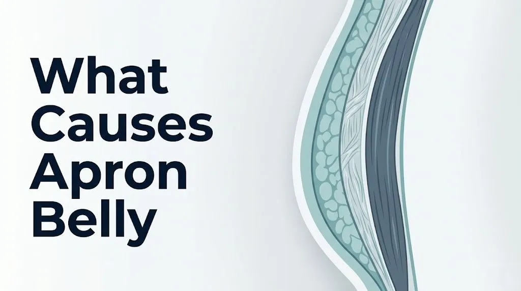
An apron belly involves three anatomical layers of the anterior abdominal wall: the skin surface, the subcutaneous adipose tissue beneath it (divided into a superficial fatty layer called Camper’s fascia and a deeper membranous layer called Scarpa’s fascia), and the underlying musculofascial envelope formed by the rectus abdominis, external oblique, internal oblique, and transversus abdominis muscles.
In a person without significant central adiposity, Scarpa’s fascia holds the lower abdominal subcutaneous tissue in place. As adipose volume increases, the mechanical load on Scarpa’s fascia and the linea alba increases proportionally. When that load exceeds the tensile capacity of the fascial support system, the tissue descends toward the pubic symphysis and beyond.
The size of the resulting panniculus varies considerably. Clinicians use a panniculus grading scale (grades 1 through 5) to describe severity, from a panniculus that barely covers the pubic hairline at grade 1 to one that descends past the knees at grade 5. Most people who describe having an apron belly are experiencing grade 1 or grade 2, which covers the pubic area but remains above the upper thigh.
People who have had cesarean section deliveries may notice an additional structural element: the C-section scar creates a line of internal adhesion that can cause the tissue above it to appear to protrude more prominently as a separate fold, sometimes called a C-section shelf. This is not purely an adipose phenomenon but involves fascial adhesion at the surgical site.
Subcutaneous Fat Versus Visceral Fat: Which Type Forms an Apron Belly
Subcutaneous adipose tissue is the primary structural component of an apron belly. It sits between the skin and the abdominal muscle fascia and is the fat you can physically grasp with your hand.
Visceral adipose tissue is anatomically distinct: it accumulates inside the peritoneal cavity, surrounding the intestines, liver, and other abdominal organs. The primary visceral fat depot is the greater omentum, a fatty apron of tissue that hangs from the transverse colon. Visceral fat produces the taut, hardened abdominal protrusion that cannot be pinched easily, sometimes called a “hard belly.” It does not hang the way subcutaneous fat does.
A person’s apron belly may involve both tissue types, but the hanging, soft, pliable fold itself is subcutaneous in nature. The distinction matters clinically because these two fat types carry different metabolic risk profiles. Research published in the New England Journal of Medicine has established that visceral adipose tissue is metabolically more active and more strongly associated with cardiovascular disease, type 2 diabetes, and inflammatory marker elevation than subcutaneous fat is. However, a large subcutaneous panniculus does signal that significant visceral adiposity is also likely present in people with generalized obesity.
Visceral fat cannot be measured by feel alone. A computed tomography (CT) scan of the abdomen or a dual-energy X-ray absorptiometry (DEXA) scan with body composition analysis is required to accurately quantify visceral adipose tissue volume. Waist circumference measurement, while imprecise, is the standard clinical screening proxy: the American Heart Association identifies waist circumference greater than 35 inches in women and greater than 40 inches in men as a threshold for elevated cardiovascular metabolic risk.
Key Takeaway: Apron belly is formed by subcutaneous fat, not visceral fat, but the two fat compartments often coexist and visceral fat carries the higher metabolic disease risk of the two.
Weight Gain and Obesity as the Primary Cause of Apron Belly
Sustained excess caloric intake is the most common cause of apron belly, operating through the physiological process of adipogenesis: the differentiation of preadipocytes into mature, lipid-storing adipocytes in subcutaneous and visceral fat depots.
When caloric surplus is chronic, adipocytes in the subcutaneous abdominal layer expand first by hypertrophy (increasing in size) and then, once hypertrophy capacity is reached, by hyperplasia (increasing in number). The lower abdominal subcutaneous depot is hormonally primed to accumulate fat more readily than the extremities in most adults, particularly those with android (central) fat distribution patterns.
According to the Centers for Disease Control and Prevention, adult obesity prevalence in the United States reached 41.9% in the most recent National Health and Nutrition Examination Survey period. Central obesity, the subtype most directly associated with panniculus formation, is even more prevalent in adults over 40 years of age regardless of total body weight classification.
Body weight threshold for apron belly development varies between individuals based on abdominal wall muscle tone, height, fat distribution genetics, and prior pregnancies. Some people develop a grade 1 panniculus at a body mass index of 27, while others with stronger fascial support and different fat distribution genetics do not develop one until a body mass index of 35 or higher. Body mass index alone is a poor predictor: it measures total weight relative to height but does not capture fat distribution.
People taking certain medications, including corticosteroids, atypical antipsychotic medications, and some antidepressants in the tricyclic class, experience drug-induced weight gain with a central distribution pattern that can produce or worsen an apron belly even without major changes in diet.
Pregnancy and Postpartum Changes That Cause Apron Belly
Pregnancy causes apron belly through two overlapping mechanisms: the mechanical stretching and weakening of the anterior abdominal wall over nine months of uterine expansion, and the hormonal changes of pregnancy that promote subcutaneous fat accumulation in the lower abdomen as an energy reserve for lactation.
During pregnancy, the linea alba, the midline fibrous connective tissue raphe that runs between the two columns of the rectus abdominis, stretches laterally to accommodate the growing uterus. The skin and subcutaneous tissue of the anterior abdominal wall also stretch significantly. After delivery, while the uterus returns to its pre-pregnancy size over six to eight weeks, the stretched skin, fascia, and subcutaneous tissue do not necessarily regain their pre-pregnancy architecture.
The American College of Obstetricians and Gynecologists acknowledges that postpartum abdominal contour changes are influenced by the degree of tissue stretching, the number of prior pregnancies, the age of the person at delivery, the weight gained during pregnancy, and genetic factors governing skin elasticity and fascial tensile recovery.
Multiple pregnancies compound these changes. Each successive pregnancy stretches already-altered tissue, and the cumulative effect on abdominal wall architecture is greater than a single pregnancy alone. Women who experienced significant gestational weight gain that was not subsequently lost retain both the adipose volume and the structural changes simultaneously.
Postpartum women should note that abdominal appearance at six weeks postpartum is not representative of the one-year postpartum baseline. The National Institutes of Health notes that full musculofascial recovery of the postpartum abdominal wall, where it occurs, typically requires six to twelve months and is substantially supported by progressive abdominal rehabilitation exercise under the guidance of a pelvic floor physical therapist.
Diastasis Recti and Abdominal Wall Separation Contributing to Apron Belly
Diastasis recti is the separation of the two columns of the rectus abdominis muscle beyond the normal inter-recti distance of approximately 1 to 2 centimeters at the midline, caused by excessive stretching or thinning of the linea alba.
It is not an injury in the traditional sense but a structural change to the midline fascia. When the linea alba loses its tensile integrity, the medial support structure of the anterior abdominal wall is compromised. The contents of the abdominal cavity, including the viscera and any subcutaneous adipose tissue, push forward and downward through the widened gap rather than being contained by a unified fascial wall.
A 2021 study published in the Journal of Clinical Medicine estimated that diastasis recti is present in approximately 39% of women at six months postpartum, with higher prevalence in women who had multiple pregnancies or who carried large babies. The condition is also present in some individuals with central obesity who have never been pregnant, as sustained intra-abdominal pressure from adipose volume can produce midline fascial thinning through the same mechanical loading process.
Diastasis recti does not always produce pain. Many people with the condition notice a domed or conical midline protrusion when performing a sit-up or when rising from a lying position, sometimes described as seeing a ridge or tent-pole shape at the abdomen midline. This structural change, combined with any subcutaneous adipose excess, produces or worsens the apron belly appearance.
Diagnosis is confirmed by a physical examination performed by a primary care physician, obstetrician-gynecologist, or pelvic floor physical therapist, typically by measuring the inter-recti distance manually or by ultrasound imaging of the linea alba.
Key Takeaway: Diastasis recti is present in roughly 39% of women at six months postpartum and directly worsens apron belly by removing the midline fascial support that normally holds abdominal contents in place.
Hormonal Causes of Apron Belly: Estrogen, Cortisol, and Insulin
Three hormones have the strongest evidence base for driving central fat accumulation and apron belly formation independently of total caloric intake: cortisol, estrogen, and insulin.
Cortisol, the primary glucocorticoid produced by the adrenal cortex under stimulation from the hypothalamic-pituitary-adrenal (HPA) axis, promotes adipogenesis directly. Abdominal subcutaneous and visceral adipocytes express glucocorticoid receptors at higher density than adipocytes in peripheral depots like the thighs and arms. Chronic cortisol elevation, whether from chronic psychological stress, disrupted sleep, or pathological hypercortisolism (Cushing’s syndrome), drives preferential fat deposition in the central abdominal region even in the absence of overall weight gain.
Insulin promotes fat storage by activating lipogenic enzymes (including fatty acid synthase and acetyl-CoA carboxylase) and suppressing lipolytic enzyme activity, particularly hormone-sensitive lipase. In people with insulin resistance, fasting insulin levels remain chronically elevated to overcome impaired receptor signaling. That persistently elevated insulin environment chronically suppresses fat release from adipocytes and chronically promotes fat storage, particularly in visceral and lower abdominal subcutaneous depots.
Estrogen’s role operates differently. When estrogen levels are adequate, they appear to direct adipose storage toward the gluteofemoral region (hips, buttocks, thighs) rather than the abdomen, through sex hormone receptors on adipocytes that differ in distribution between peripheral and central fat depots. The withdrawal of estrogen at menopause removes this directional effect, which is one reason central fat redistribution is a near-universal feature of the menopausal transition.
Men are not exempt from hormonal contributions. A 2022 analysis published in JAMA Internal Medicine found that declining testosterone in men over 45, a condition called hypogonadism, is associated with increased visceral and subcutaneous central adiposity independent of total body weight.
Menopause and Apron Belly: Why Fat Redistributes to the Abdomen
Menopause causes apron belly by removing the estrogen-mediated preferential routing of fat storage toward peripheral adipose depots, allowing central abdominal adiposity to accumulate even when total body weight remains unchanged.
This is not a weight gain phenomenon in isolation. Studies tracking body composition through the menopausal transition have documented central fat mass increases of 5 to 8 percent over the perimenopausal period in women who maintained stable body weight throughout. The clinical implication is direct: a postmenopausal woman may gain an apron belly without gaining pounds on the scale, because fat is redistributing internally from peripheral to central depots rather than being added in total volume.
The mechanism operates through estradiol receptors on adipocytes. Estradiol stimulates the enzyme lipoprotein lipase more strongly in gluteofemoral than in abdominal adipocytes, making peripheral fat depots preferentially capture circulating triglycerides during the reproductive years. As estradiol declines in perimenopause and becomes negligible postmenopause, this directional preference disappears. Abdominal adipocytes, which also have higher local 11-beta-hydroxysteroid dehydrogenase type 1 activity (the enzyme that converts inactive cortisone to active cortisol within adipose tissue), become the dominant fat storage site.
According to the Endocrine Society, perimenopausal weight management is substantially more complex than premenopausal weight management because the caloric deficit required to prevent central fat accumulation in the perimenopausal period is greater than the deficit needed during reproductive years, while the physiological signals that drive appetite and fat storage are simultaneously shifting.
Postmenopausal women evaluating abdominal changes should discuss body composition assessment, not just scale weight, with their primary care physician or obstetrician-gynecologist. A DEXA scan can distinguish fat redistribution from fat gain and help guide clinical decision-making.
Polycystic Ovary Syndrome and Central Adiposity
Polycystic ovary syndrome (PCOS) causes central adiposity through two converging mechanisms: hyperandrogenism and insulin resistance, both of which independently promote fat accumulation in the abdominal depot.
PCOS is defined clinically by the Rotterdam criteria, which require at least two of three findings: oligo-ovulation or anovulation, clinical or biochemical evidence of hyperandrogenism, and polycystic ovarian morphology on ultrasound. Insulin resistance is present in an estimated 65 to 70% of women with PCOS according to the Endocrine Society’s clinical practice guidelines, regardless of body weight. Even women with PCOS who are classified as normal weight show higher rates of central abdominal fat accumulation than weight-matched women without the condition.
Hyperandrogenism (elevated testosterone and other androgens) in PCOS shifts fat distribution from the gynoid (peripheral) pattern toward the android (central) pattern. Androgens activate receptors on central abdominal adipocytes that promote differentiation and lipid storage preferentially at that location. This androgenic fat distribution effect is the same mechanism seen in biological males, which is why men as a group tend to accumulate central fat at lower body weight thresholds than women of reproductive age.
The insulin resistance in PCOS compounds this by chronically suppressing lipolysis, as detailed in the hormonal causes section. The result is a double-drive mechanism for apron belly: androgenic central fat direction combined with insulin-mediated fat retention.
People with PCOS who notice new or worsening central adiposity despite stable dietary habits should raise this concern with their endocrinologist or obstetrician-gynecologist. Evaluation of fasting insulin, fasting glucose, and lipid panel is clinically appropriate, and some people benefit from insulin-sensitizing therapies managed by their physician.
Key Takeaway: PCOS drives apron belly formation through both hyperandrogenism (which directs fat centrally) and insulin resistance (which prevents its release), making central fat accumulation common even in normal-weight women with the condition.
Cushing’s Syndrome and Other Endocrine Disorders Causing Apron Belly
Cushing’s syndrome is a pathological condition of cortisol excess that produces a specific pattern of central fat accumulation, including a pronounced abdominal panniculus, a dorsal cervical fat pad (buffalo hump), and rounded facial fullness (moon face), in the absence of significant peripheral fat gain.
The underlying mechanism is the sustained, pathologically elevated cortisol acting on glucocorticoid receptors in central abdominal adipocytes, driving adipogenesis far beyond what chronic psychological stress alone produces. Cushing’s syndrome arises from a pituitary adenoma producing excess ACTH (Cushing’s disease), an adrenal tumor producing autonomous cortisol, an ectopic ACTH-producing tumor, or long-term exogenous corticosteroid use (iatrogenic Cushing’s syndrome).
The Endocrine Society’s clinical practice guidelines on Cushing’s syndrome recommend screening in any patient presenting with unexplained central obesity combined with at least one additional clinical feature, including hypertension, easy bruising, skin striae (purple or violaceous stretch marks wider than 1 cm), proximal muscle weakness, or osteoporosis at an unexpected age.
Iatrogenic Cushing’s syndrome from oral, inhaled, injected, or topical corticosteroid use is underrecognized as a cause of apron belly. Many people using these medications for asthma, inflammatory bowel disease, rheumatoid arthritis, or skin conditions accumulate central fat without understanding the drug mechanism driving it.
Hypothyroidism is a separate endocrine contributor. Thyroid hormone deficiency slows basal metabolic rate and can cause modest weight gain with a central distribution, though the abdominal adiposity in hypothyroidism is typically less pronounced than in Cushing’s syndrome. Any person with a new or worsening apron belly who also notices fatigue, cold intolerance, constipation, or dry skin should have thyroid-stimulating hormone (TSH) levels checked by their primary care physician.
Rapid Weight Loss, Loose Skin, and Apron Belly Formation
Significant weight loss, whether from bariatric surgery, a sustained caloric deficit diet, or GLP-1 receptor agonist therapy, does not automatically resolve an apron belly and in many cases makes the visual appearance of the fold more pronounced.
Here is why: adipose tissue volume reduces as weight is lost, but the skin envelope and fascial architecture of the abdominal wall do not shrink proportionally. Skin elasticity depends on collagen and elastin fibers in the dermis. When skin has been significantly stretched for extended periods, as it is in chronic obesity or large-volume pregnancy, those fibers sustain structural changes that limit their ability to retract even after the volume underneath is reduced.
The result is what clinicians call excess skin panniculus or post-weight-loss apron belly: a fold of skin and residual subcutaneous tissue that hangs at the lower abdomen not primarily because of ongoing fat accumulation but because of the stretched, now-redundant skin envelope. This presentation is anatomically and clinically distinct from the obesity-related panniculus, and lifestyle interventions like diet and exercise have limited ability to address it because there is genuinely insufficient tissue elasticity for retraction.
According to the American Society of Plastic Surgeons, panniculectomy (surgical removal of the overhanging panniculus) and abdominoplasty (tummy tuck, which also tightens the underlying musculofascial layer) are the two surgical procedures used for post-weight-loss excess skin. These are elective procedures with surgical risks including infection, seroma, wound dehiscence, and deep vein thrombosis, and they require evaluation by a board-certified plastic surgeon with specific post-bariatric body contouring experience.
Skin retraction after weight loss is better in younger people with shorter duration of obesity and better dermal elasticity. Older adults, those with a longer history of significant obesity, and those with nutritional deficiencies affecting collagen synthesis (including vitamin C and protein deficiencies) retract less completely.
Lipedema and Lymphedema: Non-Obesity Causes of Lower Abdominal Tissue Changes
Lipedema is a chronic, progressive disorder of abnormal adipose tissue deposition that is distinct from obesity and does not respond to caloric restriction or exercise the way obesity-related adiposity does.
Lipedema primarily affects women and typically involves a symmetric, bilateral deposition of adipose tissue in the lower extremities (hips, buttocks, thighs, and lower legs) with relative sparing of the feet and hands. In moderate to advanced stages, lipedema can produce a significant abdominal and lower abdominal adipose draping that resembles an apron belly. Critically, this tissue does not reduce proportionally with caloric restriction, and people with lipedema are frequently and incorrectly told by clinicians that their condition is simply obesity and that they are not following dietary advice when the fat does not respond.
The exact mechanism of lipedema is not fully established, but research suggests it involves abnormal adipogenesis driven by hormonal sensitivity (lipedema predominantly manifests or worsens at puberty, pregnancy, and menopause), impaired adipose microcirculation, and possible involvement of the lymphatic capillaries within the affected tissue. A 2022 review published in the journal Obesity Reviews described lipedema as a clinically underdiagnosed condition with an estimated global prevalence between 6 and 11% in adult women.
Lymphedema is a separate condition involving impaired lymphatic drainage that produces tissue swelling due to fluid and protein accumulation rather than abnormal adipose deposition. Lower abdominal lymphedema can produce tissue changes that superficially resemble an apron belly but involve a different tissue type and require entirely different management.
Anyone whose abdominal or lower extremity tissue swelling or deposition has not responded to sustained, genuine dietary restriction should discuss the possibility of lipedema or lymphedema with an endocrinologist or a physician specializing in lymphatic conditions, rather than assuming non-compliance with lifestyle advice.
Key Takeaway: Lipedema affects an estimated 6 to 11% of adult women and produces fat accumulation that does not respond to caloric restriction, making it a critically misdiagnosed non-obesity cause of apron belly.
Genetic and Family History Factors in Apron Belly Development
Genetic factors are a legitimate contributor to apron belly development, operating through inherited patterns of fat distribution, metabolic rate, adipocyte number, and abdominal wall structural characteristics.
The heritability of body fat distribution, meaning the proportion of variation in fat patterning explained by genetic rather than environmental factors, is estimated at 30 to 60% based on twin and family studies reviewed by the National Institutes of Health. This means that a meaningful proportion of your tendency toward central versus peripheral fat accumulation is encoded in your genome, independent of your diet or activity level.
Specific genes involved in central adiposity risk include variants in the FTO gene (associated with increased total adiposity and central distribution), the MC4R gene (associated with obesity predisposition), and genes regulating glucocorticoid receptor sensitivity (which may amplify cortisol’s central fat-promoting effects in genetically susceptible individuals). Research published in Nature Genetics has identified over 200 genetic loci associated with measures of adiposity including waist circumference and waist-to-hip ratio.
Genetics also influences abdominal wall structure. Fascial tensile strength, skin elasticity, and connective tissue characteristics are partially heritable, meaning some people are genetically predisposed to fascial laxity that allows panniculus descent at lower adipose volumes than people with stronger fascial architecture.
Family history functions as a practical screening tool. If a first-degree biological relative developed significant central obesity at the same age or life stage, the person’s own risk is elevated. This does not make apron belly development inevitable or unmodifiable, but it does mean that early attention to weight management and hormonal health monitoring is proportionally more important for that individual than for someone without a family history.
Health Risks Associated With Apron Belly: Metabolic Syndrome, Skin Complications, and Musculoskeletal Effects
An apron belly is not only a structural or cosmetic finding. It carries clinically relevant health risks that extend across multiple organ systems.
Metabolic syndrome is the most significant systemic risk. The presence of a substantial panniculus is a reliable clinical indicator of the adiposity pattern associated with metabolic syndrome, a cluster of conditions including central obesity, elevated fasting blood glucose, high triglycerides, low HDL cholesterol, and hypertension that together substantially elevate cardiovascular disease risk. The American Heart Association and the National Heart, Lung, and Blood Institute jointly define metabolic syndrome as requiring three or more of these five components, with central obesity as the most common entry point.
Visceral adipose tissue releases pro-inflammatory cytokines, including interleukin-6 (IL-6) and tumor necrosis factor-alpha (TNF-alpha), and free fatty acids directly into the portal circulation. This hepatic fatty acid load drives hepatic insulin resistance and non-alcoholic fatty liver disease, independent of total body weight.
Intertrigo is the most common local skin complication. The apron belly fold creates a warm, moist, occluded environment between the panniculus and the pubic and inguinal skin underneath it. Skin maceration in this environment leads to inflammatory intertrigo, which frequently becomes infected with Candida albicans (producing a red, itchy, sometimes oozing rash with satellite lesions) or with bacteria including Staphylococcus aureus. Recurrent or poorly controlled infections under the panniculus fold can progress to cellulitis, which may spread into the abdominal wall.
Musculoskeletally, a large anterior abdominal panniculus shifts the center of gravity forward, increasing lumbar lordosis (the inward curve of the lower back) and placing increased mechanical load on the L4-L5 and L5-S1 intervertebral discs and the sacroiliac joints. This is a recognized contributing factor to chronic lower back pain in people with significant central adiposity, as documented in research published in the European Spine Journal.
When to See a Doctor About Apron Belly: Clinical Thresholds and Provider Types
Most people with an apron belly do not need urgent medical evaluation, but several specific circumstances require prompt attention from a healthcare provider.
See your primary care physician within two to four weeks if:
- Your apron belly developed rapidly (over weeks to months) without a clear dietary or lifestyle explanation. Rapid unexplained central fat gain warrants evaluation for Cushing’s syndrome, hypothyroidism, or medication-induced adiposity.
- You have a first-degree family member with early-onset type 2 diabetes, cardiovascular disease, or metabolic syndrome and you have not had a metabolic screening in the past year.
- Your abdominal fat accumulates despite genuine caloric restriction sustained over three or more months, particularly if the tissue feels firm or painful rather than soft and pliable.
- You are postpartum and notice a midline ridge, dome, or bulge when rising from lying down, which may indicate diastasis recti requiring pelvic floor physical therapy referral.
- You have skin in the panniculus fold that is persistently red, weeping, itchy, or malodorous despite good hygiene, indicating intertrigo requiring dermatological treatment.
See your primary care physician or an endocrinologist promptly if you have central weight gain combined with any of the following: purple or wide stretch marks, easy bruising without injury, proximal muscle weakness, new-onset high blood pressure, or facial rounding. These combinations raise clinical concern for Cushing’s syndrome, which requires biochemical testing (24-hour urinary free cortisol, low-dose dexamethasone suppression test, late-night salivary cortisol).
Consider a referral to a pelvic floor physical therapist for postpartum abdominal wall rehabilitation if you have confirmed or suspected diastasis recti. A pelvic floor physical therapist with obstetric rehabilitation experience can guide progressive abdominal wall strengthening that is safe for the fascial healing process.
Consider a referral to an endocrinologist or a lipedema specialist if you suspect your lower abdominal tissue accumulation may represent lipedema rather than standard obesity, particularly if the tissue is painful on pressure, does not reduce with diet and exercise, and is distributed symmetrically on the lower body.
Emergency Symptoms: When Apron Belly Complications Require Immediate Care
Certain complications associated with an apron belly can escalate to medical emergencies. Do not wait to see if these resolve on their own.
Call 911 or go to the nearest emergency room immediately if you experience:
- Rapidly spreading redness, warmth, and swelling in the skin of the lower abdomen or panniculus fold, accompanied by fever, chills, or severe pain. This clinical picture may indicate necrotizing fasciitis (flesh-eating bacterial infection), a rare but rapidly fatal surgical emergency that can develop in chronically moist, macerated skin folds.
- Severe abdominal pain under or within the panniculus fold that is disproportionate to what the skin surface shows, which can indicate subcutaneous infection spreading into deeper fascial layers.
- Fever above 103°F (39.4°C) with any area of skin breakdown, open wound, or discharge under the panniculus fold.
- Sudden difficulty breathing combined with chest pain or leg swelling, which may indicate pulmonary embolism in a person with central obesity and limited mobility.
These presentations can indicate necrotizing fasciitis, deep space infection, or pulmonary embolism, all of which require emergency surgical or medical assessment, not an urgent care appointment or a home care observation period.
Key Takeaway: Skin infection in a panniculus fold can escalate to necrotizing fasciitis, a surgical emergency. Any infection under the apron belly fold with fever, rapidly spreading redness, or pain disproportionate to visible skin findings warrants emergency evaluation immediately.
Frequently Asked Questions About Apron Belly Causes
What is the main cause of apron belly?
The main cause of apron belly is excess subcutaneous adipose tissue accumulation in the anterior abdominal wall, most commonly driven by sustained caloric surplus and weight gain over time.
Hormonal factors including cortisol, insulin resistance, and estrogen decline can direct fat preferentially to this region even in people whose total body weight has not changed substantially.
Structural changes from pregnancy, particularly diastasis recti and abdominal wall stretching, are the second most common contributing cause in people who have carried pregnancies.
Can apron belly go away without surgery?
An apron belly caused primarily by adipose tissue can reduce with significant caloric deficit and weight loss, though the reduction is not always proportional to weight lost because of regional fat distribution differences.
When significant skin laxity is present after major weight loss, the residual excess skin panniculus typically cannot retract without surgical intervention such as panniculectomy or abdominoplasty.
A board-certified plastic surgeon with post-bariatric body contouring experience is the appropriate provider to evaluate whether surgical intervention is clinically indicated.
Why do I have an apron belly if I’m not overweight?
An apron belly can develop in people who are not classified as overweight through several mechanisms including diastasis recti (midline fascial separation after pregnancy), hormonal conditions like PCOS or Cushing’s syndrome, lipedema (abnormal fat deposition that does not track with caloric intake), and genetic predisposition to central fat distribution at lower body weight thresholds.
Normal weight does not exclude central adiposity: the National Institutes of Health recognizes that body mass index is not a reliable measure of fat distribution or central adiposity risk.
Evaluation by a primary care physician with body composition assessment and hormonal screening is appropriate when a visible panniculus exists without obvious weight-related cause.
Is apron belly the same as visceral fat?
Apron belly is not the same as visceral fat. An apron belly is formed by subcutaneous adipose tissue, which sits between the skin and the abdominal muscle fascia and produces the soft, hanging fold.
Visceral fat accumulates inside the peritoneal cavity around the internal organs, producing a taut, non-pliable abdominal protrusion.
People with apron belly often also have significant visceral fat, but the two tissue compartments are anatomically distinct and carry different metabolic risk profiles, with visceral fat being the more metabolically harmful of the two.
What causes apron belly after pregnancy?
Apron belly after pregnancy is caused by a combination of abdominal wall stretching during the months of uterine expansion, subcutaneous fat accumulation driven by pregnancy hormones, gestational weight gain, and in many cases diastasis recti (separation of the rectus abdominis at the midline linea alba).
After a cesarean section, internal fascial adhesions at the scar line can create an additional fold above the incision line, adding a structural element beyond fat accumulation alone.
The American College of Obstetricians and Gynecologists recognizes that postpartum abdominal recovery is influenced by the number of prior pregnancies, age at delivery, amount of gestational weight gain, and individual variation in skin elasticity and fascial recovery capacity.
Can hormones cause apron belly?
Yes, hormones are a direct physiological cause of apron belly formation through several named mechanisms. Cortisol drives adipogenesis in central abdominal adipocytes via glucocorticoid receptor activation; estrogen withdrawal at menopause removes the directional routing of fat storage toward peripheral depots; and chronic insulin elevation in insulin-resistant states promotes fat accumulation in the abdomen by suppressing lipolysis.
These mechanisms operate independently of total caloric intake, meaning hormonal-driven apron belly can develop or worsen without significant changes in diet.
Any person with an unexplained apron belly, particularly one that developed rapidly or without dietary changes, should discuss hormonal evaluation with their primary care physician or endocrinologist.
Closing
What causes apron belly is not a single answer. It is a convergence of anatomy, endocrinology, and structural mechanics: subcutaneous fat accumulation, fascial support failure, hormonal redirection of fat storage, postpartum abdominal wall changes, and in some cases distinct medical conditions like lipedema or Cushing’s syndrome. Knowing which category your situation falls into is the difference between a lifestyle approach that can genuinely help and years of dietary restriction that will not move the needle because the cause is hormonal or structural rather than caloric.
The most practical next step is a conversation with your primary care physician that includes body composition context, not just scale weight. Ask specifically about waist circumference measurement, a metabolic screening panel (fasting glucose, insulin, lipids, thyroid-stimulating hormone), and whether diastasis recti assessment or a hormonal workup is appropriate for your specific history. That information changes the clinical path forward in ways a general weight loss recommendation cannot.
You now have a specific, mechanistically accurate picture of what causes apron belly, which causes are amenable to lifestyle changes, which require medical treatment, and which complications need immediate evaluation. That specificity is what allows you to have a genuinely productive conversation with the right type of provider.