What Causes Chest Pain in a Woman: Complete 2026 Guide
Chest pain in a woman can stem from over a dozen distinct causes, ranging from a blocked coronary artery to an inflamed esophagus to a surge of anxiety, and the biological reason women experience and report chest pain differently from men is rooted in sex-specific vascular physiology, hormonal modulation of the cardiovascular system, and neural pain-referral patterns that standard chest pain checklists often fail to capture. Understanding what causes chest pain in a woman requires moving well beyond “is it my heart” to a full clinical picture that accounts for her hormonal status, age, cardiovascular history, and the specific character of the pain itself.
According to the American Heart Association, cardiovascular disease kills more women each year in the United States than all forms of cancer combined, yet research consistently shows that women receive cardiac diagnoses later, are less likely to be admitted for evaluation, and are more likely to have their symptoms attributed to anxiety or gastrointestinal causes before a cardiac workup is ordered. The landmark NIH-funded Women’s Ischemia Syndrome Evaluation (WISE) study demonstrated that nearly half of women evaluated for chest pain and found to have normal coronary angiograms still had evidence of myocardial ischemia caused by coronary microvascular dysfunction, a finding that fundamentally changed how cardiologists evaluate chest pain in women.
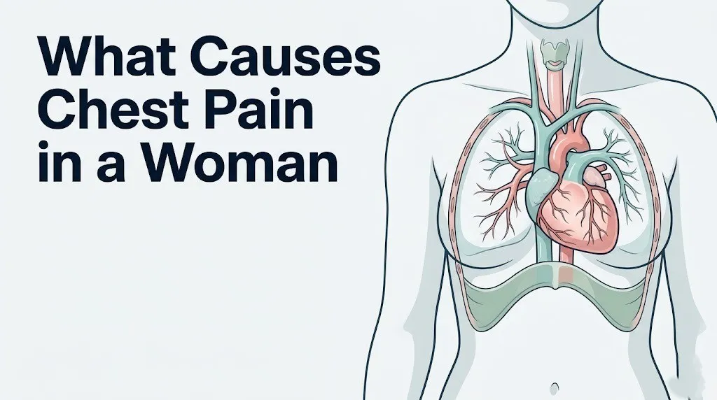
This guide covers every clinically documented cause of chest pain in women, the specific physiological mechanism behind each one, which causes are most common and which are rare but serious, how hormonal status and age modify the picture, and exactly which symptoms require you to call 911 right now rather than make a doctor’s appointment.
What Causes Chest Pain in a Woman
Chest pain in a woman is caused by one of five broad categories of conditions: cardiovascular (involving the heart and coronary arteries), pulmonary (involving the lungs, pleura, or pulmonary vasculature), gastrointestinal (involving the esophagus, stomach, or diaphragm), musculoskeletal (involving the chest wall, ribs, or cartilage), or systemic (including anxiety, autoimmune disease, and hormonal fluctuation).
No single cause dominates all age groups or hormonal stages equally. A 32-year-old woman on oral contraceptives has a meaningfully different probability profile for chest pain causes than a 58-year-old postmenopausal woman with hypertension. Age, reproductive hormone status, contraceptive use, autoimmune disease history, and lifestyle factors all shift which cause is most likely in any individual woman.
| Cause Category | Most Common Specific Causes | Relative Frequency in Women |
|---|---|---|
| Cardiovascular | Angina, SCAD, microvascular dysfunction, heart attack | Increases sharply after menopause |
| Gastrointestinal | GERD, esophageal spasm, hiatal hernia | Extremely common across all ages |
| Musculoskeletal | Costochondritis, intercostal muscle strain | Very common; often post-exertional |
| Pulmonary | Pleuritis, PE, pneumothorax, pulmonary hypertension | PE risk elevated in pregnancy/OCP use |
| Psychiatric/Autonomic | Panic disorder, generalized anxiety disorder | Common in women under 45 |
| Autoimmune/Inflammatory | Pericarditis from lupus, myocarditis | Women-predominant conditions |
The character, location, timing, and associated symptoms of chest pain provide the first clinical clues about which category is most likely. Pain that worsens with deep breathing or position change points toward pleural or musculoskeletal causes. Pain that occurs predictably after meals suggests gastrointestinal origin. Pain triggered by exertion and relieved by rest points toward cardiac ischemia. Pain that arrives suddenly with a racing heart and sense of doom during a stressful moment often points toward autonomic or anxiety-driven causes, though cardiac origin must still be excluded.
Quick Tip:
- Cardiac chest pain in women frequently presents without the classic “crushing pressure” and instead feels like fatigue, jaw tightness, nausea, or upper back discomfort.
- Any new chest pain lasting more than 15 minutes without a clear positional or dietary trigger warrants medical evaluation the same day.
- Women with diabetes are particularly likely to have atypical or silent cardiac presentations because diabetic autonomic neuropathy blunts the pain signal from ischemic myocardium.
Why Chest Pain Presents Differently in Women Than Men
Chest pain in women presents differently from men because of fundamental sex-based differences in coronary artery anatomy, hormonal modulation of vascular function, autonomic nervous system tone, and the types of cardiac disease that most commonly affect each sex.
The most cited reason is hormonal. 17-beta estradiol binds to estrogen receptors on the vascular endothelium, the inner lining of blood vessels, and activates endothelial nitric oxide synthase (eNOS). This enzyme produces nitric oxide, which relaxes and dilates blood vessels, suppresses platelet aggregation, and inhibits the oxidation of low-density lipoprotein (LDL) cholesterol into the form that initiates atherosclerotic plaque formation. Before menopause, this estrogen-eNOS-nitric oxide pathway gives women a degree of cardiovascular protection not present in men of the same age. After menopause, estrogen declines sharply and this protection dissolves, which is why coronary artery disease in women peaks 10 years later than in men on average, according to the National Heart, Lung, and Blood Institute (NHLBI).
A 2003 study published in JAMA by Milner and colleagues found that women with acute myocardial infarction were significantly more likely than men to present without chest pain at all, and more likely to report back pain, jaw pain, nausea, shortness of breath, and unusual fatigue as their primary symptoms. This means that using classic “crushing substernal chest pressure” as the primary screening criterion for cardiac events in women is clinically inadequate.
Women are also more likely than men to have obstructive plaque distributed differently throughout the coronary tree. Rather than one focal severe blockage in a major coronary artery (the pattern most common in men), women more often develop diffuse plaque erosion and coronary microvascular disease. Standard coronary angiography, which identifies focal obstructions, can appear entirely normal in a woman experiencing real myocardial ischemia from microvascular disease. This is not anxiety. This is not a functional complaint. This is a recognized and distinct pathophysiological mechanism.
The pain-referral pattern also differs. Cardiac ischemia refers pain through thoracic sympathetic afferent fibers at spinal levels T1 through T4. In women, the ratio of sympathetic to parasympathetic autonomic tone differs from men, and the threshold at which visceral pain reaches conscious awareness varies. This contributes to why women more often report diffuse, aching, or pressure-like discomfort across the upper chest, back, and jaw rather than the localized left-sided pressure that dominates textbook descriptions.
Heart Attack as a Cause of Chest Pain in Women
A heart attack, clinically called an acute myocardial infarction (AMI), occurs when blood flow through one or more coronary arteries is suddenly reduced or stopped, causing the downstream myocardial tissue to become ischemic (oxygen-deprived) and begin dying within minutes. In women, this process is triggered by the same fundamental mechanisms as in men, but the specific pathological pathway that causes the blockage is often different.
Men most commonly experience AMI from the rupture of a lipid-rich atherosclerotic plaque within a major coronary artery, followed by platelet aggregation and thrombosis that blocks blood flow. Women are more likely to experience AMI from plaque erosion (where the plaque surface is disrupted without full rupture), coronary artery vasospasm, or coronary microvascular dysfunction, particularly before menopause. According to a study published in the Journal of the American College of Cardiology, plaque erosion accounts for a larger proportion of acute coronary events in women under 50 compared to men of the same age.
The chest pain of AMI in women ranges from the familiar substernal pressure to a sensation of tightness, squeezing, or burning across the upper chest. Many women describe it as “not what they expected.” Accompanying symptoms that should elevate concern for cardiac origin include:
- Shortness of breath that occurs at rest or with minimal activity
- Nausea or vomiting without a gastrointestinal explanation
- Diaphoresis (sudden cold sweating) unrelated to heat or physical exertion
- Pain, pressure, or heaviness radiating to the left jaw, neck, left arm, or between the shoulder blades
- Sudden extreme fatigue, sometimes described as the worst tiredness of a woman’s life
- Lightheadedness or pre-syncope (near-fainting)
The Centers for Disease Control and Prevention (CDC) reports that heart disease remains the leading cause of death for women in the United States, accounting for approximately one in five female deaths annually. Despite this, research published in Circulation has documented that women wait longer to call for emergency services after symptom onset and are evaluated less aggressively on arrival, partly because atypical presentations do not trigger the same clinical urgency as classic presentations.
Younger women under 50 who experience AMI deserve particular clinical attention. Pre-menopausal AMI is relatively rare with traditional risk factors, and when it occurs, spontaneous coronary artery dissection or drug use (particularly cocaine or methamphetamine) must be actively excluded.
Key Takeaway: Women are more likely than men to have a heart attack without classic chest pressure, often presenting instead with jaw pain, back pain, nausea, or extreme fatigue. Any of these symptoms appearing together at rest or with minimal exertion warrants emergency evaluation, not a scheduled appointment.
Coronary Microvascular Dysfunction and Chest Pain in Women
Coronary microvascular dysfunction (CMD) is a condition in which the tiny arterioles and capillaries of the coronary microvasculature (vessels smaller than 500 micrometers in diameter) fail to dilate normally in response to increased myocardial oxygen demand, causing real ischemia in the absence of any blockage visible on standard coronary angiography.
CMD is far more common in women than in men. The NHLBI-funded WISE study, which enrolled over 900 women with chest pain and non-obstructive coronary arteries on angiography, found that nearly half had evidence of impaired coronary blood flow reserve consistent with CMD. This finding has transformed how cardiologists approach chest pain evaluation in women, because a normal angiogram does not rule out ischemic disease in a woman the way it more reliably does in a man.
The physiological mechanism behind CMD in women involves impaired endothelial function at the microvascular level. The coronary microvasculature relies heavily on endothelium-derived nitric oxide for its vasodilatory response. When this mechanism is impaired, as occurs with estrogen deficiency after menopause, diabetes, hypertension, or chronic inflammation, the microvessels cannot dilate adequately during physical or emotional stress. The myocardium downstream experiences oxygen deprivation. Pain signals travel through the same T1-T4 thoracic sympathetic afferents as classic angina, producing chest tightness, pressure, or aching that is clinically real but mechanistically different from large-vessel disease.
Women with CMD typically experience:
- Chest discomfort or tightness during exertion or emotional stress that resolves with rest
- Episodes that may be less predictable than classic stable angina
- Normal or near-normal stress test results on standard treadmill EKG (because the test does not adequately assess microvascular function)
- Normal coronary angiography despite persistent symptoms
Diagnosing CMD requires specialized testing. Coronary flow reserve can be measured with a stress echocardiogram, cardiac magnetic resonance imaging (cardiac MRI) with perfusion imaging, or an invasive coronary functional assessment during catheterization. A standard stress treadmill EKG is often insufficient to capture microvascular ischemia.
Postmenopausal women and women with diabetes are at the highest risk for CMD. Women with a history of chest radiation for breast cancer are also at elevated risk for CMD due to radiation-induced microvascular damage.
Spontaneous Coronary Artery Dissection: Chest Pain in Women
Spontaneous coronary artery dissection (SCAD) is a rare but underrecognized cardiac emergency in which a tear develops in the inner wall of a coronary artery, allowing blood to enter and separate the layers of the arterial wall, forming an intramural hematoma that compresses the true lumen of the artery and restricts blood flow. SCAD is not caused by atherosclerosis and does not fit the traditional risk profile for heart disease.
Research from the Mayo Clinic SCAD Research Program, published in Circulation, has consistently shown that approximately 90 percent of SCAD cases occur in women. The median age at presentation is approximately 42 years, and many affected women have no traditional cardiovascular risk factors. SCAD accounts for an estimated 25 to 35 percent of acute myocardial infarctions in women under 50, making it one of the most clinically important female-specific cardiac diagnoses.
The chest pain of SCAD is typically sudden in onset, severe, and may be accompanied by shortness of breath, nausea, and diaphoresis. It is indistinguishable from other causes of AMI based on symptoms alone. SCAD must be diagnosed by coronary imaging, usually coronary angiography with optical coherence tomography (OCT) or intravascular ultrasound (IVUS), because the intramural hematoma can be missed on standard angiography if the true lumen appears only partially narrowed.
The mechanisms that make women vulnerable to SCAD are not fully understood, but research has identified several strong associations:
- Fibromuscular dysplasia (FMD): A non-inflammatory arterial disease that affects arterial walls, found in approximately 72 percent of SCAD patients in the Mayo registry
- Peripartum state: SCAD risk peaks in the week to month following childbirth, with postpartum hormonal shifts believed to alter arterial wall integrity
- Intense physical exertion or emotional stress: sympathetic surge may precipitate dissection in susceptible arteries
- Connective tissue disorders: Marfan syndrome and Ehlers-Danlos syndrome hypermobile type have been associated with SCAD
Any woman under 50 presenting to an emergency room with acute severe chest pain, particularly in the postpartum period or following intense physical exertion, should have SCAD actively considered as a potential diagnosis. Standard atherosclerosis risk-reduction advice does not apply to SCAD management, and treatment protocols differ from conventional AMI management.
Hormonal and Menstrual Cycle Causes of Chest Pain in Women
Hormonal fluctuations throughout the menstrual cycle, pregnancy, and the postpartum period can cause or amplify chest pain in women through several distinct physiological mechanisms.
During the premenstrual phase, progesterone levels peak and then decline sharply. Progesterone affects smooth muscle tone throughout the body, including in the esophagus, where it reduces lower esophageal sphincter (LES) pressure. A relaxed LES allows gastric acid to reflux more easily into the esophageal lumen, producing the burning, pressure-like chest discomfort of acid reflux. Women who notice chest pain or heartburn consistently worsening in the days before their period are likely experiencing progesterone-mediated LES relaxation. Research published in the American Journal of Gastroenterology has confirmed cyclical worsening of GERD symptoms in association with the luteal phase of the menstrual cycle.
Beyond the GI mechanism, some women experience Prinzmetal angina, also called vasospastic angina, in which a coronary artery undergoes intermittent spasm rather than progressive narrowing. Estrogen normally modulates vascular smooth muscle tone through nitric oxide pathways. During phases of the cycle when estrogen drops relative to progesterone, vasomotor instability can increase. Prinzmetal angina produces real chest pain, typically at rest or at night, and can be severe enough to cause ST-elevation on an EKG during an episode.
Oral contraceptives affect chest pain risk through a different mechanism: venous thromboembolic risk. Combined oral contraceptive pills containing synthetic estrogen increase coagulation factor synthesis in the liver (specifically factors II, VII, VIII, and X), shifting the coagulation balance toward thrombosis. Women using combined OCP, particularly those who smoke, have significantly elevated risk of deep vein thrombosis (DVT) and pulmonary embolism (PE), both of which can cause acute chest pain. According to ACOG, the absolute risk remains low in non-smoking, healthy women, but increases substantially with smoking, obesity, prolonged immobility, or personal or family history of clotting disorders.
During pregnancy, a separate set of mechanisms applies. The physiological hypercoagulable state of pregnancy (increased fibrinogen, factor VIII, and von Willebrand factor; decreased protein S) elevates PE risk across all trimesters, with the highest risk in the third trimester and postpartum period. Additionally, as the uterus grows and the diaphragm is displaced upward, gastroesophageal reflux nearly universally worsens, and musculoskeletal chest wall discomfort from rib expansion and ligamentous relaxation is common.
Key Takeaway: Women who notice chest pain or heartburn worsening in a predictable pattern before their period may be experiencing progesterone-mediated esophageal relaxation, but new-onset chest pain at any phase of the menstrual cycle that is severe, radiating, or accompanied by shortness of breath requires cardiac exclusion before a hormonal explanation is accepted.
Chest Pain During Menopause and Perimenopause
Chest pain during menopause and perimenopause is caused by a convergence of cardiovascular risk escalation, vasomotor instability, and musculoskeletal changes that all emerge as ovarian estrogen production declines, typically between ages 45 and 55.
The most physiologically significant change is the loss of estrogen’s cardioprotective effect on the vascular endothelium. As described earlier, estrogen activates eNOS, which produces vasodilatory nitric oxide. Without adequate estrogen, endothelial function deteriorates, vascular stiffness increases, and the threshold for atherosclerotic plaque development lowers. LDL cholesterol tends to rise during perimenopause in many women, while HDL cholesterol and vascular compliance decrease. This is the primary reason cardiovascular disease risk rises sharply in postmenopausal women to approach and eventually exceed that of age-matched men.
Vasomotor symptoms, the hot flashes and night sweats of perimenopause, are driven by fluctuating estrogen levels affecting the hypothalamic thermoregulatory center. These episodes involve sudden activation of the sympathetic nervous system, producing rapid heart rate, flushing, sweating, and a sensation of chest pressure or tightness. For many women, these episodes are alarming and genuinely difficult to distinguish from cardiac symptoms without assessment. A hot flash that includes racing heart, sweating, and chest tightness in a perimenopausal woman is often hormonal, but because perimenopausal women are simultaneously entering their window of elevated cardiac risk, this distinction must be made clinically rather than assumed.
Chest pain during perimenopause that occurs predictably with exertion, radiates to the jaw or arm, or persists beyond 20 minutes requires cardiac evaluation with a primary care physician or cardiologist regardless of the woman’s awareness of perimenopausal status. An office EKG, high-sensitivity troponin level, and clinical history are the appropriate starting points. Perimenopausal and postmenopausal women with diabetes face compounded risk, because diabetic autonomic neuropathy can blunt typical cardiac pain signals, making silent ischemia more likely.
Pulmonary Causes of Chest Pain in Women
Several lung and pulmonary vascular conditions cause chest pain in women, and some carry a higher incidence in women or present with female-specific triggers that affect risk substantially.
Pulmonary embolism (PE) is one of the most dangerous causes of chest pain and occurs when a blood clot travels from a vein (most commonly the deep veins of the legs or pelvis) to the pulmonary arteries, obstructing blood flow to lung tissue. The chest pain of PE is characteristically pleuritic: sharp, worse with deep breathing or coughing, and often unilateral (one-sided). PE causes chest pain through two mechanisms. First, when lung tissue infarcts (dies from lack of blood supply), the parietal pleura overlying it becomes inflamed. The parietal pleura is innervated by the phrenic nerve (C3-C5) and intercostal nerves, making this pain well-localized and respiration-dependent. Second, acute massive PE increases resistance in the pulmonary circulation, causing the right ventricle to strain, which produces a dull, central cardiac-type pain from right ventricular ischemia.
Women-specific risk factors for PE include:
- Combined oral contraceptive pill use (2 to 4 times increased risk, rising to 30 times in women who smoke)
- Pregnancy and the postpartum period (5 to 10 times increased risk)
- Hormone replacement therapy containing estrogen
- Prolonged immobility during travel or hospitalization
- Inherited thrombophilias (factor V Leiden mutation, prothrombin gene mutation, antiphospholipid syndrome)
Pleuritis (inflammation of the pleural lining) produces sharp, unilateral, breathing-related chest pain through direct irritation of parietal pleural pain receptors by inflammation. It can occur secondary to viral or bacterial pneumonia, autoimmune disease (particularly lupus), or pulmonary infarction from PE. Pneumothorax, the abnormal accumulation of air between the lung and the chest wall, produces sudden onset unilateral sharp chest pain with shortness of breath. Spontaneous pneumothorax occurs more often in tall, slender individuals; secondary pneumothorax occurs in women with underlying lung disease.
Pulmonary arterial hypertension (PAH) disproportionately affects women, with women accounting for approximately 70 to 80 percent of idiopathic PAH cases, according to the National Heart, Lung, and Blood Institute. PAH causes exertional chest pain, shortness of breath, and fatigue as the right ventricular workload increases to overcome elevated pulmonary artery pressure. Early PAH is frequently misdiagnosed because standard chest x-rays and EKGs can appear normal.
Gastrointestinal Causes of Chest Pain in Women
Gastrointestinal conditions are among the most frequent causes of chest pain in women across all age groups, and the physiological reason they produce chest pain rather than abdominal pain is a phenomenon called shared visceral afferent innervation.
Think of it this way: the esophagus and the heart share the same postal address in the spinal cord. Pain signals from the esophagus travel through vagal afferent fibers and thoracic sympathetic fibers that enter the spinal cord at T1 through T4, the same spinal levels that receive cardiac pain signals. The brain cannot reliably distinguish whether pain arriving at these levels came from the esophagus or the heart, which is why esophageal chest pain can be clinically indistinguishable from angina without diagnostic testing. This is not a quirk of perception; it is a fundamental feature of visceral pain neuroanatomy.
Gastroesophageal reflux disease (GERD) is the most common gastrointestinal cause of chest pain in women. GERD occurs when the lower esophageal sphincter (LES), a muscular ring at the junction of the esophagus and stomach, fails to close properly, allowing acidic gastric contents to reflux into the esophageal lumen. Gastric acid irritates the esophageal mucosa, activating mucosal chemoreceptors and nociceptors that send pain signals through the shared T1-T4 pathway. The resulting chest pain is often described as burning, located behind the sternum, and worsening after meals, when lying down, or when bending forward.
Esophageal motility disorders, including diffuse esophageal spasm and achalasia, cause chest pain through abnormal coordinated or sustained contractions of esophageal smooth muscle. Esophageal spasm produces chest pain that can be severe, sudden, and radiate to the back or jaw, closely mimicking cardiac angina. Some patients describe it as a “squeezing” or “gripping” sensation identical to what textbooks attribute to cardiac ischemia. One practical clue: esophageal spasm pain is sometimes relieved by nitroglycerin (the same drug used for cardiac angina) because nitroglycerin relaxes smooth muscle generally, not just in coronary arteries.
A hiatal hernia, in which part of the stomach protrudes upward through the diaphragmatic hiatus into the thoracic cavity, can cause postprandial chest pain, fullness, and heartburn. Large hiatal hernias can compress adjacent structures and worsen reflux mechanics. Hiatal hernia is more common in women and increases in prevalence with age and higher body mass index.
Key Takeaway: Esophageal and cardiac chest pain are physiologically indistinguishable based on symptoms alone because they share the same spinal pain-referral pathway at T1-T4. A woman whose chest pain improves with antacids has likely identified a gastrointestinal cause, but this does not eliminate cardiac risk if any exertional, radiating, or associated symptoms are present.
Musculoskeletal Causes of Chest Pain in Women
Musculoskeletal causes of chest pain are genuinely common, generally benign, and produce a characteristic pattern of tenderness that distinguishes them from most cardiac or pulmonary causes.
Costochondritis is inflammation of the costochondral junctions, the cartilage connections between the ribs and the sternum. Ribs 2 through 5 are the most commonly affected levels. The pain is typically sharp or aching, located along the anterior chest wall, reproducible by pressing on the affected junction (parasternal tenderness on palpation is the hallmark finding), and often worsened by deep breathing, coughing, or upper body movement. The inflammatory mechanism involves prostaglandin-mediated pain signaling at the costochondral joint, activated by overuse, viral illness, or idiopathic causes. Tietze syndrome is a related but distinct condition in which swelling accompanies the inflammation; this is more common in young adults and frequently involves only one junction, usually at the second or third rib.
Costochondritis does not produce radiation to the jaw or arm, does not cause sweating or nausea, and does not worsen with exertion while resolving at rest (the cardiac pattern). The presence of reproducible chest wall tenderness on palpation is the most reliable clinical distinguishing feature. However, it is worth noting that cardiac chest pain and costochondritis can coexist, and chest wall tenderness does not rule out a concurrent cardiac process.
Intercostal muscle strain from heavy lifting, repetitive arm motion, coughing, or direct trauma causes chest pain through mechanical irritation and microinjury of the intercostal muscle fibers between ribs. The pain is typically well-localized, worsens with specific movements, and is relievable with rest and position change.
Women with fibromyalgia frequently experience chest wall pain as part of their syndrome. Fibromyalgia produces widespread musculoskeletal pain through central sensitization, a process involving altered pain-processing in the dorsal horn of the spinal cord and the brain’s descending pain-modulation pathways. The chest wall is one of many regions affected. Fibromyalgia has a strong female predominance, with women accounting for approximately 75 to 90 percent of diagnosed cases, according to the American College of Rheumatology.
The practical clinical point: if pressing on the chest wall with two fingertips reproduces the exact pain a woman is experiencing, musculoskeletal origin is highly likely. If the chest pain occurs during exertion but not with palpation, cardiac origin remains on the table until excluded.
Anxiety and Panic Disorder as Causes of Chest Pain in Women
Anxiety and panic disorder are legitimate physiological causes of chest pain in women, not psychological dismissals, and understanding the mechanism makes this clear.
During a panic attack, the brain activates the sympathetic nervous system in a threat-response pattern. The adrenal medulla releases epinephrine and norepinephrine into the bloodstream. Epinephrine binds to cardiac beta-1 adrenergic receptors, producing tachycardia (rapid heart rate) and increased myocardial contractility. It also binds to alpha-1 adrenergic receptors in peripheral blood vessels, causing vasoconstriction. The combination of tachycardia, vasoconstriction, and increased cardiac workload produces genuine anterior chest tightness, palpitations, and shortness of breath, all physically caused by circulating catecholamines, not by imagination.
Simultaneously, hyperventilation driven by anxiety reduces carbon dioxide levels in the blood, producing hypocapnia. Hypocapnia causes cerebral vasoconstriction (contributing to dizziness and lightheadedness) and can trigger coronary artery vasospasm, theoretically contributing to real transient ischemia in susceptible individuals.
Women are diagnosed with panic disorder at approximately twice the rate of men, according to the National Institute of Mental Health (NIMH). Panic-related chest pain is more common in women in their 20s and 30s. However, the clinical danger is that anxiety chest pain is frequently used to dismiss cardiac symptoms in younger women who are statistically less likely to have coronary artery disease. A young woman presenting with chest pain, racing heart, and shortness of breath should have cardiac causes excluded before anxiety is accepted as the primary explanation.
The distinguishing features that favor panic-related chest pain over cardiac origin:
- Pain begins and peaks rapidly (often within 10 minutes) and resolves spontaneously within 20-30 minutes
- Associated with a clear emotional trigger, confined space, or specific feared situation
- Accompanied by numbness or tingling in hands and feet (hypocapnia-related peripheral vasoconstriction)
- Reproducible with situations that trigger anxiety but absent at rest during calm periods
These features are suggestive, not diagnostic. EKG, troponin level, and clinical assessment remain the appropriate tools to exclude cardiac causes.
Autoimmune and Inflammatory Causes of Chest Pain in Women
Several autoimmune and inflammatory conditions that disproportionately affect women can cause chest pain through direct involvement of the pericardium, myocardium, or pleura.
Pericarditis is inflammation of the pericardium, the two-layered fibrous sac surrounding the heart. The chest pain of pericarditis is characteristically sharp, pleuritic (worse with breathing and coughing), and relieved by sitting forward (because this position reduces pressure on the inflamed pericardium). Pain often radiates to the left trapezius ridge because the pericardium shares innervation with the phrenic nerve, which also innervates the left shoulder region. A pericardial friction rub (a scratching sound heard with a stethoscope) is the classic auscultatory finding. Pericarditis can be caused by viral infection, but in women under 50, an autoimmune workup is standard because systemic lupus erythematosus (SLE) is a frequent underlying cause.
SLE, an autoimmune disease affecting multiple organ systems, has a pronounced female predominance: approximately 9 out of 10 people with SLE are women, according to the National Institute of Arthritis and Musculoskeletal and Skin Diseases (NIAMS). Cardiac involvement in SLE (called lupus carditis) can include pericarditis, myocarditis, and Libman-Sacks endocarditis. Any woman with a history of SLE who develops new chest pain should receive urgent cardiac evaluation.
Antiphospholipid syndrome (APS) is another female-predominant autoimmune condition that causes chest pain through a different mechanism: hypercoagulability. APS produces antibodies (antiphospholipid antibodies) that activate coagulation pathways, dramatically increasing the risk of arterial and venous thrombosis. In women with APS, acute chest pain can represent pulmonary embolism, coronary artery thrombosis, or both simultaneously.
Myocarditis, inflammation of the myocardium itself, can follow viral infection (particularly coxsackievirus B) or occur as part of an autoimmune process. Chest pain in myocarditis resembles that of both pericarditis and AMI, and troponin levels may be elevated. Cardiac MRI is the non-invasive diagnostic standard for confirming myocarditis.
Key Takeaway: Women with a history of lupus, antiphospholipid syndrome, or other systemic autoimmune diseases who develop new chest pain need urgent evaluation that specifically includes cardiac and pulmonary assessment, not just a general rheumatology review, because these conditions carry direct mechanisms for life-threatening cardiac and pulmonary events.
Risk Factors That Increase Chest Pain Likelihood in Women
Understanding which risk factors apply to which causes of chest pain helps both women and their clinicians prioritize the most likely explanation and the most urgent assessment pathway.
For cardiovascular chest pain, the established risk factors in women include:
- Postmenopausal status (loss of estrogen-mediated vascular protection)
- Hypertension (damages endothelium, accelerates atherosclerosis)
- Type 2 diabetes mellitus (increases CMD risk, causes atypical pain presentation, accelerates atherosclerosis)
- Current smoking (endothelial toxin, accelerates plaque formation, coronary vasospasm trigger)
- Hyperlipidemia (elevated LDL drives atherogenesis)
- Obesity (body mass index above 30; associated with systemic inflammation, hypertension, dyslipidemia)
- Family history of premature coronary artery disease (first-degree relative under 65 for women)
- Autoimmune disease (systemic lupus, rheumatoid arthritis: vascular inflammation accelerates atherosclerosis)
- History of preeclampsia or gestational hypertension (strongly associated with long-term cardiovascular disease risk, according to the AHA)
For pulmonary embolism risk specifically in women:
- Combined oral contraceptive use, especially with smoking or thrombophilia
- Pregnancy or postpartum period (highest risk in first 6 weeks after delivery)
- Prolonged immobility (air travel, bed rest, postoperative state)
- Inherited thrombophilia (factor V Leiden, prothrombin G20210A mutation, protein C or S deficiency)
- Antiphospholipid syndrome
For SCAD specifically:
- Age 40 to 55 (perimenopausal)
- Postpartum state (within 4 weeks of delivery)
- Fibromuscular dysplasia diagnosis
- Connective tissue disorder (Marfan syndrome, Ehlers-Danlos syndrome)
- History of extreme physical exertion immediately before symptom onset
Women with multiple risk factors from multiple categories face a more complex diagnostic picture. A 48-year-old perimenopausal woman who smokes, takes oral contraceptives, and has a history of SLE carries simultaneous elevated risk for SCAD, PE, atherosclerotic coronary disease, and lupus pericarditis. Her chest pain evaluation must account for all of these possibilities.
How Chest Pain in Women Is Diagnosed
Diagnosing the cause of chest pain in a woman involves a structured clinical evaluation that must account for sex-specific cardiac presentation patterns and the possibility of non-obstructive causes that standard tests may underdiagnose.
The initial evaluation in an emergency setting typically follows this sequence:
- 12-lead electrocardiogram (EKG/ECG): Performed within 10 minutes of arrival for any woman presenting with chest pain of suspected cardiac origin. Identifies ST-elevation (STEMI), ST-depression, T-wave changes, arrhythmias, and signs of right heart strain (suggestive of PE). A normal EKG does not exclude AMI, NSTEMI, CMD, or SCAD.
- High-sensitivity cardiac troponin assay: Troponin I and troponin T are proteins released when myocardial cells die. Elevated troponin indicates myocardial damage. Serial measurements (at 0 and 3 hours, or 0 and 1 hour with high-sensitivity assays) are the standard protocol. Women have lower baseline troponin levels than men, and some guidelines recommend sex-specific reference ranges, though this is not yet universally adopted.
- Chest X-ray: Assesses cardiac size, pulmonary vasculature, pneumothorax, and signs of pulmonary edema or consolidation.
- Blood tests: Including complete blood count (CBC), basic metabolic panel, lipid panel, D-dimer (if PE is suspected), coagulation studies, and inflammatory markers (C-reactive protein, erythrocyte sedimentation rate) if autoimmune or inflammatory causes are under consideration.
- Echocardiogram: Ultrasound imaging of the heart. Identifies regional wall motion abnormalities (areas of heart muscle not contracting normally due to ischemia), pericardial effusion (fluid around the heart from pericarditis), and right ventricular dilation (suggesting PE).
- Stress testing: Exercise or pharmacological stress testing with imaging (stress echocardiogram or stress nuclear perfusion scan) is used to detect inducible ischemia in women with stable chest pain and moderate cardiac risk. Standard treadmill EKG stress testing without imaging is less sensitive for CMD detection in women.
- CT pulmonary angiography (CTPA): The gold standard imaging test for pulmonary embolism. Ordered when PE probability is intermediate or high based on clinical scoring (Wells criteria, revised Geneva score).
- Coronary CT angiography or invasive coronary angiography: Used when obstructive coronary artery disease needs to be excluded or confirmed. For suspected SCAD, invasive angiography with OCT or IVUS provides the most accurate characterization.
Women presenting to primary care with chronic or recurrent chest pain that has not been evaluated should be assessed by a primary care physician who will order an EKG, troponin if recent-onset, and arrange appropriate referral: to a board-certified cardiologist for suspected cardiac causes, gastroenterologist for GERD or esophageal motor disorders, pulmonologist for suspected pulmonary causes, or rheumatologist for suspected autoimmune pericarditis.
Emergency Symptoms: When to Call 911 or Go to the ER
Certain causes of chest pain in women represent immediately life-threatening emergencies. Do not wait to see if these symptoms resolve on their own.
Call 911 or go to the nearest emergency room immediately if you experience:
- Sudden severe chest pain, pressure, tightness, or squeezing, especially at rest or without a clear musculoskeletal or dietary trigger
- Chest pain with shortness of breath, even if the pain itself is mild
- Chest pain radiating to the left jaw, neck, left arm, left shoulder, or upper back between the shoulder blades
- Chest pain accompanied by sudden sweating, cold clamminess, or nausea and vomiting
- Chest pain with lightheadedness, dizziness, or fainting (syncope)
- Chest pain with a very rapid, irregular, or pounding heartbeat
- Sudden sharp chest pain that worsens dramatically with every breath (possible pulmonary embolism, pneumothorax, or pleuritis with acute origin)
- Chest pain in a woman who is currently pregnant or within 4 weeks of delivery (elevated SCAD and PE risk)
- Any chest pain in a woman with a known history of SLE, antiphospholipid syndrome, or heart disease that is new, changed in character, or more severe than prior episodes
- Chest pain with confusion, difficulty speaking, or sudden severe headache (possible aortic dissection involving cerebral vessels or associated neurological event)
These presentations can indicate acute myocardial infarction, SCAD, pulmonary embolism, aortic dissection, tension pneumothorax, or cardiac tamponade. Every one of these requires emergency medical assessment, not an urgent care appointment, a telehealth call, or a scheduled visit.
Chest pain that is reproducible by pressing on the chest wall, that has been present unchanged for weeks without worsening, that is clearly tied to meals and relieved by antacids, or that occurs exclusively with known panic episodes in a woman with a documented anxiety disorder may be reasonably evaluated at a scheduled primary care appointment. When in doubt, err toward emergency evaluation. The cost of an unnecessary ER visit is insignificant compared to the cost of a delayed AMI or PE diagnosis.
Key Takeaway: A woman experiencing chest pain with shortness of breath, sweating, jaw or arm pain, or any symptom that is sudden and severe should call 911 immediately. Women’s heart attacks do not always announce themselves with classic crushing pressure, and waiting to see if symptoms improve is one of the most dangerous decisions a woman can make in this situation.
Frequently Asked Questions About Chest Pain in Women
Can chest pain in women be a sign of something other than a heart attack?
Yes, chest pain in women is caused by non-cardiac conditions in the majority of cases, including GERD, esophageal spasm, costochondritis, pulmonary embolism, panic disorder, pericarditis, and hormonal changes.
The challenge is that non-cardiac and cardiac causes can produce identical symptoms, which is why any new or unexplained chest pain in a woman warrants professional evaluation to exclude cardiac origin before an alternative explanation is accepted.
A primary care physician or emergency medicine physician can use an EKG, troponin level, and clinical history to efficiently narrow the differential diagnosis.
What does a heart attack feel like for a woman?
A heart attack in a woman often does not feel like the classic crushing left-sided chest pressure depicted in standard descriptions, and may instead present as jaw pain, unusual fatigue, back pain between the shoulder blades, nausea, dizziness, or shortness of breath with mild chest discomfort.
The 2003 JAMA study by Milner and colleagues documented that women with confirmed acute myocardial infarction were substantially more likely than men to report atypical symptoms as their primary or only complaint.
Any combination of these symptoms, especially at rest or during minimal activity, warrants emergency evaluation rather than watchful waiting.
Can hormones cause chest pain in women?
Yes, hormonal fluctuations cause chest pain in women through several distinct mechanisms, including progesterone-mediated lower esophageal sphincter relaxation (worsening acid reflux before menstruation), estrogen-related vasomotor instability during perimenopause (producing vasospastic angina and hot flash-related chest tightness), and the hypercoagulable effect of synthetic estrogen in combined oral contraceptives (increasing pulmonary embolism risk).
The specific hormonal mechanism differs depending on which phase of a woman’s reproductive life she is in and which medications she takes.
A woman who notices cyclical chest pain should discuss the pattern with her primary care physician or obstetrician-gynecologist to identify which mechanism is most likely and whether further cardiac evaluation is warranted.
When should a woman go to the ER for chest pain?
A woman should go to the ER immediately (or call 911) for chest pain that is sudden and severe, accompanied by shortness of breath, radiating to the jaw or arm, associated with sweating or nausea, or occurring in the setting of pregnancy, recent delivery, or known cardiovascular disease.
Chest pain that is positional, reproducible by pressing on the chest wall, and tied to a known dietary or anxiety trigger in a woman with no cardiac risk factors can be scheduled for a same-day or next-day primary care appointment, not an emergency visit, but only if none of the above emergency features are present.
When there is genuine uncertainty, choosing the emergency room is always the safer decision.
What is coronary microvascular dysfunction and why does it affect women more?
Coronary microvascular dysfunction is impaired dilation of the tiny arterioles and capillaries of the coronary circulation, causing real myocardial ischemia in the absence of obstructive blockages visible on standard coronary angiography.
It affects women more than men because estrogen normally activates endothelial nitric oxide synthase (eNOS) in microvascular endothelial cells, maintaining their vasodilatory capacity; when estrogen declines at menopause, this mechanism weakens and microvascular function deteriorates.
The NHLBI-funded WISE study found evidence of impaired coronary flow consistent with CMD in nearly half of women evaluated for chest pain with normal angiograms, confirming that a normal angiogram does not rule out ischemic heart disease in women.
Can anxiety cause chest pain that feels like a heart attack in women?
Yes, anxiety and panic disorder cause genuine physical chest pain in women through sympathetic nervous system activation, circulating epinephrine, and hypocapnia from hyperventilation, all of which produce real tachycardia, vasoconstriction, and anterior chest tightness.
The pain of a panic attack can be severe, sudden, and subjectively indistinguishable from a cardiac event, which is why cardiac causes must be formally excluded before anxiety is identified as the primary explanation for a woman’s chest pain, particularly if she is over 40 or has cardiac risk factors.
Once cardiac causes have been excluded by a physician with appropriate testing, cognitive behavioral therapy (CBT) and targeted pharmacological management under a psychiatrist’s or primary care physician’s guidance have strong evidence for reducing panic disorder frequency and severity.
Closing
Chest pain in a woman is never a single-answer question. The cause depends on her age, hormonal status, cardiovascular history, autoimmune disease history, contraceptive use, and the specific character of the pain itself. What this guide establishes is that women’s chest pain is physiologically distinct from men’s in ways that matter clinically: the cardiac causes women face most commonly (microvascular dysfunction, SCAD, vasospastic angina) are different from the focal obstructive coronary disease that dominates male-centric cardiac guidelines, and the non-cardiac causes (GERD, costochondritis, panic, pericarditis) require the same systematic exclusion of cardiac origin before they can be accepted as the explanation.
The single most practical action any woman with unexplained chest pain can take is to seek evaluation from a primary care physician or emergency medicine physician rather than attempting to self-diagnose based on symptom patterns. Bring a clear description of when the pain started, what it feels like, how long it lasts, what makes it better or worse, and any associated symptoms. That information, combined with an EKG and troponin level, gives a clinician the foundation to work from.
Your body’s signals are specific and meaningful. A woman who understands what causes chest pain and which features distinguish serious causes from benign ones is far better equipped to act quickly when it matters most.