What Causes Bone Loss in Teeth? Key Causes Explained
What causes bone loss in teeth is, in the vast majority of cases, chronic bacterial infection of the gum tissue that activates the immune system to destroy the bone supporting each tooth root, a process called alveolar bone resorption. The destruction is driven not by bacteria directly eating bone, but by the body’s own inflammatory response to bacterial toxins, which is why bone loss can progress silently for years before a person notices any symptoms.
This matters more than most people realize. According to the National Institute of Dental and Craniofacial Research (NIDCR), severe periodontal disease affecting the bone and connective tissue around teeth is present in approximately 8.5% of adults aged 20 to 64 in the United States, and that figure rises to nearly 70% among adults over 65 when all stages of periodontal disease are included. Bone lost to periodontal disease does not regenerate without clinical intervention.
This article covers every major cause of dental bone loss with the specific cellular and molecular mechanism behind each, the populations most at risk for each cause, how each is identified clinically, and the signs that require same-day emergency evaluation rather than a routine dental appointment.
What Causes Bone Loss in Teeth: An Overview
What causes bone loss in teeth is primarily chronic bacterial infection of the periodontal tissues, though tooth extraction, dental abscess, systemic diseases, medications, and hormonal changes also produce alveolar bone loss through distinct but sometimes overlapping mechanisms.
No single cause produces bone loss identically in every person. The rate, pattern, and severity of bone loss depend on the cause, the individual’s immune response, their systemic health status, and whether the underlying driver is being actively treated or left unaddressed.
| Cause | Mechanism | Population Most Affected | Urgency |
|---|---|---|---|
| Periodontitis | RANKL-driven osteoclast activation by bacterial LPS | Adults over 35, smokers, diabetics | Periodontist referral within weeks |
| Tooth extraction | Loss of mechanical load, osteoclast-mediated resorption | Anyone after extraction | Socket preservation within days |
| Dental abscess | Localized bacterial invasion of periapical bone | Any adult with untreated decay | Same-day if swelling or fever present |
| Osteoporosis | Systemic low bone density affecting jaw | Postmenopausal women | Endocrinologist and periodontist coordination |
| Bisphosphonates | Suppressed osteoclast turnover causing necrosis | Patients on IV or oral bisphosphonates | Oral surgeon evaluation before any extraction |
| Diabetes | AGE accumulation, impaired neutrophil function | Adults with uncontrolled type 2 diabetes | Improved glycemic control plus periodontist |
| Smoking | Anaerobic microenvironment, impaired vasculature | Active smokers | Cessation plus periodontal treatment |
| Peri-implantitis | Biofilm-driven bone loss around implant threads | Implant patients with poor oral hygiene | Periodontist or implant surgeon |
| Aggressive periodontitis | Neutrophil dysfunction, genetic susceptibility | Adults under 35 with rapid bone loss | Urgent periodontist referral |
The single most actionable thing to understand from this overview: bone loss from any of these causes progresses faster when left untreated than when caught early and professionally managed. No amount of home care reverses established bone loss.
The Anatomy of Alveolar Bone and the Periodontal Complex
Alveolar bone is the specialized bone of the jaw that forms the tooth sockets, and understanding its unique anatomy explains why it is uniquely vulnerable to the causes of bone loss that affect no other bone in the human body.
The tooth-supporting structure consists of four interconnected tissues. The alveolar bone proper is the thin, dense bone lining the interior of each tooth socket. It is perforated by numerous small canals (Volkmann’s canals) through which blood vessels and nerve fibers connect the bone to the periodontal ligament (PDL), a network of collagen fiber bundles that suspends the tooth root within its socket. The outer surface of the tooth root is covered by cementum, a mineralized connective tissue to which the PDL fibers attach. The gingiva (gum tissue) covers the outer surface and forms a seal at the junctional epithelium, the cuff of epithelial cells adhering to the tooth at the base of the gingival sulcus.
Think of this structure like a fence post set in concrete. The concrete is the alveolar bone, the post is the tooth root, and the PDL fibers are the mesh of embedded reinforcing wires holding the post precisely in position. When the concrete begins to dissolve from below due to bacterial infection, the post eventually loosens, even if the wires are intact. That loosening is exactly what tooth mobility from bone loss represents.
The alveolar bone proper is the most metabolically active bone in the body, continuously remodeling in response to occlusal (biting) forces through the PDL. This high metabolic activity makes it respond faster to pathological stimulation than cortical bone elsewhere in the skeleton, which is why alveolar bone loss can be dramatic within months in severe periodontitis. According to the Journal of Periodontology, alveolar bone turnover rates in health exceed those of most skeletal sites, and this same responsiveness makes it the first oral structure to be destroyed in periodontal disease.
Adolescents and young adults generally have denser alveolar bone than older adults, but individuals with genetic susceptibility to aggressive periodontitis can lose bone at rates that exceed even what older adults typically experience.
Periodontitis as the Primary Cause of Bone Loss in Teeth
Periodontitis causes bone loss in teeth by triggering a cascade in which bacterial pathogens activate the host immune system to produce cytokines that stimulate osteoclast differentiation and activity, resulting in the progressive destruction of the alveolar bone proper and the periodontal ligament through the RANKL/OPG pathway.
The specific pathogens most consistently implicated are Porphyromonas gingivalis, Tannerella forsythia, and Treponema denticola, collectively called the “red complex” in periodontal microbiology. These gram-negative anaerobic bacteria release lipopolysaccharide (LPS) from their outer membrane. LPS is recognized by toll-like receptors on host macrophages and dendritic cells, triggering these immune cells to produce interleukin-1 beta (IL-1 beta), interleukin-6 (IL-6), and tumor necrosis factor alpha (TNF-alpha).
These cytokines then stimulate PDL fibroblasts, gingival fibroblasts, and osteoblast precursors to produce receptor activator of nuclear factor kappa-B ligand (RANKL). RANKL binds to its receptor RANK on osteoclast precursor cells, driving their differentiation into mature, bone-resorbing osteoclasts. Normally, osteoprotegerin (OPG), produced by osteoblasts, acts as a decoy receptor for RANKL, intercepting it before it can activate osteoclasts. In active periodontitis, the RANKL/OPG ratio shifts dramatically toward RANKL dominance, and the protective brake fails.
The result is that mature osteoclasts attach to the surface of the alveolar bone and secrete hydrogen ions (via proton pumps) and matrix metalloproteinases (MMPs) that dissolve both the mineral hydroxyapatite and the collagen scaffold of the bone, creating irreversible bone destruction that can be measured in millimeters on dental radiographs.
Research published in the Journal of Clinical Periodontology has confirmed that patients with Stage III and Stage IV periodontitis (the classifications established in the 2017 World Workshop on the Classification of Periodontal and Peri-Implant Diseases) show radiographic bone loss exceeding 33% of the root length, with Stage IV cases involving tooth mobility and potential tooth loss if untreated.
Key Takeaway: Periodontitis destroys alveolar bone not through direct bacterial attack but by hijacking the host immune system via the RANKL/OPG pathway, which is why bone loss continues even when pain is absent and why controlling the bacterial load is the foundation of all periodontal treatment.
Tooth Extraction and Bone Loss: The Resorption Timeline
Tooth extraction causes bone loss by removing the mechanical load transmitted through the periodontal ligament to the alveolar bone, eliminating the physiological stimulus that maintains bone density and triggers a predictable resorption sequence that begins within days of the extraction.
The alveolar bone proper exists because of the tooth it supports. Without the PDL generating compression and tension signals during biting and chewing, osteoclasts are no longer counterbalanced by osteoblastic bone formation, and the alveolar ridge begins resorbing. Research cited by the American Association of Oral and Maxillofacial Surgeons (AAOMS) indicates that the buccal (cheek-side) cortical plate of the extraction socket loses approximately 50% of its width in the first three months after extraction, with the most rapid resorption occurring in the first four to eight weeks.
The resorption pattern follows a predictable sequence:
- Blood clot forms in the socket within hours of extraction, providing the initial scaffolding for healing.
- Granulation tissue replaces the clot by approximately one week.
- Woven bone (immature bone) begins forming from the socket walls inward by weeks two to four.
- Buccal cortical plate height begins to collapse as PDL-derived osteoblast precursors disappear.
- By three months, the socket fills but the ridge height and width are permanently reduced without intervention.
- By twelve months, the alveolar ridge volume loss averages 40 to 60% without socket preservation procedures.
Socket preservation, a procedure in which a bone graft material is placed into the socket at the time of extraction, substantially reduces but does not completely prevent this resorption. Individuals planning dental implants should discuss socket preservation with an oral surgeon or periodontist before the extraction takes place, not after bone loss has already occurred.
Older adults over 65 experience faster extraction-site bone resorption than younger adults due to lower baseline bone density and reduced osteoblast activity. This population should have socket preservation planned proactively if implant placement is anticipated.
Dental Abscess and Localized Infection Causing Bone Loss
A dental abscess causes localized bone loss by allowing bacteria from infected pulp tissue or a deep periodontal pocket to invade the adjacent periapical bone (the bone surrounding the root tip), triggering a localized inflammatory response that destroys bone as it attempts to contain the infection.
There are two types of dental abscess relevant to bone loss. A periapical abscess originates from infected dental pulp, typically following untreated dental decay that reaches the pulp chamber. Bacteria travel through the root canal system and exit at the apical foramen (the root tip), seeding infection into the periapical bone. A periodontal abscess originates within a deep periodontal pocket and destroys bone laterally along the root surface rather than at the tip.
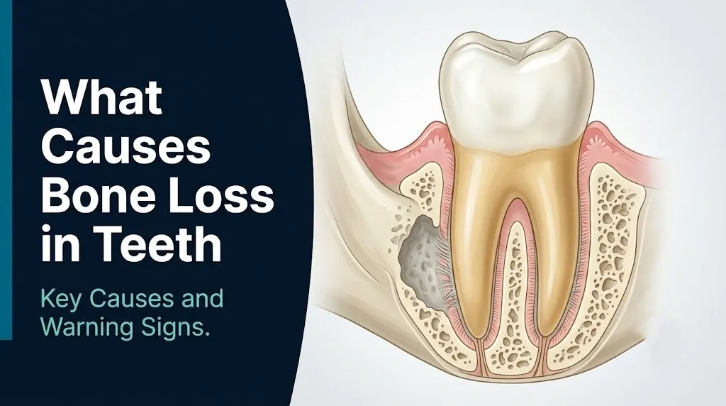
Both types produce localized areas of bone destruction visible on periapical radiographs as circular or ovoid radiolucencies (dark areas) around the root tip or lateral root surface. According to the Mayo Clinic, dental abscesses do not resolve without treatment and will progressively expand the zone of bone destruction as the infection continues to spread.
The critical warning for anyone with a known or suspected dental abscess: untreated abscesses can spread from the alveolar bone into the fascial spaces of the head and neck. This spreading infection can cause Ludwig’s angina (a bilateral infection of the floor of the mouth that can compress the airway) or progress inferiorly into the chest, causing descending necrotizing mediastinitis. Both are life-threatening conditions requiring emergency surgical intervention.
Individuals with diabetes are at elevated risk for rapidly spreading dental infections because hyperglycemia impairs neutrophil function, reducing the immune system’s ability to contain bacterial invasion at the site. A person with poorly controlled diabetes who develops facial swelling from a dental source must be evaluated by an emergency medicine physician the same day.
Smoking and Bone Loss Around Teeth
Smoking causes bone loss around teeth by creating an anaerobic oral microenvironment that selectively favors the growth of the same gram-negative anaerobic pathogens that drive periodontitis, while simultaneously impairing the vascular and immune responses the body needs to control the resulting infection.
Tobacco smoke reduces oxygen tension in the gingival sulcus and periodontal pocket, creating conditions that actively benefit Porphyromonas gingivalis and other obligate anaerobes. Research published in the Journal of Dental Research has confirmed that smokers harbor significantly higher proportions of the red complex periodontal pathogens in their subgingival plaque than non-smokers at equivalent levels of oral hygiene.
The vascular effects compound the microbiological problem. Nicotine causes sustained vasoconstriction of the small blood vessels supplying the gingiva, which reduces the redness and bleeding that normally signal gum inflammation to a patient and a clinician. Smokers’ gums often appear deceptively healthy while harboring deep periodontal pockets with significant bone loss underneath. This masking effect means that smoking-related periodontal disease is frequently diagnosed at a more advanced stage than in non-smokers because the obvious warning signs are suppressed.
According to the American Academy of Periodontology (AAP), smokers are two to three times more likely to develop periodontitis than non-smokers, and the bone loss associated with smoking-related periodontitis tends to be more severe and more diffuse across the mouth than in non-smokers with equivalent plaque levels. Periodontal treatment also responds less predictably in active smokers because the impaired vasculature limits the healing response in the gingival and periodontal tissues.
Former smokers who have quit for more than five years show substantially improved periodontal treatment outcomes compared to active smokers, supporting smoking cessation as a clinically important adjunct to any periodontal management plan.
Key Takeaway: Smoking both accelerates periodontitis-driven bone loss and masks the clinical warning signs (gum redness and bleeding) that would normally prompt earlier evaluation, which is why smokers should have periodontal probing depth assessments at every dental visit regardless of how their gums appear visually.
Diabetes and Dental Bone Loss
Diabetes causes and accelerates bone loss in teeth through two distinct pathways: impaired neutrophil function that reduces the immune system’s ability to control periodontal bacterial infection, and the accumulation of advanced glycation end-products (AGEs) in periodontal tissues that amplifies the inflammatory cytokine response driving alveolar bone destruction.
In individuals with poorly controlled type 2 diabetes, chronically elevated blood glucose levels cause glucose to react non-enzymatically with proteins and lipids in the body’s tissues, forming AGEs. In the periodontal ligament and alveolar bone, AGE accumulation increases the expression of pro-inflammatory cytokines including TNF-alpha and IL-6 by binding to specific cell surface receptors (RAGE, receptor for AGEs). This amplified cytokine environment accelerates the RANKL-driven osteoclast activation already described in the periodontitis section, producing faster and more severe bone loss at equivalent levels of bacterial infection.
The Journal of Clinical Periodontology has published research establishing a bidirectional relationship between diabetes and periodontitis: not only does poorly controlled diabetes worsen periodontal bone loss, but active periodontitis independently worsens glycemic control by increasing systemic inflammatory load, elevating insulin resistance. This bidirectional relationship means that treating periodontal disease can produce measurable improvements in HbA1c levels, with some meta-analyses showing reductions of approximately 0.4% in HbA1c following intensive periodontal treatment.
Individuals with a glycated hemoglobin (HbA1c) level above 7% have substantially higher periodontal pocket depths and more severe bone loss than those with HbA1c below 7%, even with identical oral hygiene practices. A person with type 2 diabetes being treated by an endocrinologist should have their periodontal status assessed annually by a general dentist or periodontist, and any periodontitis diagnosed should be communicated to their endocrinologist as a contributing factor to glycemic instability.
Osteoporosis and Bone Loss in Teeth
Osteoporosis causes dental bone loss by reducing the overall mineral density of the jaw bones, making the alveolar bone more susceptible to the bacterial-driven resorption of periodontitis and reducing the baseline bone volume available to support tooth roots and dental implants.
Osteoporosis is defined by a bone mineral density T-score of -2.5 or lower at the femoral neck or lumbar spine. The jaw bones, while not routinely included in standard dual-energy X-ray absorptiometry (DXA) screening, reflect the same systemic reduction in bone density that affects the axial and appendicular skeleton. Research published in the Journal of Dental Research has found an association between low systemic bone mineral density and reduced alveolar bone height and density on dental radiographs, particularly in postmenopausal women.
The mechanism involves the same osteoclast/osteoblast imbalance as systemic osteoporosis. When estrogen levels decline after menopause, estrogen’s inhibitory effect on osteoclast differentiation and activity is reduced. More osteoclasts become active, and bone resorption outpaces bone formation throughout the skeleton, including in the jaw. In the oral environment, this systemic predisposition to bone loss is compounded by any concurrent periodontal bacterial infection, meaning that postmenopausal women with untreated periodontitis experience a synergistic acceleration of alveolar bone loss from two simultaneous pathways.
Women with known osteoporosis who are being treated by an endocrinologist or rheumatologist should inform their dentist and periodontist, because their jaw bone density may be lower than typical for their age, affecting prognosis for periodontal treatment and implant placement. Conversely, a dentist who notices unexpectedly severe bone loss on dental radiographs in a postmenopausal woman with minimal periodontal disease history should raise the possibility of systemic osteoporosis with the patient’s primary care physician.
Key Takeaway: Osteoporosis does not cause dental bone loss independently, but it lowers the starting bone density in the jaw, meaning any periodontitis that develops in a patient with osteoporosis produces more severe and faster bone loss than the same periodontal disease would in someone with normal bone density.
Medications That Cause Bone Loss in Teeth
Several widely prescribed medications cause or accelerate bone loss in teeth through distinct mechanisms, and patients taking these drugs require modified dental management that their prescribing physician and dentist must coordinate before any invasive dental procedure.
Bisphosphonates (including alendronate, risedronate, zoledronic acid, and pamidronate) are used to treat osteoporosis, Paget’s disease, and bone metastases from cancer. Bisphosphonates work by binding to hydroxyapatite in bone and being internalized by osteoclasts, inhibiting their resorptive function. In the jaw, this means that osteoclast-mediated bone repair after tooth extraction, implant placement, or other trauma is severely impaired. The result is a necrotic zone of non-healing bone called medication-related osteonecrosis of the jaw (MRONJ), which can present as exposed, non-healing bone in the mouth that persists for more than eight weeks.
According to the American Association of Oral and Maxillofacial Surgeons (AAOMS) position paper on MRONJ, risk is highest with intravenous bisphosphonates (zoledronic acid, pamidronate) used in oncology settings, but oral bisphosphonates used for osteoporosis also carry risk, particularly after more than four years of use. Any patient taking bisphosphonates should inform their dentist before any tooth extraction or implant surgery is planned.
Corticosteroids such as prednisone, used for inflammatory and autoimmune conditions, reduce osteoblast activity and increase osteoclast activity throughout the body. Prolonged oral corticosteroid use at doses above 5 mg/day of prednisone equivalent suppresses bone formation, accelerating bone loss in the jaw as in the rest of the skeleton.
Antiepileptic drugs, particularly phenytoin (Dilantin), cause gingival hyperplasia, an overgrowth of gingival tissue that creates false periodontal pockets, traps bacterial plaque, and promotes periodontal infection and secondary bone loss. Calcium channel blockers (nifedipine, amlodipine) used for hypertension produce a similar pattern of gingival overgrowth in a subset of patients.
Patients on any of these medications should disclose their full medication list at every dental appointment, including the dose and duration of use.
Bone Loss Around Dental Implants and Peri-Implantitis
Peri-implantitis causes bone loss around dental implants through a biofilm-driven inflammatory mechanism that is biologically similar to periodontitis around natural teeth but progresses more aggressively because implants lack the protective biological seal provided by the junctional epithelium and periodontal ligament of natural teeth.
Unlike a natural tooth root, which is covered by living cementum and connected to alveolar bone by PDL fibers, a dental implant integrates directly with bone (osseointegration) and is covered by the gingiva without the same sulcular attachment. This structural difference means that once bacterial biofilm establishes at the implant-gingiva interface, the bacteria have a more direct path to the bone surface and the immune defense mechanisms are less anatomically organized to contain the infection.
The American Academy of Periodontology defines peri-implantitis as an inflammatory condition affecting the soft and hard tissues around an osseointegrated implant, characterized by bleeding on probing, increased pocket depth, and progressive bone loss beyond the initial bone remodeling expected after implant placement. The prevalence of peri-implantitis is estimated between 22 and 65% of implant patients in various studies reviewed by the AAP, varying by patient risk factors, implant design, and maintenance protocol.
Risk factors that make peri-implantitis more likely:
- Prior or current periodontitis (the highest single risk factor, as the same bacterial community and host response that destroyed natural tooth bone can establish around implants)
- Poor oral hygiene around implant crowns
- Active cigarette smoking
- Poorly controlled diabetes
- Infrequent professional implant maintenance visits (recommended at least twice yearly)
- Implant placement in a site with inadequate bone volume requiring significant grafting
Any person with dental implants who notices bleeding, swelling, pain, or a sensation of looseness around an implant should contact the placing periodontist or implant surgeon promptly for evaluation.
Key Takeaway: Patients with a history of periodontal bone loss around natural teeth have the highest risk of peri-implantitis around dental implants, because the same bacteria and immune dysfunction that destroyed natural tooth bone will affect implants in the same oral environment without rigorous maintenance and bacterial control.
Bone Loss in Teeth in Women and Hormonal Influences
Bone loss in teeth in women is influenced by estrogen at multiple life stages, including puberty, pregnancy, and particularly menopause, because estrogen receptors on osteoblasts and immune cells in the periodontal tissues respond to estrogen fluctuations in ways that modify both bone density and the inflammatory response to periodontal bacteria.
During pregnancy, elevated progesterone and estrogen levels alter the immune response in the gingiva, promoting an exaggerated inflammatory reaction to dental plaque. This produces pregnancy gingivitis in a substantial proportion of pregnant women, a reversible condition that, if accompanied by pre-existing periodontal disease, can accelerate bone loss during the pregnancy. Research published in Obstetrics and Gynecology has found an association between severe maternal periodontitis and adverse pregnancy outcomes including preterm birth and low birth weight, though the causal relationship remains under active research.
After menopause, the decline in circulating estrogen reduces its inhibitory effect on osteoclast differentiation in both the systemic skeleton and the jaw. The National Institute of Dental and Craniofacial Research reports that postmenopausal women show higher rates of tooth loss than premenopausal women of comparable oral hygiene status, and research has found an association between postmenopausal osteoporosis and radiographic alveolar bone loss in this population.
Hormone replacement therapy (HRT) has been associated in observational studies with reduced tooth loss and better alveolar bone maintenance in postmenopausal women, though the decision to use HRT involves many factors beyond dental health and must be discussed with a gynecologist or endocrinologist, not determined based on dental considerations alone.
Pregnant women with any history of periodontal disease or gum bleeding should have a periodontal evaluation in their second trimester, as periodontal treatment during the second trimester is considered safe and clinically indicated to reduce the risk of disease progression during pregnancy.
Aggressive Periodontitis and Rapid Bone Loss in Younger Adults
Aggressive periodontitis, now classified as Stage III or Stage IV, Grade C periodontitis under the 2017 classification by the American Academy of Periodontology and the European Federation of Periodontology, causes rapid and severe alveolar bone loss in patients who are otherwise systemically healthy and often younger than 35 years of age.
The defining clinical feature is bone loss that is disproportionately severe relative to the amount of plaque and calculus (tartar) present. A 25-year-old with moderate plaque and radiographic bone loss of 50% of the root length has a presentation that does not fit a standard periodontitis progression timeline and strongly suggests Grade C disease. The underlying pathophysiology involves an exaggerated monocyte and macrophage response to bacterial LPS, producing significantly higher levels of IL-1 beta and TNF-alpha per unit of bacterial stimulus than a typical immune response. Some individuals with Grade C periodontitis also show neutrophil chemotaxis defects that reduce the effectiveness of neutrophil-mediated bacterial clearance in the periodontal pocket.
Family clustering is well recognized. Research published in the Journal of Dental Research has found that first-degree relatives of individuals with Grade C periodontitis have higher rates of the condition than the general population, supporting a genetic component to the heightened immune reactivity. Specific genetic polymorphisms in the IL-1A and IL-1B gene cluster have been associated with increased periodontitis susceptibility in several study populations.
Individuals under 35 with any of the following should be evaluated by a board-certified periodontist without delay:
- More than one tooth with deep periodontal pockets (greater than 5 mm) in the absence of heavy calculus
- Unexplained tooth mobility in otherwise healthy permanent teeth
- Rapid gum recession affecting multiple teeth over months rather than years
- A parent or sibling with early tooth loss or severe gum disease
Bone Loss in Teeth Without Obvious Gum Disease
Bone loss in teeth can occur without the classic signs of gum disease such as red, swollen, or bleeding gums, particularly in smokers (whose vasoconstriction suppresses visible gum inflammation), in patients with certain systemic conditions, and through non-periodontal mechanisms including occlusal trauma and localized infection.
This is the diagnostic blind spot that most health content on this topic fails to address. A person can have Grade B or Grade C periodontitis with measurable bone loss on radiographs and periodontal probing depths of 5 to 7 mm while their gums appear completely pink and non-bleeding at a casual visual inspection. This is particularly true in smokers, as described earlier, but also in patients who perform fastidious oral hygiene that reduces surface gingival inflammation while a deeper anaerobic infection continues below the level that toothbrushes and floss can reach.
Occlusal trauma is a non-bacterial cause of bone loss that occurs when excessive biting forces, from teeth grinding (bruxism), clenching, or a poorly fitted dental restoration, are transmitted through the PDL to the alveolar bone at levels that exceed the bone’s remodeling capacity. The result is a specific pattern of bone loss called angular or vertical bone defects, where bone loss appears on one side of a tooth root rather than evenly across the crest of the alveolar ridge. Occlusal trauma does not initiate periodontitis, but it accelerates bone loss in teeth that are already affected by periodontal infection.
Dental trauma from a prior injury can cause bone loss years after the original event through root resorption, where the body’s immune system treats damaged cementum on a previously traumatized root as foreign material and resorbs the root structure and adjacent bone. This can occur with no gum disease whatsoever.
Any person who has been told they have bone loss on a dental X-ray without a clear periodontal disease diagnosis should ask their dentist specifically about probing depths at each tooth, the pattern of bone loss (horizontal versus vertical), and whether occlusal trauma or a prior dental injury could be contributing.
Symptoms and Signs of Bone Loss in Teeth
The symptoms of bone loss in teeth are often absent or subtle until the condition is advanced, which is why dental radiographs taken during routine dental examinations are the primary tool for detecting bone loss before it becomes severe enough to cause tooth mobility or spontaneous pain.
When symptoms do appear, they reflect the progressive loss of supporting structure around the tooth root:
- Gum recession (the gum line appears to move downward or upward from the tooth, exposing root surface that was previously covered)
- Teeth that appear longer than they previously did, a direct visual consequence of gum recession and underlying bone loss
- Tooth sensitivity, particularly to cold, caused by exposed root dentin on teeth where the insulating gum and bone support has receded
- Tooth mobility or looseness, occurring when bone loss has reduced the socket support to the point where the PDL can no longer keep the tooth firmly positioned
- A change in bite or the way upper and lower teeth fit together, caused by drift or tipping of teeth that have lost their bony support
- Persistent bad breath or a bad taste in the mouth, produced by the anaerobic bacterial communities in deep periodontal pockets
- Visible pus between the gum and tooth when the gum is pressed, indicating an active periodontal abscess
- Pain when biting or chewing, occurring when a tooth with reduced bone support transmits forces to sensitized PDL fibers
Bone loss detected on dental X-rays before any of these symptoms develop is the best clinical outcome because it allows treatment before irreversible structural damage has reached a critical threshold. According to the American Academy of Periodontology, adults without known risk factors should have periodontal assessments (including full-mouth probing depths) at every dental cleaning, with radiographic bone assessment at intervals determined by their individual risk status.
How Dental Bone Loss Is Diagnosed
Dental bone loss is diagnosed through a combination of clinical periodontal probing, dental radiographs, and in advanced cases, cone beam computed tomography (CBCT), with each tool providing distinct and complementary information about the location, severity, and pattern of bone loss.
The diagnostic process typically follows this sequence when a dentist suspects bone loss:
- Full-mouth periodontal probing: A calibrated probe is inserted into the gingival sulcus at six points around each tooth and the depth in millimeters is recorded. Normal depth is 1 to 3 mm. Depths of 4 mm or greater indicate a periodontal pocket, and depths of 6 mm or more indicate advanced disease with probable bone loss. The pattern of depths (generalized vs. localized, deep vs. shallow) guides the radiographic examination.
- Bleeding on probing assessment: Bleeding when the probe is inserted indicates active gingival inflammation. Sites that bleed on probing are more likely to be losing attachment and bone than non-bleeding sites.
- Periapical radiographs: These small, detailed X-rays of individual teeth or groups of teeth show the bone level around each root relative to the cementoenamel junction (CEJ), the visible reference point on the tooth. Bone loss is measured as the distance from the CEJ to the current bone crest. Normal bone height is within 1 to 2 mm of the CEJ.
- Panoramic radiograph (OPG): A full-mouth overview X-ray that provides a broader picture of bone level across all teeth. Less detailed than periapical films but useful for identifying gross bone loss patterns and pathology.
- Cone beam computed tomography (CBCT): A three-dimensional imaging modality that shows bone architecture in all planes, including buccal and lingual bone that standard 2D radiographs cannot capture. Used for complex cases, implant planning, and situations where the bone loss pattern is unclear on 2D films.
- Bone sounding: A procedure performed under local anesthesia in which the probe penetrates through the gingiva to directly contact the alveolar bone crest, providing the most precise measurement of bone level at a specific site.
Patients scheduled for a periodontal evaluation should bring a list of all current medications, their complete medical history (including any systemic conditions like diabetes, osteoporosis, or cancer), and any prior dental radiographs if they are visiting a new dental provider.
When Dental Bone Loss Becomes a Dental Emergency
Certain presentations of dental bone loss and dental infection require immediate emergency evaluation. Do not wait to see if these resolve on their own.
Call 911 or go to the nearest emergency room immediately if you experience:
- Neck swelling or swelling under the chin or floor of the mouth accompanied by any dental pain or known dental infection (this can indicate Ludwig’s angina, a life-threatening bilateral infection of the floor-of-mouth fascial spaces that can compress and close the airway within hours)
- Difficulty swallowing, difficulty opening the mouth (trismus), or a sensation that the throat is closing, in the setting of any dental pain or known dental abscess (these are signs of fascial space infection spreading toward the airway)
- Difficulty breathing associated with facial or neck swelling from a dental source
- High fever (above 39 degrees Celsius, 102.2 degrees Fahrenheit) with severe dental pain and facial swelling (indicates systemic spread of dental infection requiring intravenous antibiotic therapy and possible surgical drainage)
- Exposed, non-healing bone visible in the mouth that has been present for more than eight weeks in a patient currently taking or who has previously taken bisphosphonate medications (meets the clinical criteria for MRONJ requiring evaluation by a board-certified oral and maxillofacial surgeon)
- Rapidly spreading redness or swelling across the face, jaw, or neck following any dental procedure or known dental infection
These presentations indicate life-threatening infection with potential airway compromise, sepsis, or mediastinal spread, and all require emergency surgical and medical assessment, not an urgent care appointment or a call to a dental office the following morning.
Frequently Asked Questions About Bone Loss in Teeth
Can bone loss in teeth be reversed or stopped?
Established bone loss in teeth cannot be reversed by the body on its own, but further bone loss can be stopped with professional periodontal treatment that controls the bacterial infection driving the destruction.
Bone regeneration procedures including guided bone regeneration (GBR) with bone graft materials can restore some lost bone volume in selected cases, but the outcome depends on defect geometry, overall health, and smoking status.
Any person with diagnosed dental bone loss should be evaluated by a periodontist to determine whether their specific pattern of bone loss is amenable to regenerative treatment or requires stabilization through debridement alone.
What does bone loss around teeth feel like?
Dental bone loss typically causes no pain in its early and middle stages, which is why it is often discovered on dental X-rays before a patient has noticed any symptoms.
When symptoms do develop, they include tooth sensitivity (particularly to cold air and cold liquids on exposed root surfaces), a sense that teeth look longer, and eventually tooth looseness or a change in how teeth fit together when biting.
Pain from a dental abscess causing localized bone destruction can be sharp, throbbing, and constant, particularly when biting, and is distinctly different from the silent progression of chronic periodontitis-related bone loss.
How fast does dental bone loss progress?
Bone loss from chronic periodontitis typically progresses at approximately 0.1 to 0.3 mm per year in untreated moderate disease, though Grade C (aggressive) periodontitis can produce several millimeters of bone loss within a single year.
The rate depends on the specific bacteria present, the individual’s immune response, smoking status, glycemic control in diabetics, and whether professional treatment is reducing the bacterial load.
A dentist or periodontist can estimate the rate of bone loss by comparing current radiographs to prior films taken at a known time interval, which is one reason maintaining a consistent dental record over years is clinically useful.
Can you have bone loss in teeth without gum disease?
Dental bone loss can occur without the classic visible signs of gum disease, particularly in smokers whose vasoconstriction suppresses gum redness and bleeding while deep infection continues below the gum line.
Non-periodontal causes including dental abscess, occlusal trauma from bruxism, dental trauma with root resorption, and medication-related osteonecrosis can all produce bone loss in the absence of widespread gum disease.
Any unexplained bone loss on dental radiographs warrants a complete periodontal probing assessment, an occlusal evaluation, a medication review, and if needed, referral to a periodontist or oral surgeon for further assessment.
What happens if dental bone loss is left untreated?
Untreated dental bone loss leads to progressive tooth mobility, eventual tooth loss, and in the case of dental abscess or infection, potential spread of infection into the fascial spaces of the head and neck.
Chronic periodontitis left untreated for years can result in loss of multiple teeth and loss of sufficient bone volume to support dental implants without extensive grafting procedures.
Research published in the Journal of Clinical Periodontology has confirmed that the systemic inflammatory burden of untreated periodontitis is associated with worsened glycemic control in diabetics and has been studied as a possible contributing factor in cardiovascular disease risk, though causal relationships in the latter remain under investigation.
What type of dentist treats bone loss in teeth?
A board-certified periodontist is the dental specialist trained specifically to diagnose and treat periodontal bone loss, peri-implantitis, and conditions affecting the bone and soft tissue supporting teeth.
A general dentist manages early-stage periodontal disease (Stage I and Stage II) and should refer to a periodontist when probing depths exceed 5 mm, when radiographic bone loss is present, or when the response to initial treatment is inadequate after 8 to 12 weeks.
For bone loss caused by dental abscess requiring surgery, extraction with socket preservation, or MRONJ, a board-certified oral and maxillofacial surgeon is the appropriate specialist.
Protecting What Cannot Be Rebuilt
The defining clinical reality about dental bone loss is that the bone destroyed by periodontitis, dental abscess, or any other cause cannot be fully regenerated by the body alone. What can be restored through professional care is the conditions that allow the remaining bone to stay stable and, in selected cases, partially rebuilt through surgical procedures.
The most practical step for anyone concerned about bone loss is a dental appointment that includes full-mouth periodontal probing and updated radiographs. That appointment will tell you more in 30 minutes than any amount of home observation. If the assessment finds bone loss, ask specifically about the stage and grade of any periodontitis, whether a periodontist referral is indicated, and what the radiographic progression rate has been compared to prior films.
Bone lost to periodontal disease is gone, but bone loss that is caught and treated does not have to continue. That distinction is what makes early professional evaluation the single most actionable thing this article can point you toward.